Introduction
Atherosclerosis is characterized by atherosclerotic
plaques that result from inflammatory cell infiltration, lipid
peroxidation, extracellular matrix deposition and other factors
(1). Moreover, atherosclerosis
remains a major cause of long-term morbidity and mortality
worldwide as it is a prerequisite to a large number of
cardiovascular diseases (CVDs) (2); for example, the prevalence of CVDs
was predicted to reach 43.9% in the United States in 2030 (3), and in 2015 alone, 17.7 million people
died from CVD worldwide (4). While
a coronary artery bypass graft is widely performed for the
revascularization of occluded vessels, it cannot be used to treat
the damage caused by atherosclerosis (5).
Endothelial cell dysfunction is considered the main
cause of atherosclerosis (6).
Endothelial cell dysfunction is a complex process initiated by
phenotypic changes in endothelial cells that allows for the
permeation of lipoproteins into subendothelial cells, as well as
their physicochemical modification and trapping by macrophages,
which ultimately become the foam cells that constitute the
highly-thrombotic necrotic core of atherosclerosis plaque (7–9).
Normal endothelial structure and function are important for
maintaining vascular homeostasis. Moreover, a central feature of
endothelial cell dysfunction is a reduced bioavailability of nitric
oxide, which leads to abnormal physiological responses of the
endothelial cells to stimuli, altered metabolism, oxidative stress
and damage, and the recruitment of immune cells that accelerate
atherosclerosis (8–10). Alterations in hemodynamics, immune
responses and interventional therapy may also result in endothelial
damage, dysfunction or death. Furthermore, risk factors for
atherosclerosis, such as hypertension, alcoholism, smoking and
diabetes, alter the function of endothelial cells (11).
Endothelial progenitor cells (EPCs) are relevant to
the prognosis of cardiovascular diseases. Previous research has
shown that EPCs play a vital role in the occurrence and development
of atherosclerosis (12). Clinical
trials and experimental studies have suggested that EPCs can form
functional blood vessels in vivo (13–15),
thus providing important cellular resources for the therapy of
cardiovascular diseases via their direct involvement in
angiogenesis and secretion of protective paracrine factors
(16–19). EPC transplantation has also
achieved positive results in the treatment of acute lung injury
(20), cerebral ischemia (21), acute renal ischemia-reperfusion
injury (22) and aneurysm
(23) in animal models, suggesting
that EPCs may have a clinical application in atherosclerosis.
Cell transplantation is one of the most widely
studied biological approaches for improving atherosclerosis and
other vascular diseases, with good clinical application prospects
(24). Previous studies have shown
that vascular endothelial repair is achieved via the migration and
proliferation of adjacent endothelial cells (25–27).
Moreover, Asahara et al (28) found that EPCs are bone
marrow-derived CD34+ cells, which can differentiate and
proliferate into mature endothelial cells, thus constituting an
essential part of the vascular system. Atherosclerosis is a disease
caused by an imbalance between vascular endothelial injury and
repair (29). Previous studies
have revealed that the bone marrow, vascular wall, adipose tissue,
spleen, liver and intestine can release EPCs (30–32).
Furthermore, EPCs can be specifically targeted to the site of
endothelial injury, participate in the repair of damaged vascular
endothelium and promote angiogenesis in ischemic tissues (33). However, transplantation of
autologous EPCs still has several limitations, including a limited
supply of expanded EPCs, impaired function, activity of
transplanted cells and low survival rate of transplanted cells in
an ischemic host environment (34,35).
In addition, EPCs in the bone marrow, peripheral blood and
umbilical cord blood are highly immunogenic and can cause rejection
(36–38). Currently, EPCs can only be used for
autologous transplantation (39,40).
Furthermore, factors such as coronary heart disease, hypertension,
diabetes mellitus, emphysema, acute lung injury, hyperlipidemia,
liver fibrosis, systemic sclerosis, old age and long-term smoking,
can decrease the number and function of autologous EPCs, which
results in a reduced proportion of injected cells that successfully
accumulate at the sites of vascular damage (41). Therefore, it is important to
develop novel pro-angiogenic strategies to improve the efficacy of
EPC transplantation.
Ultrasmall superparamagnetic iron oxide
nanoparticles (USPIONs) with diameters <50 nm have an iron oxide
core that is stabilized by a monomer or polymer coating (42–44).
USPIONs possess a highly reactive surface, uniform particle size
distribution, beneficial suspension properties and the possibility
of additional coating modification by conjugation with a drug
(42–44). Moreover, USPIONs are being
developed for cell processing (45), automated DNA extraction (46), detection of pathogens (47), drug delivery and neuroimaging. In
addition, USPIONs are also being developed for imaging of tumors
and metastases in the liver, spleen and bone marrow, and perfusion
imaging of atherosclerotic plaques and thrombosis (42,43,48–50).
While MRI technology, EPC-mediated therapeutic
angiogenesis and vascular repair have advanced, the mechanisms of
migration, adhesion, proliferation and angiogenic properties of
EPCs remain unknown. Thus, identifying materials for the labeling
of live cells is important for target tracing of living cells and
promotion of tissue angiogenesis. To the best of our knowledge, no
previous studies have examined USPION labeling of rabbit EPCs by
MRI. Therefore, in the present study, labeled EPCs were
transplanted into a rabbit arteriosclerosis model, and MRI was used
to assess the effect of EPC transplantation and to examine the
application of nanoparticles in vivo.
Materials and methods
Cell culture
Rabbit EPCs were extracted according to a previous
report (51) using 40 rabbits
(age, 2–3 months; sex, 20 males and 20 females; weight, 2.0-2.5 kg;
Model Animal Research Center of Nanjing University). After mixing 5
ml rabbit anti-coagulated whole blood with sterile PBS (1:1 ratio),
5 ml rabbit lymphocyte separation medium (Tianjin HaoYang
Biological Manufacture, Co., Ltd.) was added to 5 ml of the above
mixture (1:1 ratio). Density gradient centrifugation was performed
at 367 × g for 20 min at room temperature. The intermediate
albuginea layer was extracted and washed with PBS three times, and
the M199 medium (Prospec-Tany Techno Gene Ltd.) was added to count
the suspended cells. Mononuclear cells at 5×10⁶/ml were
seeded on human fibronectin-coated (5 g/cm2; BD
Biosciences) culture dishes. EPCs were cultured in M199 culture
medium containing 20% fetal calf serum (FCS; Gibco; Thermo Fisher
Scientific, Inc.), 8 ng/ml vascular endothelial growth factor
(VEGF; Prospec-Tany Techno Gene Ltd.) and 8 ng/ml basic fibroblast
growth factor (bFGF; Prospec-Tany Techno Gene Ltd.) at 37°C with 5%
CO2. After 48 h, non-adherent cells were removed, and
the culture medium was changed every day to assess the morphology
of the cells.
Flow cytometry
On the 10th day of culture, adherent cells were
collected and adjusted to 1×10⁶/ml with PBS. The
proportions of cells positive for CD133, CD31, CD34 and vascular
endothelial growth factor receptor 2 (VEGFR2) were determined by
flow cytometry. Briefly, the cells were blocked with rabbit serum
(cat. no. SL034; Beijing Solarbio Science & Technology Co.,
Ltd.) at 37°C for 30 min. Cells were washed with PBS and incubated
with FITC-CD34 (1:100; cat. no. bs-0646R-FITC; BIOSS), PE-CD133
(1:100; cat. no. bs-0395R-PE; Beijing Biosynthesis Biotechnology,
Co., Ltd.), FITC-CD31 (1:100; cat. no. bs-0195R-FITC; Beijing
Biosynthesis Biotechnology, Co., Ltd.) and PE-VEGF receptor 2
(1:100; cat. no. bs-10412R-PE; Beijing Biosynthesis Biotechnology,
Co., Ltd.) primary antibodies at 4°C for 20 min.
Rabbit auricular whole blood (1 ml) was collected
and mixed with 2 ml erythrocyte lysate (BD Biosciences). The
mixture was placed at room temperature for 5 min and centrifuged at
367 × g for 20 min at room temperature. After discarding the
supernatant, the cell pellets were adjusted to 1×10⁶/ml
with PBS. CD34+ and VEGFR+ circulating EPCs
were detected by flow cytometry. An isotype-matched antibody
(1:100; cat. no. bs-0295R-FITC; Beijing Biosynthesis Biotechnology,
Co., Ltd.) was used as the control. Samples were analyzed on a
fluorescence-activated cell sorting instrument (FACSCalibur; BD
Biosciences) and Cell Quest v6.0.1 software (BD Biosciences).
Angiogenesis
Matrigel (BD Biosciences; 500 µl) was added into 4
ml M199 medium. Adherent cells at 1×10⁶/ml were
collected and seeded on Matrigel-coated plates. After incubation at
37°C for 24, 48 and 72 h, angiogenesis was observed under a light
microscope (magnification, ×20; Carl Zeiss AG).
Cell labeling with USPION and PLL
Polylysine (PLL; 0.5 ml; Sangon Biotech Co., Ltd.)
was diluted 10 times with sterilized water. USPIONs (Taiwan
Advanced Nanotech, Inc.; 0.2 ml) were prepared at a final
concentration of 1 µg/µl. The diameters of the USPION particles and
USPION particles conjugated with PLL (USPION-PLL) were measured by
scanning electron microscopy (magnification, ×40; JSM-7800 Prime;
JEOL, Ltd.).
USPION concentrations for labeling were 0, 1, 2, 4,
8, 16, 32, 64, 128 and 256 µg/ml. Sterile cover slides (diameter,
115 mm) were placed in 24-well culture plates (Corning, Inc.).
After 12 h of incubation at 37°C of different USPION concentrations
with 2×10⁷/ml EPCs in 24-well culture plates, the
unlabeled USPIONs were removed by washing three times with PBS.
Then, M199 medium supplemented with 20% FCS was added, and the
cells were cultured at 37°C for 12 h. Cells in the control group
were cultured in M199 medium at 37°C for 12 h. Cells were incubated
with 0.4% Trypan blue for 2–3 min at room temperature to detect the
activity of labeled cells and determine the optimal USPION
concentration. Cover slides placed in the dish were stained with
Prussian blue (40 g/l; cat. no. BP-DL122; Nanjing SenBeiJia
Biological Technology Co., Ltd.) for 30 min at room temperature
primarily, and then another 15 min for secondary staining. Labeling
efficiency was observed under a confocal microscope (magnification,
×20; Carl ZeissAG) with image acquisition.
Cell viability
An MTT assay was used to assess the proliferative
activity of EPCs after labeling. A total of 1×10⁵ EPCs
per well were seeded into 96-well plates. Then, 6-wells in the
optimal labeling concentration and unlabeled groups were assessed
at 1, 2, 3, 4, 5, 6 and 7 days. After incubation with 20 µl MTT
solution at 37°C (Nanjing KeyGen Biotech Co., Ltd.; 5 g/l) for 4 h,
the culture medium was discarded and 100 µl DMSO was added into
each well. Formazan crystals were dissolved by shaking for 10 min.
Absorption was measured on a microplate reader at 490 nm (Bio-Rad
Laboratories, Inc.).
Cell adhesion
Adherent labeled EPCs cultured for 3 days were
collected and suspended in M199 medium. Then, 500 cells/well were
added into 24-well plates. Adherent cells were cultured in M199
medium containing 20% FCS (0.5 ml) in an incubator with 5%
CO2 at 37°C for 1 h. In total, 20 visual fields were
randomly selected for cell counting using a light microscope
(magnification, ×20).
Scanning electron microscopy
EPCs were labeled with USPIONs at the optimal
labeling concentration for 24 h and fixed with 2.5% neutral
glutaraldehyde solution. Scissors was used to remove 1–3 muscle
blocks with a cross-section of ~1×1 mm. Extracted muscle blocks
were attached to glass slides to make them stretch. The slides were
then fixed with 2.5% neutral glutaraldehyde solution (10 ml 25%
glutaraldehyde solution; 50 ml 0.2 M phosphate buffer; 40 ml
distilled water) at 4°C for >15 min. Electron microscopy was
performed using a JSM-7800 Prime microscope (magnification, ×200;
JEOL, Ltd.). Elemental analysis of labeled cells was performed on
an energy dispersive spectrometer (JEOL, Ltd.). Data were analyzed
using the Gatan Microscopy Suite software version 2.11 (Gatan,
Inc.; Thermo Fisher Scientific, Inc.).
MRI
Labeled cells (2×10⁶) in the experimental
and control groups were collected and diluted in 0.5% agarose to
1×10⁶ cells/ml. T2 map scanning was performed by MRI
(1.5 T; Philips Medical Systems B.V) with an 8-channel head
circular. The imaging parameters were: Field-of-view (FOV), 9.0 mm;
echo time (TE), 105–1,500 ms; repetition time (TR), 2,300 ms; echo
number, 32 and number of signals averaged (NSA)=1. The final T2
value was the average of three central T2 values in each test
tube.
Atherosclerosis rabbit model
In total, 40 rabbits (Model Animal Research Center
of Nanjing University; age, 2–3 months; sex, 20 males and 20
females; weight, 2.0-2.5 kg) were divided into the model and
control groups, with 20 animals per group. The animals were housed
at 20–25°C and 40–60% humidity, under a 12-h light/dark cycle, with
free access to food and water. The rabbits in the model and control
groups were fed high-fat (18% extra fat vs. the standard diet) and
standard diets for 12 weeks, respectively. In the 6th and 12th
weeks, 2 ml rabbit auricular venous blood was collected from the
ear margin vein and placed at room temperature for 30 min. Blood
was sampled from an ear margin vein. Serum was extracted by
centrifugation at 200 × g for 10 min at 4°C for the measurement of
total cholesterol (TC; cat no. RJ21458), triglycerides (TG; cat.
no. RJ21057), low-density lipoproteins (LDL; cat. no. RJ21040) and
high-density lipoproteins (HDL; cat. no. RJ21068) using ELISAs
(Shanghai Renjie Biotechnology Co., Ltd.).
After 12 weeks, the model and control groups were
anesthetized by intramuscular injection with 5 mg/kg Lumianning
(China Animal Husbandry Animal Health Products Co., Ltd.) and an
intravenous injection of 15 mg/kg propofol and scanned using CT and
MRI with the animals in the supine position. Vascular lesions were
located based on CT scan results. The vessels in the lesion area
were separated in the model group. The rabbits were euthanized by
intramuscular injection with 16 mg/kg Lumianning and intravenous
injection with 80 mg/kg propofol; the rabbits died immediately due
to cardiac blood conduction and respiratory inhibition, and the
death was painless as xylazine (one of the main ingredients of
Lumianning alongside dihydroetorphine hydrochloride) is an
anesthetic (52–56). In the control group, the vessels in
the corresponding area were fixed with 3% formaldehyde at 4°C for
24 h, embedded in paraffin and sectioned into 5-µm sections.
Hematoxylin and eosin (H&E) staining was subsequently performed
and stained cells were visualized using a light microscope
(magnification, ×20). The sections were stained with 0.2%
hematoxylin for 3–8 min at room temperature and then stained with
0.5 g/100 ml eosin for 1–3 min at room temperature.
Immunohistochemistry was then performed. The
sections were prepared as above and were blocked with 5% goat serum
(cat. no. 0060-01; SouthernBiotech) for 10 min at room temperature.
Subsequently, the sections were incubated with the following
primary antibodies for 2 h at room temperature: Anti-CD280 (1:100;
cat. no. b-6412R; Beijing Biosynthesis Biotechnology Co., Ltd.) and
anti-matrix metalloproteinase (MMP)-2 (1:200; cat. no. b-0412R;
Beijing Biosynthesis Biotechnology Co., Ltd.). Following the
primary antibody incubation, the sections were incubated with a
horseradish peroxidase-conjugated goat anti-rabbit IgG secondary
antibody (1:3,000; cat. no. bs-10295-g-HRP; Beijing Biosynthesis
Biotechnology Co., Ltd.) for 30 min at room temperature. The
morphological changes of the vessels were observed using light
microscopy (magnification, ×400). The study was approved by the
Animal Care and Use Committee of The Affiliated Drum Tower Hospital
of Nanjing University Medical School, Nanjing Drum Tower
Hospital.
Cell transplantation
In the transplantation group, 10 rabbits were
injected with 1×10⁷ labeled EPCs into the injured
arterial lumen with a 1 ml syringe. In the control group, 10
rabbits were injected with unlabeled EPCs. Then, 1, 7 and 16 days
after cell transplantation, the rabbits were scanned in the supine
position on a 1.5 T MRI with head and neck phased-array coils. The
imaging sequences were: T2 weighted image (T2WI) spin-echo
sequence, TR=2,300 ms; TE=120.0 ms and FOV=9.0 mm. Local signal
intensity was observed in the control and transplantation
groups.
Then, 1, 7 and 16 days after transplantation, 1
rabbit per group was sacrificed after MRI scanning as previously
described. The atherosclerotic artery (3 cm) was separated, fixed
with 10% formaldehyde at 4°C for 24 h and embedded in paraffin.
After transverse sectioning (5 µm) of the blood vessel, it was
fixed at room temperature with 3% paraformaldehyde for 30 min,
washed with PBS three times and stained with Prussian blue (40 g/l)
at room temperature for 30 min. The effect of labeled cell
transplantation was observed under a confocal microscope
(magnification, ×20; Carl Zeiss AG).
Statistical analysis
Data were analyzed using SPSS 17.0 (IBM Corp).
Measurement data are presented as the mean ± SD. A comparison of
group means was performed using an unpaired Student's t-test.
P<0.05 was considered to indicate statistically significant
difference.
Results
Characteristics of the EPCs
It was found that EPCs cultured in
fibronectin-coated plates in culture medium changed from a
globe-like shaped cells to a thin structure, with some cells
acquiring a fusiform pebble-like shape at day 10 (Fig. 1A). Furthermore, a Matrigel model
was used to investigate whether cultured cells could be induced to
form capillary-like structures in vitro. The present results
suggested that EPCs could form a capillary-like structure after 48
h (Fig. 1B). Moreover, flow
cytometry of EPCs demonstrated that the expression levels of the
EPC surface markers CD133 (Fig. 1C and
E), CD31 (Fig. 1F and H), CD34
(Fig. 1D and G) and VEGFR2
(Fig. 1H) were 98.1, 83.4, 3.4 and
52.1%, respectively. These results were consistent with those of
surface markers in mature endothelial cells (57,58).
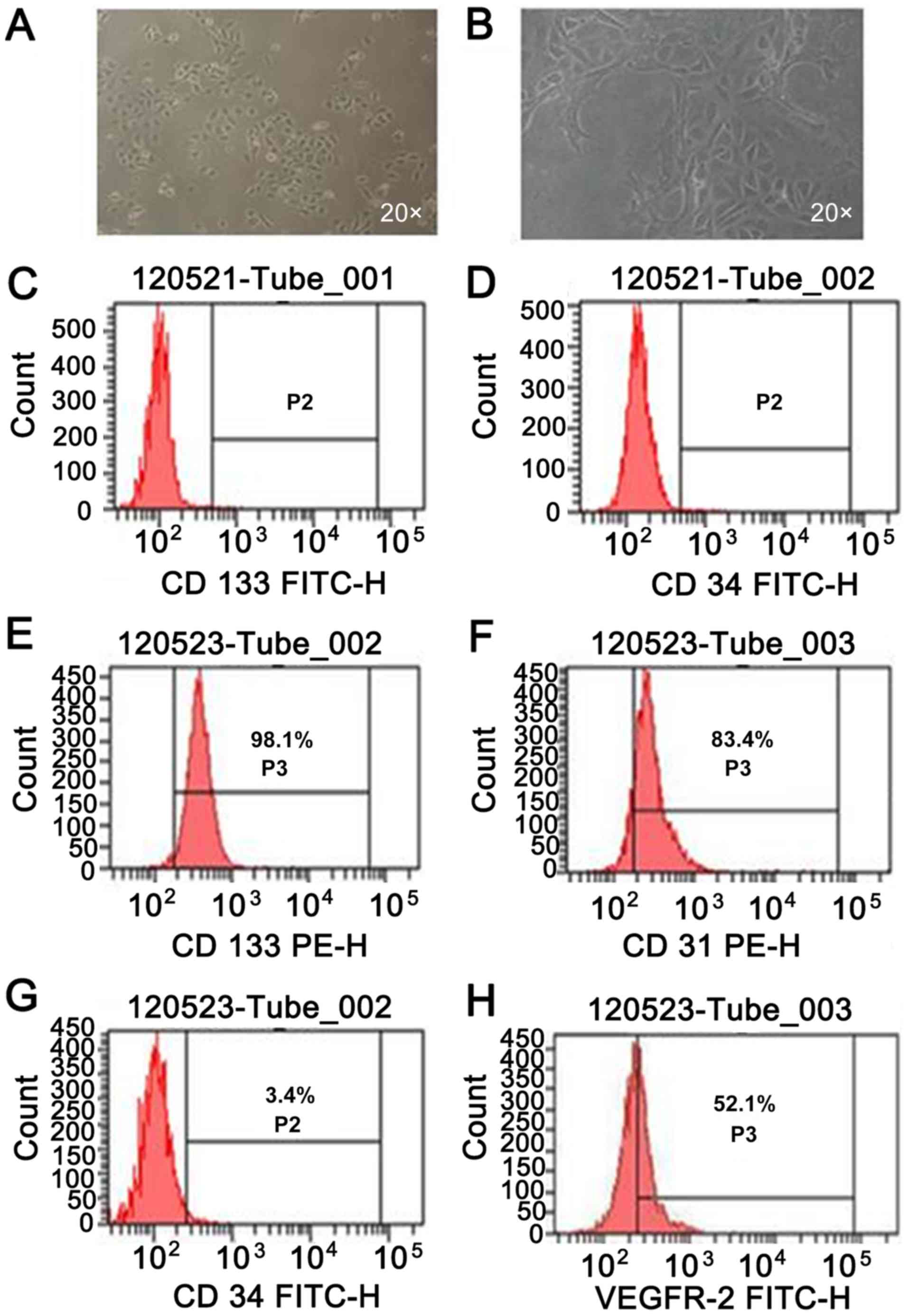 | Figure 1.Morphological characteristics of
rabbit EPCs. (A) Cells changed from a globe-like shape to a thin
structure, with acquiring a fusiform pebble-like shape at day 10.
Magnification, ×20. (B) EPCs formed a capillary-like structure
after 48 h on Matrigel. Magnification, ×20. (C-H) Expression levels
in EPCs of the endothelial cells surface markers (C and E) CD133,
(D and G) CD34, (F) CD31 and (H) VEGFR2 were 98.1, 3.4, 83.4 and
52.1%, respectively. EPCs, endothelial progenitor cells; FACS,
fluorescence-activated cell sorting; VEGFR-2, vascular endothelial
growth factor receptor 2. |
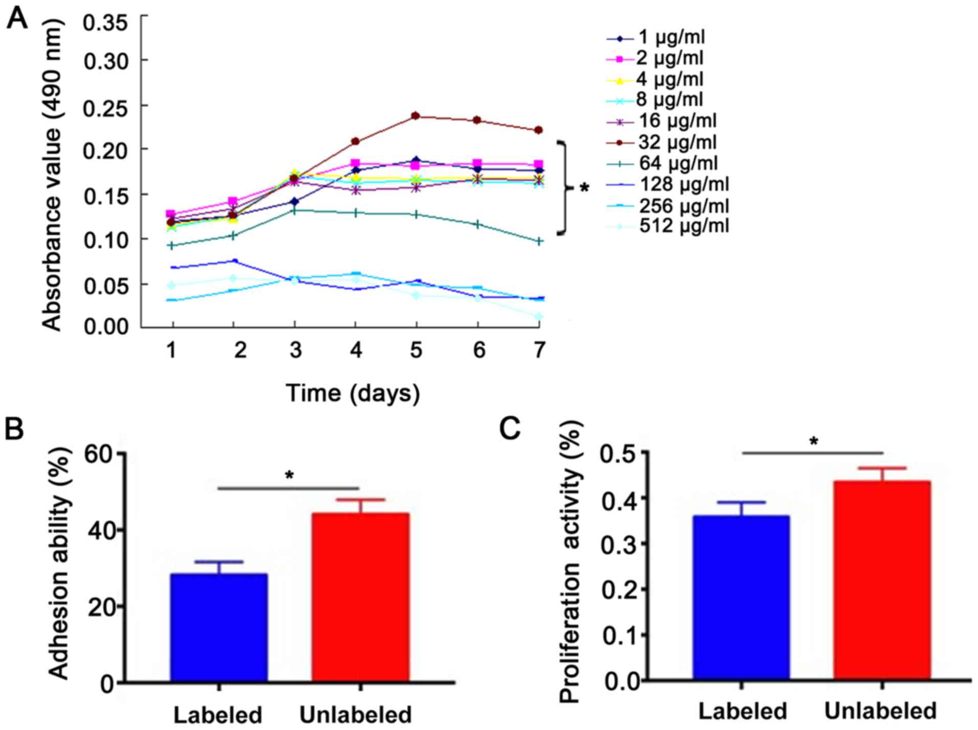
USPION-labeled EPCs retain adhesion
and proliferative abilities
To examine cell proliferation and viability of EPCs
labeled with different USPION concentrations, an MTT assay was
performed. It was demonstrated that cell viability decreased with
increasing USPION labeling time and concentration. While no
significant differences were found in absorbance (490 nm) values
among the 1, 2, 4, 8 and 16 µg/ml groups (Fig. 2A), cell proliferation and viability
were suppressed in the 64, 128, 256 and 512 µg/ml groups. It was
also found that the optimal USPION concentration was 32 µg/ml, with
a cell viability of 78.3±12.2% (P<0.05 vs. 64 µg/ml group;
Fig. 2A). The adhesion rate of
EPCs in the labeled group was only 28.16±3.45% at 1 h after passage
with 32 µg/ml USPION, which was much lower compared with the
unlabeled group (53.98±3.71%; P<0.05). The present results
suggested that the proliferative ability of EPCs in model rabbits
was significantly lower compared with the control group
(0.588±0.032 vs. 0.644±0.031; P<0.05; Fig. 2B and C).
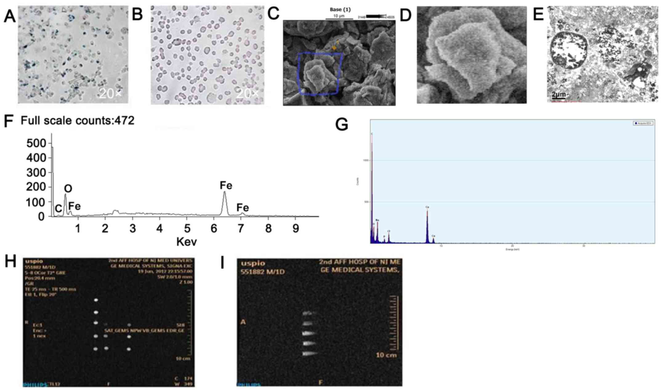
Labeling of EPCs is successful
The prevalence of USPION content in the labeled EPCs
was demonstrated by Prussian blue staining and scanning electron
microscopy. Prussian blue staining identified blue iron particles
in the cytoplasm of labeled EPCs at a 32 µg/ml USPION labeling
(Fig. 3A), while no blue iron
particles were found in the unlabeled EPCs (Fig. 3B). Scanning electron microscopy
results indicated that USPION-PLLs were uniform in size and 10–20
nm in diameter (Fig. 3C and D).
After labeling of EPCs, it was found that USPIONs were irregularly
distributed in the cytoplasm and uniform in size, and no
nanoparticles were found in the nucleus (Fig. 3E). Energy spectrum element analysis
results suggested that USPION-PLL nanoparticles were mainly
composed of iron (Fig. 3F), but no
iron was found in the labeled cells after 3 days of culture
(Fig. 3G). In vitro MRI
results indicated that the T2WI signal intensity of USPION-labeled
EPCs decreased with increasing USPION concentration compared with
unlabeled cells. However, the T2WI signal intensity of 32 µg/ml
USPION-labeled EPCs showed relatively good signals compared with
the other groups (Fig. 3H and I),
suggesting successful labeling of EPCs with USPIONs.
Success of the atherosclerotic rabbit
model
Serum TC, LDL and HDL levels in rabbits fed high-fat
diet were increased significantly at 12 weeks (P<0.01; Fig. 4A-D). CT images showed that the
blood vessel wall in the control group was smooth and intact with
no abnormal phenotypes, such as protuberances in the vessel lumen.
However, atherosclerotic plaques attached to the aortic wall were
found in the modeling group, which were protruding into the
vascular lumen (Fig. 4E and F).
H&E staining results demonstrated that the vascular endothelium
in the control group was intact with uniformly distributed elastic
fibers. Moreover, no foam cell or lipid deposition was observed
under light microscopy in the control group. However, obvious
intimal hyperplasia and irregular arterial lumen were observed in
the model group, with lipid plaques intruded into the lumen and a
large number of foam cells aggregated in the plaques (Fig. 4G and H). Compared with the control
group, matrix metalloproteinase-2 expression was positive in the
atherosclerotic plaques of the modeling group, mainly in the
cytoplasm of foam cells and endothelial cells in the plaques
(Fig. 4I and J). In addition,
positive expression of CD280 was also observed in the foam cells of
atherosclerotic plaques in the modeling group (Fig. 4K and L). Collectively, the present
results indicated the successful establishment of the
atherosclerotic rabbit model in this study.
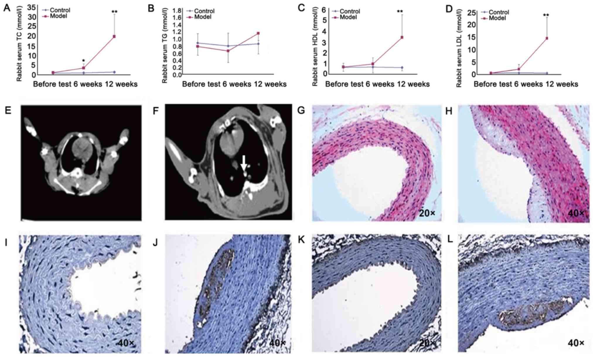 | Figure 4.Trend charts of rabbit blood lipid
indices in the model and control groups. Serum (A) TC, (B) TG, (C)
HDL and (D) LDL levels in atherosclerosis model and control
rabbits. (E) Rabbit aortic artery in the control group. (F)
Atherosclerotic plaque in the rabbit aortic artery in the modeling
group, as indicated by the arrow. (G) Continuous and intact intima
of rabbit aortic artery in the control group. Magnification, ×20.
(H) Sclerotic plaque protrusions in arterial vessels in model
rabbits. Magnification, ×40. (I) Continuous and intact intima of
rabbit aorta in the control group without MMP-2 expression.
Magnification, ×40. (J) Atherosclerotic plaques in model rabbits
appear brown after anti-MMP-2 immunohistochemical staining.
Magnification, ×40. (K) Continuous and intact intima of rabbit
aorta in the control group without CD280 expression. Magnification,
×20. (L) Foamy brown signals were observed in atherosclerotic
plaques in model rabbits after anti-CD280 immunohistochemical
staining. Magnification, ×40. *P<0.05, **P<0.01 vs. control
group. TC, total cholesterol; TG, triglycerides; LDL, low-density
lipoproteins; HDL, high-density lipoproteins (HDL); MMP, matrix
metalloproteinases. |
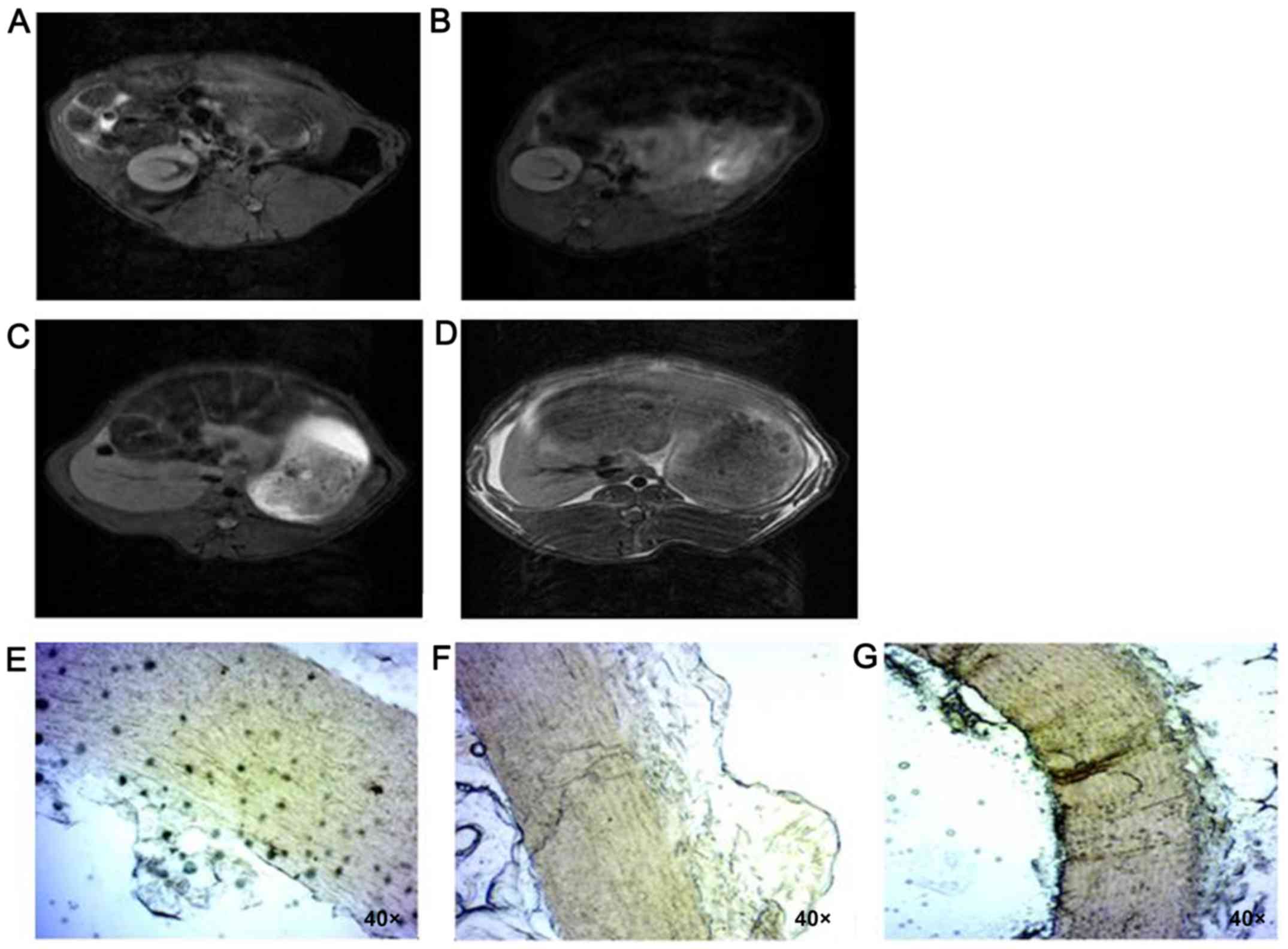
Transplantation of USPION-labeled EPCs
promotes atherosclerosis repair
Compared with the control group, the injured vessels
in the model group showed no significant difference in
contralateral vessels after the transplantation of USPION-labeled
EPCs. Moreover, there were low signals in the lumen, suggesting a
fluent blood flow in the lumen (Fig.
5A-D). Histopathological analysis results suggested that 1 day
after transplantation of labeled cells, blue-stained granules were
observed in the intima of vascular lesions in model rabbits, thus
indicating that the labeled EPCs were successfully implanted in the
damaged vessels (Fig. 5E).
Moreover, no blue-stained cells were found in pathological sections
7 days after transplantation of the labeled cells (Fig. 5F). In the control group, no
blue-stained cells were observed after transplantation of unlabeled
cells throughout the assay (Fig.
5G).
Discussion
In the present study, it was found that
USPION-labeled EPCs could promote angiogenesis both in vitro
and in vivo. Thus, the present results may facilitate the
development of a novel therapeutic tools for atherosclerotic
vessels.
Stem cell transplantation therapy has become a hot
topic in biomedical research, but its clinical application is still
limited despite breakthroughs (59–61).
Currently, the greatest challenge facing EPC transplantation is how
to improve in vivo survival, identify the outcomes of the
transplanted cells and monitoring the effectiveness of the
treatment (62). The conventional
tracing method is used to performed regular observation using
histopathological sections, but this method cannot dynamically
monitor EPCs involved in the repair and regeneration processes of
damaged vessels in real-time in vivo (62).
The development of molecular MRI technology provides
new approaches for studying the therapeutic effects of EPCs
transplantation by non-invasive dynamic monitoring (63). Previous studies investigating
molecular imaging contrast agents based on USPION in cells are the
most common (42,43,48–50).
It has been shown that USPION-labeled EPCs can reach the treatment
target under the action of external magnetic fields and can
participate in repairing damaged blood vessels (64). Moreover, USPION is highly
sensitive, and has no short-term or long-term biological side
effects on the labeled cells (65–67).
SPIO particles constitute an important method for
evaluating plaque instability in vivo (68–70).
Furthermore, EPCs labeled with SPIO particles have been widely used
in the treatment of diseases, including atherosclerosis (71). This method has also been used to
evaluate plaque inflammatory response in patients with carotid
atherosclerosis (43,69). The relatively large size of SPIO
particles may result in improved uptake rate of cells (72,73)
and the longer half-life of blood pool USPIONs allows them to be
frequently used for in vivo vessel labeling, as shown in
animals and patients with several diseases, including vessel
disorders (74,75). In patients, the half-life of blood
pool USPIONs is >24 h (76),
whereas that of SPIOs is <6 min (77). The present study investigated the
potential effect of USPION-labeled EPCs homing to injured
endothelium and whether local atherosclerosis could be prevented by
this EPC administration. High-resolution MRI can detect plaque
components in vivo, such as intra-plaque or subintimal
hemorrhage, fibrous cap and lipid-rich plaque core (78–80).
Thus, using USPIONs to enhance MRI could be used to show plaque
inflammation in vivo (81,82).
Previous studies have focused on the transplantation
of EPCs for the treatment of vascular diseases (18,25,38–40,62–64,83).
Werner et al (84) revealed
that spleen-derived endogenous or transfused progenitor cells home
to the site of vascular injury, which results in enhanced
reendothelialization and regeneration of endothelial cells, thus
indicating a critical and novel therapeutic approach for the
management of vascular injury at the early stages of
atherosclerosis. Moreover, the results from this previous study
(84) were consistent with those
from the present study.
In addition, Ma et al (51) found that atherosclerosis is
inhibited by SPIO-labeled EPCs, suggesting that these cells may
play a role in the restoration of endothelial injury and
atherosclerosis prevention. However, George et al (85) showed an opposite result, that the
transfer of bone marrow cells or EPCs may cause increased
atherosclerotic lesion size, and that EPC transfer may potentially
influence plaque stability. Therefore, the discrepancy between
these studies may be due to the different animal models used and
different time points. In the study by George et al
(85), an apolipoprotein E
knockout mouse model, with atherosclerotic plaques already formed
in the abdominal aorta and carotid artery, was used.
The diameter of the USPIONs used in the present
study was 6–10 nm, which is smaller than that of SPIO particles
(44). Encapsulated USPIONs using
polylysine form electrostatic complexes to stimulate the
endocytosis of EPCs, and USPIONs can enter the cytoplasm (65–67).
The present results suggested that the adhesion and proliferation
of EPCs were not significantly different compared with unlabeled
cells, and that the biological function of these cells was
maintained, which was in line with results from a previous study
(23). Furthermore, the half-life
of USPION in the plasma is longer than that of SPIO, it is highly
biocompatible and can produce a large paramagnetism effect when
subjected to a magnetic field (65–67).
Moreover, USPION can form a low signal region for imaging on
spin-echo and gradient-echo MRI sequences (44).
In previous imaging studies of in vitro cell
populations, it has been shown that the change rate of signal
intensity on T2WI is largest after cells are labeled with USPIONs
(83,86–88).
In the present study, MRI results suggested that the change rate of
signals for labeled EPCs was the most significant on T2WI. However,
T2WI is associated with disadvantages such as large magnetic
susceptibility artifacts on the gas-tissue interface (89). The present results also indicated
that no signal change was visible in each MRI imaging sequence and
that a small number of blue-stained particles were observed in the
intima 1 day after transplantation, while no blue-stained particles
were found at 7 and 16 days. These results may be due to the fact
that damaged local intima was repaired or that there was
hyperplasia of the smooth muscle cells, meaning EPCs could not
remain in the intima (90,91). The time of local incubation was
short, the cells may have been expelled from the affected blood
vessels due to the blood flow and migrated to other vascular
segments (92). Furthermore, after
digestion and before transplantation, the cells remained in an
in vitro environment and therefore a large number of cells
died, which would affect cell adhesion to the vascular wall. In
addition, the number of transplanted cells in the present study too
small. Moreover, cells cannot stay locally and can undergo immune
rejection (36–38).
As the number of experimental animals was small in
the present study, further studies are required to assess the
feasibility of diagnosing and treating atherosclerotic
cardiovascular disease using the homing effect of EPCs. Currently,
when using animal models, the presence of EPCs can only be
confirmed by biopsy or sacrificing the animals to obtain tissues of
corresponding target organs for pathological diagnosis. Thus, there
is a lack of dynamic monitoring of the transplanted cells, meaning
that the clinical application is limited. Therefore, there is an
urgent need to use a non-invasive imaging labeling of the cells in
order to trace in vivo cell distribution and to evaluate the
cell retention time, differentiation and proliferation in the
target organs. In addition, tracing labeled cells can lead to
optimization of the window for cell transplantation, provide ideal
feedback of cellular localization, and improve the understanding of
the dose or number of cells required for therapeutic
treatments.
In conclusion, magnetic labeling of cells with
USPIONs may be an effective novel approach for monitoring the
temporal and spatial migration of EPCs into vessels. Furthermore,
the present results may facilitate the development of cell-based
therapeutics for atherosclerosis.
Acknowledgements
Not applicable.
Funding
This work was supported by the Nanjing Municipal
Health Bureau Youth Science and Technology Talents Launching
Project (grant no. QYK10145).
Availability of data and materials
The datasets used and/or analyzed during the current
study are available from the corresponding author on reasonable
request.
Authors' contributions
HW and KZ conceived the idea, designed the
experiments and supervised the study. LC, HS, TT, and LL performed
the experiments, data analysis and data interpretation. JL
performed the CT and MRI scans. HW and LC wrote the manuscript. All
authors read and approved the final manuscript.
Ethics approval and consent to
participate
The authors declare that the study was approved by
The Ethics Committee of Drum Tower Hospital Affiliated to Medical
College of Nanjing University.
Patient consent for publication
Not applicable.
Competing interests
The authors declare that they have no competing
interests.
Glossary
Abbreviations
Abbreviations:
|
USPION
|
ultrasmall superparamagnetic iron
oxide nanoparticle
|
|
EPCs
|
endothelial progenitor cells
|
|
USPION-PLL
|
USPION-poly-l-lysine
|
|
SPIO
|
superparamagnetic iron oxide
|
|
T2WI
|
T2 weighted image
|
References
|
1
|
Zhang Y, Koradia A, Kamato D, Popat A,
Little PJ and Ta HT: Treatment of atherosclerotic plaque:
Perspectives on theranostics. J Pharmacy Pharmacol:. 71:3327–1043.
2019. View Article : Google Scholar
|
|
2
|
World Health Organization (WHO), World
Heart Federation, Organization WS, . Global atlas on cardiovascular
disease prevention and control Policies, strategies and
interventions. WHO; Geneva: 2011
|
|
3
|
Benjamin EJ, Blaha MJ, Chiuve SE, Cushman
M, Das SR, Deo R, de Ferranti SD, Floyd J, Fornage M, Gillespie C,
et al: Heart disease and stroke statistics-2017 update: A report
from the american heart association. Circulation. 135:e146–e603.
2017. View Article : Google Scholar : PubMed/NCBI
|
|
4
|
World Health Organization (WHO), .
Cardiovascular diseases (CVDs). WHO; Geneva: 2020, https://www.who.int/en/news-room/fact-sheets/detail/cardiovascular-diseases-(cvds)April
6–2020
|
|
5
|
Biancari F, Anttila V, Dell'Aquila AM,
Airaksinen JKE and Brascia D: Control angiography for perioperative
myocardial Ischemia after coronary surgery: Meta-analysis. J
Cardiothorac Surg. 13:242018. View Article : Google Scholar : PubMed/NCBI
|
|
6
|
Athyros VG, Tziomalos K, Katsiki N and
Mikhailidis DP: Novel data on the pathogenesis of atherosclerosis,
treatment targets, and new therapeutic interventions in
lipid-related cardiovascular risk factors. Curr Pharm Des.
20:6215–6219. 2014. View Article : Google Scholar : PubMed/NCBI
|
|
7
|
Rabelink TJ, de Boer HC and van Zonneveld
AJ: Endothelial activation and circulating markers of endothelial
activation in kidney disease. Nat Rev Nephrol. 6:404–414. 2010.
View Article : Google Scholar : PubMed/NCBI
|
|
8
|
Gimbrone MA Jr and Garcia-Cardena G:
Endothelial cell dysfunction and the pathobiology of
atherosclerosis. Circ Res. 118:620–636. 2016. View Article : Google Scholar : PubMed/NCBI
|
|
9
|
Cahill PA and Redmond EM: Vascular
endothelium-Gatekeeper of vessel health. Atherosclerosis.
248:97–109. 2016. View Article : Google Scholar : PubMed/NCBI
|
|
10
|
Theodorou K and Boon RA: Endothelial cell
metabolism in atherosclerosis. Front Cell Dev Biol. 6:822018.
View Article : Google Scholar : PubMed/NCBI
|
|
11
|
Kwon Y, Norby FL, Jensen PN, Agarwal SK,
Soliman EZ, Lip GY, Longstreth WT Jr, Alonso A, Heckbert SR and
Chen LY: Association of smoking, alcohol, and obesity with
cardiovascular death and ischemic stroke in atrial fibrillation:
The atherosclerosis risk in communities (ARIC) study and
cardiovascular health study (CHS). PLoS One. 11:e01470652016.
View Article : Google Scholar : PubMed/NCBI
|
|
12
|
Ruan C, Shen Y, Chen R, Wang Z, Li J and
Jiang Y: Endothelial progenitor cells and atherosclerosis. Front
Biosci (Landmark Ed). 18:1194–1201. 2013. View Article : Google Scholar : PubMed/NCBI
|
|
13
|
Yoder MC: Human endothelial progenitor
cells. Cold Spring Harb Perspect Med. 2:a0066922012. View Article : Google Scholar : PubMed/NCBI
|
|
14
|
Peters EB: Endothelial progenitor cells
for the vascularization of engineered tissues. Tissue Eng Part B
Rev. 24:1–24. 2018. View Article : Google Scholar : PubMed/NCBI
|
|
15
|
Li DW, Liu ZQ, Wei J, Liu Y and Hu LS:
Contribution of endothelial progenitor cells to neovascularization
(Review). Int J Mol Med. 30:1000–1006. 2012. View Article : Google Scholar : PubMed/NCBI
|
|
16
|
Mudyanadzo TA: Endothelial progenitor
cells and cardiovascular correlates. Cureus.
10:e33422018.PubMed/NCBI
|
|
17
|
Grisar JC, Haddad F, Gomari FA and Wu JC:
Endothelial progenitor cells in cardiovascular disease and chronic
inflammation: From biomarker to therapeutic agent. Biomark Med.
5:731–744. 2011. View Article : Google Scholar : PubMed/NCBI
|
|
18
|
Dzau VJ, Gnecchi M, Pachori AS, Morello F
and Melo LG: Therapeutic potential of endothelial progenitor cells
in cardiovascular diseases. Hypertension. 46:7–18. 2005. View Article : Google Scholar : PubMed/NCBI
|
|
19
|
Qiu Y, Zhang C, Zhang G and Tao J:
Endothelial progenitor cells in cardiovascular diseases. Aging Med.
1:204–208. 2018. View Article : Google Scholar
|
|
20
|
Gao X, Chen W, Liang Z and Chen L:
Autotransplantation of circulating endothelial progenitor cells
protects against lipopolysaccharide-induced acute lung injury in
rabbit. Int Immunopharmacol. 11:1584–1590. 2011. View Article : Google Scholar : PubMed/NCBI
|
|
21
|
Chen ZZ, Jiang XD, Zhang LL, Shang JH, Du
MX, Xu G and Xu RX: Beneficial effect of autologous transplantation
of bone marrow stromal cells and endothelial progenitor cells on
cerebral ischemia in rabbits. Neurosci Lett. 445:36–41. 2008.
View Article : Google Scholar : PubMed/NCBI
|
|
22
|
Chen B, Bo CJ, Jia RP, Liu H, Wu R, Wu J,
Ge YZ and Teng GJ: The renoprotective effect of bone marrow-derived
endothelial progenitor cell transplantation on acute
ischemia-reperfusion injury in rats. Transplant Pro. 45:2034–2039.
2013. View Article : Google Scholar
|
|
23
|
Li ZF, Fang XG, Yang PF, Huang QH, Zhao
WY, Liang C, Zhao R and Liu JM: Endothelial progenitor cells
contribute to neointima formation in rabbit elastase-induced
aneurysm after flow diverter treatment. CNS Neurosci Ther.
19:352–357. 2013. View Article : Google Scholar : PubMed/NCBI
|
|
24
|
Cui K, Ma X, Yu L, Jiang C, Fu C, Fu X, Yu
X, Huang Y, Hou S, Si C, et al: Autologous bone marrow mononuclear
cell transplantation delays progression of carotid atherosclerosis
in rabbits. Mol Neurobiol. 53:4387–4396. 2016. View Article : Google Scholar : PubMed/NCBI
|
|
25
|
Zhang M, Malik AB and Rehman J:
Endothelial progenitor cells and vascular repair. Curr Opin
Hematol. 21:224–228. 2014. View Article : Google Scholar : PubMed/NCBI
|
|
26
|
Rajendran P, Rengarajan T, Thangavel J,
Nishigaki Y, Sakthisekaran D, Sethi G and Nishigaki I: The vascular
endothelium and human diseases. Int J Biol Sci. 9:1057–1069. 2013.
View Article : Google Scholar : PubMed/NCBI
|
|
27
|
Kruger-Genge A, Blocki A, Franke RP and
Jung F: Vascular endothelial cell biology: An update. Int J Mol
Sci. 20:44112019. View Article : Google Scholar
|
|
28
|
Asahara T, Murohara T, Sullivan A, Silver
M, van der Zee R, Li T, Witzenbichler B, Schatteman G and Isner JM:
Isolation of putative progenitor endothelial cells for
angiogenesis. Science. 275:964–967. 1997. View Article : Google Scholar : PubMed/NCBI
|
|
29
|
Pirro M, Stingeni L, Vaudo G, Mannarino
MR, Ministrini S, Vonella M, Hansel K, Bagaglia F, Alaeddin A, Lisi
P and Mannarino E: Systemic inflammation and imbalance between
endothelial injury and repair in patients with psoriasis are
associated with preclinical atherosclerosis. Eur J Prev Cardiol.
22:1027–1035. 2015. View Article : Google Scholar : PubMed/NCBI
|
|
30
|
Groleau J, Dussault S, Haddad P, Turgeon
J, Menard C, Chan JS and Rivard A: Essential role of copper-zinc
superoxide dismutase for ischemia-induced neovascularization via
modulation of bone marrow-derived endothelial progenitor cells.
Arterioscler Thromb Vasc Biol. 30:2173–2181. 2010. View Article : Google Scholar : PubMed/NCBI
|
|
31
|
Ankeny RF, Ankeny CJ, Nerem RM and Jo H:
Maturing EPCs into endothelial cells: May the force be with the
EPCs: Focus on ‘Fluid shear stress induces differentiation of
circulating phenotype endothelial progenitor cells’. Am J Physiol
Cell Physiol. 303:C589–C591. 2012. View Article : Google Scholar : PubMed/NCBI
|
|
32
|
Psaltis PJ and Simari RD: Vascular wall
progenitor cells in health and disease. Circ Res. 116:1392–1412.
2015. View Article : Google Scholar : PubMed/NCBI
|
|
33
|
Zhang X, Mao H, Chen JY, Wen S, Li D, Ye M
and Lv Z: Increased expression of microRNA-221 inhibits PAK1 in
endothelial progenitor cells and impairs its function via
c-Raf/MEK/ERK pathway. Biochem Biophys Res Commun. 431:404–408.
2013. View Article : Google Scholar : PubMed/NCBI
|
|
34
|
Aicher A, Brenner W, Zuhayra M, Badorff C,
Massoudi S, Assmus B, Eckey T, Henze E, Zeiher AM and Dimmeler S:
Assessment of the tissue distribution of transplanted human
endothelial progenitor cells by radioactive labeling. Circulation.
107:2134–2139. 2003. View Article : Google Scholar : PubMed/NCBI
|
|
35
|
Asahara T, Kawamoto A and Masuda H:
Concise review: Circulating endothelial progenitor cells for
vascular medicine. Stem Cells. 29:1650–1655. 2011. View Article : Google Scholar : PubMed/NCBI
|
|
36
|
Nuzzolo ER, Capodimonti S, Martini M,
Iachininoto MG, Bianchi M, Cocomazzi A, Zini G, Leone G, Larocca LM
and Teofili L: Adult and cord blood endothelial progenitor cells
have different gene expression profiles and immunogenic potential.
Blood Transfus. 12 (Suppl 1):S367–S374. 2014.PubMed/NCBI
|
|
37
|
Souidi N, Stolk M, Rudeck J, Strunk D,
Schallmoser K, Volk HD and Seifert M: Stromal cells act as
guardians for endothelial progenitors by reducing their
immunogenicity after co-transplantation. Stem Cells. 35:1233–1245.
2017. View Article : Google Scholar : PubMed/NCBI
|
|
38
|
Ladhoff J, Fleischer B, Hara Y, Volk HD
and Seifert M: Immune privilege of endothelial cells differentiated
from endothelial progenitor cells. Cardiovasc Res. 88:121–129.
2010. View Article : Google Scholar : PubMed/NCBI
|
|
39
|
Fang J, Guo Y, Tan S, Li Z, Xie H, Chen P,
Wang K, He Z, He P, Ke Y, et al: Autologous endothelial progenitor
cells transplantation for acute ischemic stroke: A 4-Year Follow-Up
Study. Stem Cells Transl Med. 8:14–21. 2019. View Article : Google Scholar : PubMed/NCBI
|
|
40
|
Fan CL, Gao PJ, Che ZQ, Liu JJ, Wei J and
Zhu DL: Therapeutic neovascularization by autologous
transplantation with expanded endothelial progenitor cells from
peripheral blood into ischemic hind limbs. Acta Pharmacol Sin.
26:1069–1075. 2005. View Article : Google Scholar : PubMed/NCBI
|
|
41
|
Yin Y, Liu H, Wang F, Li L, Deng M, Huang
L and Zhao X: Transplantation of cryopreserved human umbilical cord
blood-derived endothelial progenitor cells induces recovery of
carotid artery injury in nude rats. Stem Cell Res Ther. 6:372015.
View Article : Google Scholar : PubMed/NCBI
|
|
42
|
Suzuki M, Bachelet-Violette L, Rouzet F,
Beilvert A, Autret G, Maire M, Menager C, Louedec L, Choqueux C,
Saboural P, et al: Ultrasmall superparamagnetic iron oxide
nanoparticles coated with fucoidan for molecular MRI of
intraluminal thrombus. Nanomedicine (Lond). 10:73–87. 2015.
View Article : Google Scholar : PubMed/NCBI
|
|
43
|
Usman A, Sadat U, Patterson AJ, Tang TY,
Varty K, Boyle JR, Armon MP, Hayes PD, Graves MJ and Gillard JH:
Use of ultrasmall superparamagnetic iron oxide particles for
imaging carotid atherosclerosis. Nanomedicine (Lond). 10:3077–3087.
2015. View Article : Google Scholar : PubMed/NCBI
|
|
44
|
Zhao X, Zhao H, Chen Z and Lan M:
Ultrasmall superparamagnetic iron oxide nanoparticles for magnetic
resonance imaging contrast agent. J Nanosci Nanotechnol.
14:210–220. 2014. View Article : Google Scholar : PubMed/NCBI
|
|
45
|
Fang Y, Wu Y, Liao P, Chen Z, Chen H, Yu
J, Liu Y, Li S, Su E, He N, et al: Design and application of a
high-throughput sample processing module based on magnetic beads.
Nanosci Nanotech Lett. 10:320–328. 2018. View Article : Google Scholar
|
|
46
|
Chen Z, Wu Y, Kang M, He N, Wan S, Su E
and Lijun W: Research on automated nucleic acid extraction
instrument based on magnetic nanoparticles separation. Nanosci
Nanotech Lett. 10:60–68. 2018. View Article : Google Scholar
|
|
47
|
Liu H, Dong H, Chen Z, Lin L, Chen H, Li S
and Deng Y: Magnetic nanoparticles enhanced microarray detection of
multiple foodborne pathogens. J Biomed Nanotech. 13:1333–1343.
2017. View Article : Google Scholar
|
|
48
|
Wang X, Zhang T, Zhao X, Guan Z, Wang Z,
Zhu Z, Xie Q, Wang J and Niu B: Quantification of folate
metabolites in serum using ultraperformance liquid chromatography
tandem mass spectrometry. J Chromatogr B Analyt Technol Biomed Life
Sci. 962:9–13. 2014. View Article : Google Scholar : PubMed/NCBI
|
|
49
|
Ittrich H, Peldschus K, Raabe N, Kaul M
and Adam G: Superparamagnetic iron oxide nanoparticles in
biomedicine: Applications and developments in diagnostics and
therapy. Rofo. 185:1149–1166. 2013. View Article : Google Scholar : PubMed/NCBI
|
|
50
|
Guo L, Chen H, He N and Deng Y: Effects of
surface modifications on the physicochemical properties of iron
oxide nanoparticles and their performance as anticancer drug
carriers. Chin Chem Lett. 29:1829–1833. 2018. View Article : Google Scholar
|
|
51
|
Ma ZL, Mai XL, Sun JH, Ju SH, Yang X, Ni Y
and Teng GJ: Inhibited atherosclerotic plaque formation by local
administration of magnetically labeled endothelial progenitor cells
(EPCs) in a rabbit model. Atherosclerosis. 205:80–86. 2009.
View Article : Google Scholar : PubMed/NCBI
|
|
52
|
Lipman NS, Marini RP and Erdman SE: A
comparison of ketamine/xylazine and ketamine/xylazine/acepromazine
anesthesia in the rabbit. Lab Anim Sci. 40:395–398. 1990.PubMed/NCBI
|
|
53
|
Marini RP, Li X, Harpster NK and Dangler
C: Cardiovascular pathology possibly associated with
ketamine/xylazine anesthesia in Dutch belted rabbits. Lab Anim Sci.
49:153–160. 1999.PubMed/NCBI
|
|
54
|
Baneux PJ, Garner D, McIntyre HB and
Holshuh HJ: Euthanasia of rabbits by intravenous administration of
ketamine. J Am Vet Med Assoc. 189:1038–1039. 1986.PubMed/NCBI
|
|
55
|
Cicero L, Fazzotta S, Palumbo VD, Cassata
G and Lo Monte AI: Anesthesia protocols in laboratory animals used
for scientific purposes. Acta Biomed. 89:337–342. 2018.PubMed/NCBI
|
|
56
|
Huang YH, Xu Q, Shen T, Li JK, Sheng JY
and Shi HJ: Prevention of in-stent restenosis with endothelial
progenitor cell (EPC) capture stent placement combined with
regional EPC transplantation: An atherosclerotic rabbit model.
Cardiol J. 26:283–291. 2019. View Article : Google Scholar : PubMed/NCBI
|
|
57
|
Patel J, Seppanen EJ, Rodero MP, Wong HY,
Donovan P, Neufeld Z, Fisk NM, Francois M and Khosrotehrani K:
Functional definition of progenitors versus mature endothelial
cells reveals key SoxF-dependent differentiation process.
Circulation. 135:786–805. 2017. View Article : Google Scholar : PubMed/NCBI
|
|
58
|
Flores-Nascimento MC, Alessio AM, de
Andrade Orsi FL and Annichino-Bizzacchi JM: CD144, CD146 and
VEGFR-2 properly identify circulating endothelial cell. Rev Bras
Hematol Hemoter. 37:98–102. 2015. View Article : Google Scholar : PubMed/NCBI
|
|
59
|
Wei L, Wei ZZ, Jiang MQ, Mohamad O and Yu
SP: Stem cell transplantation therapy for multifaceted therapeutic
benefits after stroke. Prog Neurobiol. 157:49–78. 2017. View Article : Google Scholar : PubMed/NCBI
|
|
60
|
Kamelska-Sadowska AM, Wojtkiewicz J and
Kowalski IM: Review of the current knowledge on the role of stem
cell transplantation in neurorehabilitation. Biomed Res Int.
2019:32908942019. View Article : Google Scholar : PubMed/NCBI
|
|
61
|
Cho J, D'Antuono M, Glicksman M, Wang J
and Jonklaas J: A review of clinical trials: Mesenchymal stem cell
transplant therapy in type 1 and type 2 diabetes mellitus. Am J
Stem Cells. 7:82–93. 2018.PubMed/NCBI
|
|
62
|
Chong MS, Ng WK and Chan JK: Concise
review: Endothelial progenitor cells in regenerative medicine:
Applications and challenges. Stem Cells Transl Med. 5:530–538.
2016. View Article : Google Scholar : PubMed/NCBI
|
|
63
|
Peng X, Li C, Bai Y, Wang X, Zhang Y, An
Y, Teng GJ and Ju S: Noninvasive evaluation of the migration effect
of transplanted endothelial progenitor cells in ischemic muscle
using a multimodal imaging agent. Int J Nanomedicine. 13:1819–1829.
2018. View Article : Google Scholar : PubMed/NCBI
|
|
64
|
Zhang BF, Jiang H, Chen J, Hu Q, Yang S
and Liu XP: Silica-coated magnetic nanoparticles labeled
endothelial progenitor cells alleviate ischemic myocardial injury
and improve long-term cardiac function with magnetic field guidance
in rats with myocardial infarction. J Cell Physiol.
234:18544–18559. 2019. View Article : Google Scholar : PubMed/NCBI
|
|
65
|
Leung K: Ultrasmall superparamagnetic iron
oxide-Leu-Ile-Lys-Lys-Pro-Phe. Molecular Imaging and Contrast Agent
Database (MICAD); Bethesda, MD: 2004
|
|
66
|
Leung K: Ultrasmall superparamagnetic iron
oxide-cyclo(Cys-Asn-Asn-Ser-Lys-Ser-His-Thr-Cys). Molecular Imaging
and Contrast Agent Database (MICAD) Bethesda, MD: 2004
|
|
67
|
Leung K: Ultrasmall superparamagnetic iron
oxide nanoparticles conjugated with Ile-Pro-Leu-Pro-Phe-Tyr-Asn.
Molecular Imaging and Contrast Agent Database (MICAD) Bethesda, MD:
2004
|
|
68
|
Zhou Q, Yang KR, Gao P, Chen WL, Yang DY,
Liang MJ and Zhu L: An experimental study on MR imaging of
atherosclerotic plaque with SPIO marked endothelial cells in a
rabbit model. J Magn Reson Imaging. 34:1325–1332. 2011. View Article : Google Scholar : PubMed/NCBI
|
|
69
|
Tang TY, Muller KH, Graves MJ, Li ZY,
Walsh SR, Young V, Sadat U, Howarth SP and Gillard JH: Iron oxide
particles for atheroma imaging. Arterioscler Thromb Vasc Biol.
29:1001–1008. 2009. View Article : Google Scholar : PubMed/NCBI
|
|
70
|
Alam SR, Stirrat C, Richards J, Mirsadraee
S, Semple SI, Tse G, Henriksen P and Newby DE: Vascular and plaque
imaging with ultrasmall superparamagnetic particles of iron oxide.
J Cardiovasc Magn Reson. 17:832015. View Article : Google Scholar : PubMed/NCBI
|
|
71
|
Moon SH, Kim SM, Park SJ, Kim H, Bae D,
Choi YS and Chung HM: Development of a xeno-free autologous culture
system for endothelial progenitor cells derived from human
umbilical cord blood. PLoS One. 8:e752242013. View Article : Google Scholar : PubMed/NCBI
|
|
72
|
Daldrup-Link HE, Rudelius M, Oostendorp
RA, Settles M, Piontek G, Metz S, Rosenbrock H, Keller U, Heinzmann
U, Rummeny EJ, et al: Targeting of hematopoietic progenitor cells
with MR contrast agents. Radiology. 228:760–767. 2003. View Article : Google Scholar : PubMed/NCBI
|
|
73
|
Matuszewski L, Persigehl T, Wall A,
Schwindt W, Tombach B, Fobker M, Poremba C, Ebert W, Heindel W and
Bremer C: Cell tagging with clinically approved iron oxides:
Feasibility and effect of lipofection, particle size, and surface
coating on labeling efficiency. Radiology. 235:155–161. 2005.
View Article : Google Scholar : PubMed/NCBI
|
|
74
|
Rausch M, Sauter A, Frohlich J, Neubacher
U, Radu EW and Rudin M: Dynamic patterns of USPIO enhancement can
be observed in macrophages after ischemic brain damage. Magn Reson
Med. 46:1018–1022. 2001. View Article : Google Scholar : PubMed/NCBI
|
|
75
|
Saleh A, Schroeter M, Jonkmanns C, Hartung
HP, Modder U and Jander S: In vivo MRI of brain inflammation in
human ischaemic stroke. Brain. 127:1670–1677. 2004. View Article : Google Scholar : PubMed/NCBI
|
|
76
|
McLachlan SJ, Morris MR, Lucas MA, Fisco
RA, Eakins MN, Fowler DR, Scheetz RB and Olukotun AY: Phase I
clinical evaluation of a new iron oxide MR contrast agent. J Magn
Reson Imaging. 4:301–307. 1994. View Article : Google Scholar : PubMed/NCBI
|
|
77
|
Weissleder R, Stark DD, Engelstad BL,
Bacon BR, Compton CC, White DL, Jacobs P and Lewis J:
Superparamagnetic iron oxide: Pharmacokinetics and toxicity. AJR Am
J Roentgenol. 152:167–173. 1989. View Article : Google Scholar : PubMed/NCBI
|
|
78
|
Ryu CW, Kwak HS, Jahng GH and Lee HN:
High-resolution MRI of intracranial atherosclerotic disease.
Neurointervention. 9:9–20. 2014. View Article : Google Scholar : PubMed/NCBI
|
|
79
|
Kerwin WS and Canton G: Advanced
techniques for MRI of atherosclerotic plaque. Top Magn Reson
Imaging. 20:217–225. 2009. View Article : Google Scholar : PubMed/NCBI
|
|
80
|
Wang E, Shao S, Li S, Yan P, Xiang Y, Wang
X, Li J, Wang G, Sun Q and Du Y: A High-resolution MRI study of the
relationship between plaque enhancement and ischemic stroke events
in patients with intracranial atherosclerotic stenosis. Front
Neurol. 9:11542018. View Article : Google Scholar : PubMed/NCBI
|
|
81
|
Smits LP, Tiessens F, Zheng KH, Stroes ES,
Nederveen AJ and Coolen BF: Evaluation of ultrasmall
superparamagnetic iron-oxide (USPIO) enhanced MRI with ferumoxytol
to quantify arterial wall inflammation. Atherosclerosis.
263:211–218. 2017. View Article : Google Scholar : PubMed/NCBI
|
|
82
|
Kaneko C, Nitta N, Tsuchiya K, Watanabe S,
Nitta-Seko A, Ohta S, Otani H, Sonoda A, Murata K and Shiomi M: MRI
study of atherosclerotic plaque progression using ultrasmall
superparamagnetic iron oxide in Watanabe heritable hyperlipidemic
rabbits. Br J Radiol. 88:201501672015. View Article : Google Scholar : PubMed/NCBI
|
|
83
|
Yao Y, Li Y, Ma G, Liu N, Ju S, Jin J,
Chen Z, Shen C and Teng G: In vivo magnetic resonance imaging of
injected endothelial progenitor cells after myocardial infarction
in rats. Mol Imaging Biol. 13:303–313. 2011. View Article : Google Scholar : PubMed/NCBI
|
|
84
|
Werner N, Junk S, Laufs U, Link A, Walenta
K, Bohm M and Nickenig G: Intravenous transfusion of endothelial
progenitor cells reduces neointima formation after vascular injury.
Circ Res. 93:e17–e24. 2003. View Article : Google Scholar : PubMed/NCBI
|
|
85
|
George J, Afek A, Abashidze A, Shmilovich
H, Deutsch V, Kopolovich J, Kopolovich J, Miller H and Keren G:
Transfer of endothelial progenitor and bone marrow cells influences
atherosclerotic plaque size and composition in apolipoprotein E
knockout mice. Arterioscler Thromb Vasc Biol. 25:2636–2641. 2005.
View Article : Google Scholar : PubMed/NCBI
|
|
86
|
Mergo PJ, Engelken JD, Helmberger T and
Ros PR: MRI in focal liver disease: A comparison of small and
ultra-small superparamagnetic iron oxide as hepatic contrast
agents. J Magn Reson Imaging. 8:1073–1078. 1998. View Article : Google Scholar : PubMed/NCBI
|
|
87
|
Czarniecki M, Pesapane F, Wood BJ, Choyke
PL and Turkbey B: Ultra-small superparamagnetic iron oxide contrast
agents for lymph node staging of high-risk prostate cancer. Transl
Androl Urol. 7 (Suppl 4):S453–S461. 2018. View Article : Google Scholar : PubMed/NCBI
|
|
88
|
Matuszewski L, Tombach B, Heindel W and
Bremer C: Molecular and parametric imaging with iron oxides.
Radiologe. 47:34–42. 2007.(In German). View Article : Google Scholar : PubMed/NCBI
|
|
89
|
Engels RRM, Israel B, Padhani AR and
Barentsz JO: Multiparametric magnetic resonance imaging for the
detection of clinically significant prostate cancer: What
urologists need to know. Part 1: Acquisition. Eur Urol. 77:457–468.
2020. View Article : Google Scholar : PubMed/NCBI
|
|
90
|
Yang JX, Pan YY, Wang XX, Qiu YG and Mao
W: Endothelial progenitor cells in age-related vascular remodeling.
Cell Transplant. 27:786–795. 2018. View Article : Google Scholar : PubMed/NCBI
|
|
91
|
Simard T, Jung RG, Motazedian P, Di Santo
P, Ramirez FD, Russo JJ, Labinaz A, Yousef A, Anantharam B,
Pourdjabbar A and Hibbert B: Progenitor cells for arterial repair:
Incremental advancements towards therapeutic reality. Stem Cells
Int. 2017:82704982017. View Article : Google Scholar : PubMed/NCBI
|
|
92
|
Lin Y, Weisdorf DJ, Solovey A and Hebbel
RP: Origins of circulating endothelial cells and endothelial
outgrowth from blood. J Clin Invest. 105:71–77. 2000. View Article : Google Scholar : PubMed/NCBI
|