Introduction
Poly (ADP ribose) polymerase 1 (PARP1) is involved
in the base excision pathway of DNA repair in most eukaryotic
cells. As a part of this pathway, PARP serves a key role in the
maintenance of DNA integrity (1).
The PARP family consists of 17 subtypes, of which PARP1 is the most
abundant and ubiquitous member that participates in various
functions performed by this family (2). Increased expression of PARP1 has been
reported to be an independent negative prognostic marker in mucosal
melanomas (3). PARP1 recruits
Kruppel-like factor 4 to activate telomerase expression in cancer
and stem cells (4). The common PARP
inhibitors (PARPis) include olaparib, veliparib and rucaparib.
Besides breast cancer gene (BRCA) mutation-associated cancer, the
benefits of PARPis in earlier treatment settings, including
neoadjuvant, adjuvant and promising combination therapeutic
strategies, such as those with other DNA damage response inhibitors
and immune checkpoint inhibitors, are of increasing interest
(5). However, to date, PARPi
treatment is based on the lethal synthesis theory and is restricted
to patients with BRCA1/2 mutation-associated breast and ovarian
cancer (6).
Melanoma is a heterogeneous disease and one of the
most aggressive forms of skin tumour with poor prognosis (7). Melanoma is associated with numerous
genetic mutations or alterations in signalling pathways (e.g.
BRAF), and there is no effective therapy for melanoma at the
metastatic stage (e.g. uveal melanoma) (8). Veliparib has been reported to be
effective in the treatment of BRAF inhibitor-resistant melanoma
cells (9). However, while a
randomized phase II study of veliparib with temozolomide
demonstrated a higher median progression-free survival for
veliparib compared with that of the placebo, the difference was not
statistically significant (10).
Besides, while olaparib increased response to dacarbazine (an
alkylating agent) in uveal melanoma (8), both veliparib and olaparib combined
increased the sensitivity of various histological subtypes of
single-nucleotide polymorphism (SNP)-carrier cancer cells to
alkylating agents, without an effect on wild-type cells (11).
Except for immunotherapy, single drugs directed to
single targets have not made much progress in the treatment of
tumours. Danshen, a common traditional Chinese medicine used in
clinical practice, contains multiple components (e.g. tanshinone I,
tanshinone IIA and tanshinone IIB) and has multiple targets
(12). The effects of tanshinone
IIA are well studied, particularly in tumours (13,14).
In addition, the role of tanshinone I in tumour proliferation,
metastasis and drug resistance has also been studied (15,16).
However, to some extent, its weak potency and poor drug-like
properties restrict its clinical development as a cancer therapy
(17).
PARPis are designed to modulate enzyme activity
without affecting the expression of PARP1. Increased PARP1 gene
expression in tumours has been shown to be associated with melanoma
ulceration and poorer overall survival (OS) (18). Our previous study in prostate cancer
demonstrated that inhibition of PARP1 expression significantly
reduced prostate cancer cell proliferation and migration
irrespective of BRCA1/2 mutations (19). However, in vascular smooth muscle
cells and endothelial cells, PARP1 inhibition may be protective
against apoptosis and/or necrosis in response to
H2O2 or tumour necrosis factor (20). Thus, the exact role of PARPis and
PARP1 expression in wild-type or SNP-containing melanoma cells
requires further elucidation. The present study explored the role
of PARPis and PARP1 in tumour progression, and screened for
compounds that significantly promoted melanoma efficacy and
modulated PARP1 expression to provide a potential basis for
assessing related drugs for targeting PARP1 in melanoma.
Materials and methods
Materials
Sunitinib, veliparib, olaparib, mefloquine,
simvastatin, dihydroartemisinin, tanshinone I, cryptotanshinone,
gossypol and docetaxel were purchased from Selleck Chemicals.
Anti-FoxO3a (cat. no. 12829), anti-α/β-tubulin (cat. no. 2148) and
anti-Bcl-2 (cat. no. 15071) antibodies were purchased from Cell
Signaling Technology, Inc. phosphorylated (p)FoxO3a were from Abcam
Inc. (cat. no. ab154786). Anti-PARP1 (cat. no. sc-8007) was
obtained from Santa Cruz Biotechnology, Inc. Tanshinone I, dioscin
and simvastatin were obtained from Shanghai YuanYe Biotechnology
Co., Ltd. DMEM, RPMI-1640, MEM and FBS were obtained from Gibco;
Thermo Fisher Scientific, Inc. MTS was purchased from Shanghai
BestBio (cat. no. BB-4204-3). The secondary fluorescent antibodies,
IRDye 800CW goat anti-mouse (cat. no. 926-32210) and IRDye 800CW
goat anti-rabbit (cat. no. 926-32211) were from were from LI-COR.
All other laboratory reagents in common use were of domestic
analytical pure grade.
OS analysis
OS data for PARP1 and PARP2 expression, and
prognosis for skin cutaneous melanoma (SKCM) and uveal melanoma
(UVM) were obtained from the Gene Expression Profiling Interactive
Analysis (GEPIA) database (http://gepia.cancer-pku.cn) by setting the percentage
of the Cut off-Low and Cut off-High according to the gene and tumor
subtype (21).
Cell lines and cultures
Melanoma cell lines (A-375) and renal tumour cell
lines (769-P, Caki-1 and ACHN) were purchased from the Cell Bank of
Type Culture Collection of the Chinese Academy of Sciences. The
cell lines were cultured in 90% DMEM (A-375 cells), RPMI-1640
(769-P and Caki-1 cells) or MEM (ACHN cells) supplemented with 10%
FBS at 37°C with 5% CO2 and saturated humidity.
Cell viability tests
The effects of tanshinone I and simvastatin, and
veliparib or olaparib alone or in combination with sunitinib or
docetaxel (24, 48 and 72 h) on A-375 cell proliferation were
assessed using the MTS assay. Briefly, after the cells were seeded
in 96-well plates (3,000-5,000 per well), the corresponding drugs
were added the next day and incubated in 37°C for 24–72 h before
the medium was replaced with an MTS mixture (10 µl/well) in 37°C
for 1–3 h. The optical values were measured using a multimode
reader (BioTek Instruments, Inc.) at 490 nm. The control group was
normalized to ‘100’, and the relative survival rates were then
calculated.
Colony formation experiment
Cell (A-375, ACHN, 769-P or Caki-1) suspensions were
seeded in 6-well dishes at ~1×10³ cells/well and
incubated in the CO2 incubator at 37°C for 24 h. The
medium was replaced with fresh medium containing the simvastatin or
tanshinone I (with a concentration of 0, 1, 3 and 10 µM). When
macroscopic clones were observed in the dish after 10–15 days, the
cells were washed twice with PBS, and then fixed and dyed with 4%
crystal violet in alcohol for 20–30 min in room temperature. After
the dye solution was washed with distilled water and air-dried,
images of the cells were captured using a camera directly, and the
number of cells in the different groups were counted manually or
using Image-Pro Plus 16.0 (Media Cybernetics, Inc.).
Morphological analysis
After the cells were treated by drugs for 24 h,
images of the cells were captured under a fluorescent microscope in
ordinary light (magnification, ×20), when the morphological
features of cells changed significantly.
Western blotting
After the A-375 cells were treated with the same
concentration (10 µM) of drugs (valiparib, sunitinib, mefloquine,
simvastatin, dihydroartemisinin, tanshinone I, cryptotanshinone,
gossypol and dioscin) for 24 h in the CO2 incubator at
37°C, cells were washed three times with PBS, and
radioimmunoprecipitation assay lysis buffer (Nanjing KeyGen Biotech
Co., Ltd.; cat. no. KGP702-100) was added to extract total protein.
The cells were then scraped and the lysate was collected. After the
cells in the eppendorf tube were inserted into the ice, they were
broken using a ultrasonic cell ddisruptorr (150W/20KHz; Sonics and
Materials, Inc.) 3–5 times (5 sec each time, with a 45% amplitude),
the lysates were centrifuged at 12,000 × g for 30 min at 4°C and
the supernatants were retained. The protein concentrations were
quantified using a bicinchoninic acid kit (Thermo Fisher
Scientific, Inc.). Protein samples were separated by SDS-PAGE with
a 10% gel and transferred to polyvinylidene difluoride membranes.
The membranes were cut and blocked with 5% skimmed milk for 1–2 h
in the room temperature and incubated overnight at 4°C with the
primary antibodies of the PARP-1 (1:200), FoxO3a (1:1,000),
p-FoxO3a (1:1,000), tubulin (1:1,000) and anti-Bcl-2 (1:1,000).
After washing with PBS, the membranes were incubated in
fluorescently labelled secondary antibodies (1:7,500) for 1–2 h at
room temperature. The membranes were then washed with PBS and
directly scanned on an LI-COR Image Studio Ver5.2 imaging system
(without other visualization reagents). The grey values of the
western blot bands were analysed using ImageJ (National Institutes
of Health; version 1.4.3.67).
Xenograft tumour model
The animal experiments were approved by the ethics
committee of the First Affiliated Hospital of Guangzhou Medical
University (Guangzhou, China). A total of 16 male BALB/c nude mice
(weight 18–22 g; age, ~4 weeks old) were obtained from the
Guangdong Medical Laboratory Animal Centre. The mice were allowed
to acclimatise for a week in a specific pathogen free environment
[temperature, 20–26°C; relative humidity, 40–70%; with a high
pressure disinfection equipment, purified water system, and
automatic switching on (at 7 a.m.) and off (at 7 p.m.) light
system]. Each cage had a drinking bottle for the water and the food
was supplied on the other side of the lid. The mice had common food
and drank water freely. A subcutaneous xenograft tumour model of
A-375 cells was established in nude mice. A-375 cells
(~2×10⁶) in a 150-µl suspension with 20% Matrigel
(Corning, Inc.; cat. no. 354248) were subcutaneously injected after
the mice were anesthetized by intraperitoneal injection of 40–60
mg/kg sodium pentobarbital. After 8 days, 15 tumour-bearing mice
were selected for subsequent experiments; one mouse was excluded
due to relatively slow tumorigenesis. The 15 mice were equally
divided into three groups: i) Control (solvent, DMSO); ii) 10 mg/kg
tanshinone I; and iii) 10 mg/kg tanshinone I and 20 mg/kg
simvastatin, and were administered intraperitoneal injection from
days 8 to 26, with the tumour size measured every 4–5 days. On day
30, mice were sacrificed with excessive intraperitoneal injection
of sodium pentobarbital (three times that of the anaesthetic dose),
and the tumour volume and weight of the nude mice were observed and
calculated. The tumour volume was estimated using the following
formula: Volume = (a × b2)/2; ‘a’ represent the longest
diameters, ‘b’ represent the shortest diameters. The humane
endpoint set for this study was that, when the maximum tumour size
of the mice was up to ~1,200 mm3, all the nude mice were
sacrificed 4 days later and then the tumor volume were
measured.
Molecular characteristics and
medicinal properties
The two-dimensional and three-dimensional structures
and other molecular characteristics of simvastatin and tanshinone I
were obtained using PubChem (https://pubchem.ncbi.nlm.nih.gov/).
Statistical analysis
All of the experiments had a minimum of three
replicates. Data are presented as the mean ± SEM and were analysed
by one-way analysis of variance followed by a Tukey's (homogeneity
of variance) or Dunnett's T3 (heterogeneity of variance) post hoc
test. All data were analysed using SPSS 16.0 (SPSS, Inc.).
P<0.05 was considered to indicate a statistically significant
difference.
Results
Background and precise processes of
this research
Our previous study demonstrated that PARP1 small
interfering RNA inhibited prostate cancer cell proliferation
(19). The present study assessed
the association between PARP1 expression and melanoma prognosis.
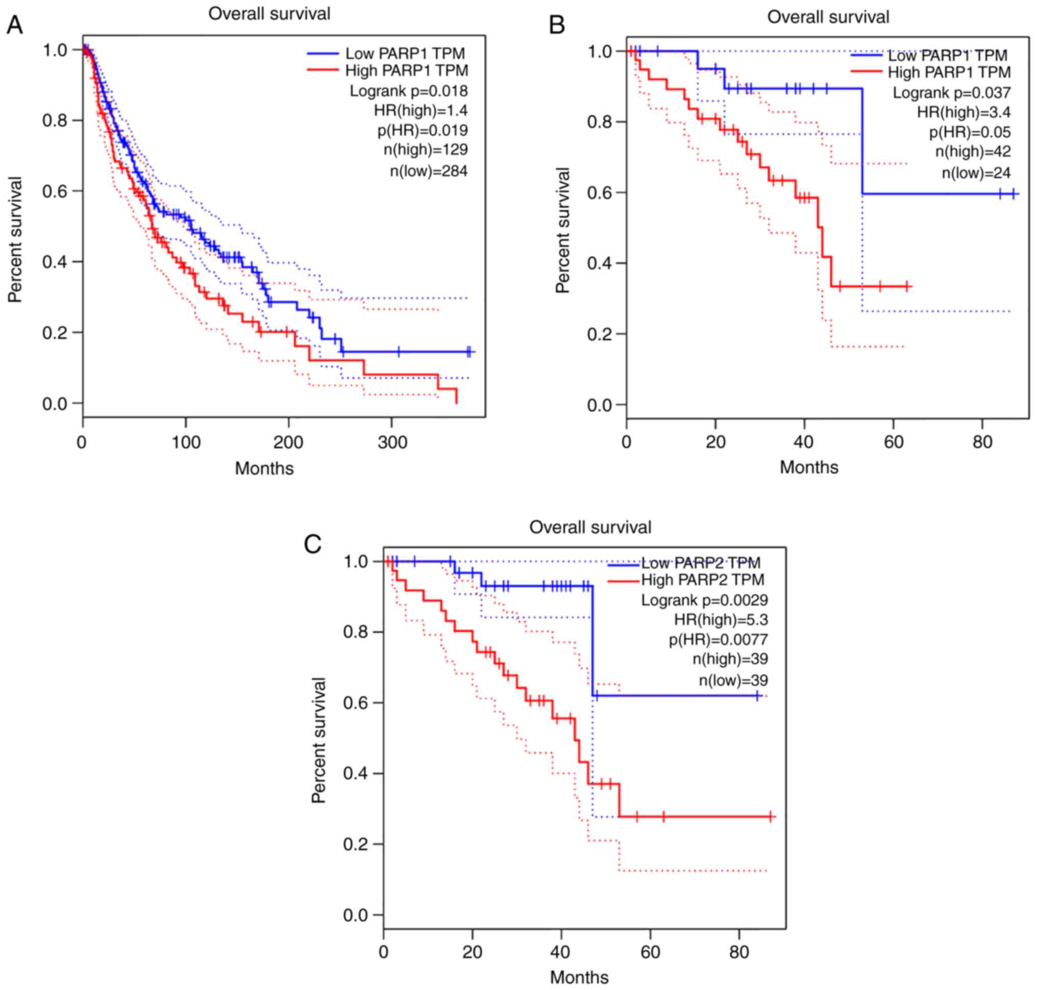
Data from the GEPIA database demonstrated that high PARP expression
was correlated with poor OS in melanoma (Fig. 1). However, no significant
differences were observed between the effects of PARPis (1–10 µM)
on melanoma cells (Fig. 2). Testing
of the combination of angiogenesis inhibitors with PARPis
demonstrated that veliparib reversed the inhibitory effect of
sunitnib, an angiogenesis inhibitor, on A-375 cells (Fig. 2). Thus, instead of PARPis, this
study focused on the role of PARP1 expression in tumours. The
present study screened for compounds that significantly inhibited
PARP1 expression and explored the role of PARP1 in melanoma
progression to provide a basis for assessing PARP1 as a tumour
target.
High PARP1 expression is associated
with melanoma OS
The relationship between PARP expression and
melanoma prognosis was analysed. Data from the GEPIA database
demonstrated that high PARP expression was associated with poor OS
in melanoma (Fig. 1). Notably, high
PARP1 expression was associated with poor OS in SKCM (Fig. 1A) and UVM (Fig. 1B). Additionally, high PARP2
expression was associated with a poor OS in UVM (Fig. 1C).
PARPis reverse the inhibitory effect
of sunitinib on PARP1 expression and melanoma cell
proliferation
After evaluating the association between PARP1 and
melanoma prognosis, the effects of PARPis on melanoma cells were
explored. The results of the present study demonstrated that
veliparib (1–10 µM) and olaparib (1–10 µM) had no significant
effect on melanoma cell survival (Fig.
2A and B). Although 30 µM veliparib had a partial inhibitory
effect on melanoma cell survival, but this concentration appears
high for an antitumour drug. In addition, the combination of PARPis
and other anti-tumour drugs was tested. As an antiangiogenic drug,
sunitinib may have activity in patients with melanoma and KIT
mutations (22). Thus, the effects
of combining sunitinib, an angiogenesis inhibitor, and PARPis on
melanoma cell growth and PARP1 expression were evaluated. The
results demonstrated that sunitinib significantly inhibited
melanoma cell proliferation and that veliparib reversed this
inhibitory effect on A-375 cells (Fig.
2C). Similarly, another PARPi, olaparib, also significantly
reversed the inhibitory effect of sunitinib on melanoma cell
proliferation (Fig. 2C). In
addition, sunitinib significantly inhibited PARP1 expression in
melanoma cells, an effect that was significantly reversed by
veliparib without significantly affecting the expression of Bcl-2,
FoxO3a or pFoxO3a (Fig. 2D).
Additionally, veliparib partly reversed the inhibitory effect of
docetaxel, a tubulin polymerization promoter, on melanoma cell
proliferation (Fig. 2E). These
results suggested that PARP1 expression may be associated with
melanoma cell proliferation or survival.
Tanshinone I and simvastatin exert
inhibitory effects on melanoma A-375 cell proliferation and PARP1
expression
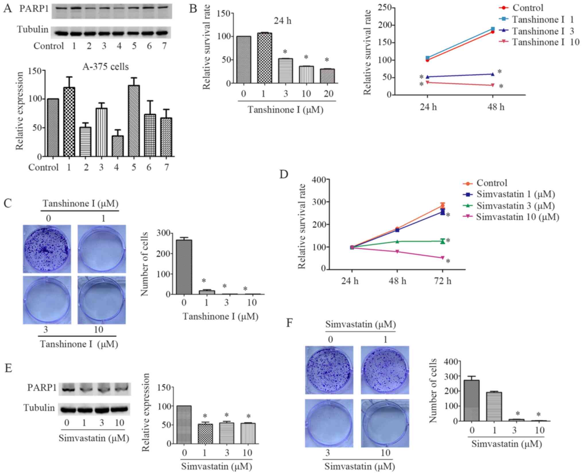
To further confirm the relationship between PARP1
expression and melanoma cells, the present study screened for drugs
that significantly inhibited PARP1 expression among common drugs
and traditional Chinese medicine monomers (mefloquine, simvastatin,
dihydroartemisinin, tanshinone I, cryptotanshinone, gossypol and
dioscin). The results demonstrated that tanshinone I and
simvastatin exerted a notable inhibitory effect on PARP1 expression
in melanoma A-375 cells (Fig.
3A).
The effects of tanshinone I and simvastatin on the
molecular biological function of melanoma cells in vitro
were investigated. Compared with that of the control group,
tanshinone I significantly inhibited A-375 cell proliferation
(Fig. 3B) and colony formation
(Fig. 3C). By comparing the
druggability of the tanshinone I and simvastatin, simvastatin,
which is already a clinically used drug, was used to assess its
effects on PARP1 expression. Compared with that of the control
group, simvastatin significantly inhibited proliferation (Fig. 3D), PARP1 expression (Fig. 3E) and colony formation (Fig. 3F) in A-375 cells.
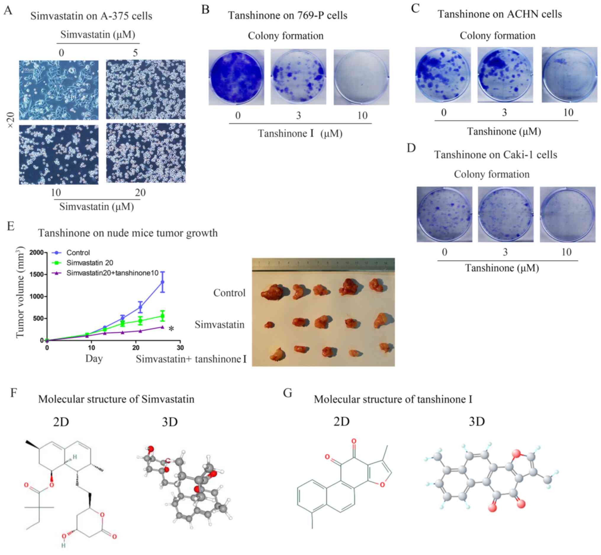
Tanshinone I and simvastatin inhibit
tumour growth in nude mice
Simvastatin notably affected the morphology of A-375
cells, which were markedly rounded with characteristic
morphological changes of apoptosis (Fig. 4A). In addition to melanoma cells,
the results also demonstrated that tanshinone I significantly
inhibited colony formation in 769-P renal cancer cells (Fig. 4B), Caki-1 cells (Fig. 4C) and ACHN cells (Fig. 4D). Subsequently, the combined effect
of tanshinone I and simvastatin on melanoma was explored in
vivo. The combination of tanshinone I and simvastatin inhibited
the growth of tumour xenografts formed by A-375 cells in nude mice
compared with that of the control group (Fig. 4E). Finally, in order to examine the
molecular characteristics and differences of the two compounds, the
two-dimensional and three-dimensional structures of simvastatin and
tanshinone I were obtained using PubChem (https://pubchem.ncbi.nlm.nih.gov/), as demonstrated in
Fig. 4F and G. Based on the
molecular characteristics and medicinal properties of tanshinone I
and simvastatin, the combination of simvastatin and tanshinone I
may exert an inhibitory effect on tumour progression.
Discussion
The current database analysis demonstrated that
PARP1 was negatively associated with the OS and prognosis of
melanoma. Sunitinib inhibited PARP1 expression and proliferation in
A-375 cells, whereas its effects were partly reversed by veliparib.
Screening revealed simvastatin and tanshinone I as compounds that
inhibited PARP1 expression, and their combination inhibited the
growth of xenograft tumours constructed using A-375 cells in nude
mice. The results of the present study identified drugs (tanshinone
I and simvastatin) that inhibited PARP1 expression, and provided
evidence that tanshinone I may improve melanoma tumour cell
sensitivity to simvastatin by regulating PARP1 expression.
The identification of specific targets to inhibit
tumour cell growth or enhance the efficacy of
chemotherapy/radiotherapy provides an important basis for the
development of anti-tumour drugs. Although PARPis have been
approved for BRCA mutation-associated cancer, their effects on
other molecular tumour subtypes and the specific mechanism
underlying the effects of PARP on tumour proliferation, invasion
and metastasis remain to be further clarified. Tanshinone I is a
monomer of Salvia miltiorrhiza and inhibits tumour cell
proliferation (12); however, its
specific mechanism remains to be determined. The present study
identified tanshinone I and simvastatin as compounds that exerted
inhibitory effects on PARP1 expression, and demonstrated that
tanshinone I improved tumour sensitivity to simvastatin.
Various tyrosine kinase inhibitors have been
approved for melanoma treatment (23); however, their effectiveness is
limited because of drug resistance. A novel benzoxazole compound
exhibited synergistic anti-tumour effects in combination with
vemurafenib (a BRAF inhibitor) and docetaxel (24). Unlike O6-alkylguanine DNA
alkyltransferase (MGMT)-deficient melanoma cells, an
antiproliferative senescent response induced by temozolomide was
enhanced by PARPi in MGMT-positive cancer cells (25), which indicates the different
functions for PARPi. KIT mutations may serve as an adverse
prognostic factor in metastatic melanoma and sunitinib may have
activity in patients with melanoma and KIT mutations (22). In the present study, sunitinib
significantly inhibited PARP1 expression. However, the combination
of a PARPi and sunitinib reduced the inhibitory effects of
sunitinib on melanoma cell growth and PARP1 expression. Notably,
inhibition of PARP1 activity in the cells by veliparib appeared to
stimulate PARP1 expression; this may be associated with the
feedback inhibition, similar to the phenomenon of phorbol
12-myristate-13-acetate on tumour necrosis factor-α converting
enzyme to some extent (26).
Cells overexpressing Bcl-2 have been reported to
exhibit a significantly improved response to salvage radiotherapy
compared with that of cells with low Bcl-2 expression (27). In the present study, a PARPi and
sunitinib affected PARP1 expression, but did not affect the
expression of Bcl-2 and pFoxO3a. The initial PARPis are analogs of
nicotinamide designed to compete with nicotinamide adenine
dinucleotide at the catalytic pocket of PARP to modulate enzyme
activity (28). Detection of PARP1
expression in this study indicated that, besides the enzymatic
activity of PARP1, PARP1 expression may be important for
carcinogenesis. Olaparib increased the response to dacarbazine, an
alkylating agent, in a patient-derived xenograft model of uveal
melanoma (8). Although veliparib
and olaparib have been reported to increase the sensitivity of
various histological subtypes of SNP carrier cancer cells to
alkylating agents, they have no effect on wild-type cells (11). The results of the present study
demonstrated that veliparib and olaparib had no significant effect
on melanoma cell survival. These results were concordant with those
reported by a previous study in which clinicians demonstrated
resistance to olaparib in patients with cancer (29). However, the sensitivity may also be
associated with the tumour type, as breast cancer cells exhibited
increased sensitivity to the same concentration of olaparib
(30).
The present study screened for compounds that
affected PARP1 expression, and preliminarily evaluated the drug
potency of these compounds and their combined effect on tumour
growth to provide a way of exploring PARP1-targeted anti-tumour
drugs. Through experimental verification, tanshinone I and
simvastatin provided a good foundation for the development of
anti-tumour drugs targeting PARP1 and their chemical structure
derivatives are anticipated. However, whether the other targets of
simvastatin and tanshinone I were involved in their effects remains
to be studied. Therefore, further studies of these compounds are
essential for identifying additional related targets for this
combined effect. In addition, although proliferation, colony
formation and morphology were assessed, the lack of direct
apoptosis and cell cycle analyses may be a limitation of the
present study.
The doses of statins required for anti-tumour
effects are 100- to 500-fold higher than those needed to lower
cholesterol levels; therefore, the use of tumour-targeted delivery
systems may greatly improve their anti-tumour efficacy (31). In this study, the combination of
tanshinone I and simvastatin improved the anti-tumour efficacy of
simvastatin, but whether the combined effect of tanshinone I and
other types of statins have the same effect requires further
assessment. In a previous study, simvastatin increased the
anti-tumour activity of paclitaxel (PTX) carried by lipid
nanoemulsions (LDE), but not of the commercial PTX (not carried by
LDE), possibly because of increased low-density lipoprotein
receptor expression by statins that bind and internalize LDE-PTX
(32). Thus, other potential
mechanisms for the effects of the combination of simvastatin and
tanshinone I on melanoma require further exploration. Simvastatin
is a prodrug for β-hydroxy β-methylglutaryl-CoA reductase, which is
activated by drug-metabolizing enzymes into metabolite in
vivo (33). Compared with the
in vivo metabolism, the drug-metabolizing enzyme activity of
cancer cells in vitro is usually lower. As the results of
the present study demonstrated that simvastatin inhibited melanoma
cells in vitro and in vivo, it may be hypothesized
that simvastatin and its metabolite exert inhibitory activity on
melanoma cells.
In conclusion, the results of the present study
demonstrated that tanshinone I and simvastatin significantly
inhibited PARP1 expression, and that tanshinone I may effectively
improve tumour cell sensitivity to simvastatin. To provide more
evidence for the function of tanshinone I on different types of
tumour cells, renal cancer cells were selected for functional
experiments. Similar to the result on melanoma cells, tanshinone I
also inhibited the colony formation of renal cancer cells. These
findings suggested that inhibiting PARP1 expression may be a
potential method for treatment of melanoma and renal cell
carcinoma. The enzyme activity and expression of PARP1 may serve a
role in tumour progression by different mechanisms.
Acknowledgements
Not applicable.
Funding
The current study was funded in part by research
grants from the Science and Technology Project of Guangzhou
Municipal Health and Family Planning Commission (grant no.
20181A011059) and the National Natural Science Foundation of China
(grant nos. 81570633 and 81803576).
Availability of data and materials
The datasets used and/or analysed during the current
study are available from the corresponding author on reasonable
request.
Authors' contributions
YZ and YL performed the majority of experiments,
analysed the data and prepared the manuscript. WenqiW designed the
project and provided expert advice to the study, and revised the
manuscript. JH analysed the survival data of melanoma obtained from
the database. YH, SZ and WeizhouW performed the in vivo
experiments. HL and XD analysed parts of the data, revised and
discussed the manuscript. All authors read and approved the final
manuscript.
Ethics approval and consent to
participate
The animal experiments were approved by the ethics
committee of the First Affiliated Hospital of Guangzhou Medical
University.
Patient consent for publication
Not applicable.
Competing interests
The authors declare that they have no competing
interests.
References
|
1
|
Kummar S, Chen A, Parchment RE, Kinders
RJ, Ji J, Tomaszewski JE and Doroshow JH: Advances in using PARP
inhibitors to treat cancer. Bmc Med. 10:252012. View Article : Google Scholar : PubMed/NCBI
|
|
2
|
Luo X and Kraus WL: On PAR with PARP:
Cellular stress signaling through poly(ADP-ribose) and PARP-1.
Genes Dev. 26:417–432. 2012. View Article : Google Scholar : PubMed/NCBI
|
|
3
|
Donizy P, Wu CL, Mull J, Fujimoto M,
Chłopik A, Peng Y, Shalin SC, Selim MA, Puig S, Fernandez-Figueras
MT, et al: Up-regulation of PARP1 expression significantly
correlated with poor survival in mucosal melanomas. Cells.
9:11352020. View Article : Google Scholar
|
|
4
|
Hsieh MH, Chen YT, Chen YT, Lee YH, Lu J,
Chien CL, Chen HF, Ho HN, Yu CJ, Wang ZQ and Teng SC: PARP1
controls KLF4-mediated telomerase expression in stem cells and
cancer cells. Nucleic Acids Res. 45:10492–10503. 2017. View Article : Google Scholar : PubMed/NCBI
|
|
5
|
Pilié PG, Gay CM, Byers LA, O'Connor MJ
and Yap TA: PARP inhibitors: Extending benefit beyond BRCA-mutant
cancers. Clin Cancer Res. 25:3759–3771. 2019. View Article : Google Scholar : PubMed/NCBI
|
|
6
|
Bian C, Zhang C, Luo T, Vyas A, Chen SH,
Liu C, Kassab MA, Yang Y, Kong M and Yu X: NADP+ is an
endogenous PARP inhibitor in DNA damage response and tumor
suppression. Nat Commun. 10:6932019. View Article : Google Scholar : PubMed/NCBI
|
|
7
|
Dinnes J, errante di Ruffano L, Takwoingi
Y, Cheung ST, Nathan P, Matin RN, Chuchu N, Chan SA, Durack A,
Bayliss SE, et al: Ultrasound, CT, MRI, or PET-CT for staging and
re-staging of adults with cutaneous melanoma. Cochrane Database
Syst Rev. 7:CD0128062019.PubMed/NCBI
|
|
8
|
de Koning L, Decaudin D, El BR, Nicolas A,
Carita G, Schuller M, Ouine B, Cartier A, Naguez A, Fleury J, et
al: PARP inhibition increases the response to chemotherapy in uveal
melanoma. Cancers (Basel). 11:7512019. View Article : Google Scholar
|
|
9
|
Fratangelo F, Camerlingo R, Carriero MV,
Pirozzi G, Palmieri G, Gentilcore G, Ragone C, Minopoli M, Ascierto
PA and Motti ML: Effect of ABT-888 on the apoptosis, motility and
invasiveness of BRAFi-resistant melanoma cells. Int J Oncol.
53:1149–1159. 2018.PubMed/NCBI
|
|
10
|
Middleton MR, Friedlander P, Hamid O, Daud
A, Plummer R, Falotico N, Chyla B, Jiang F, McKeegan E, Mostafa NM,
et al: Randomized phase II study evaluating veliparib (ABT-888)
with temozolomide in patients with metastatic melanoma. Ann Oncol.
26:2173–2179. 2015. View Article : Google Scholar : PubMed/NCBI
|
|
11
|
Abecassis I, Sedgewick AJ, Romkes M, Buch
S, Nukui T, Kapetanaki MG, Vogt A, Kirkwood JM, Benos PV and Tawbi
H: PARP1 rs1805407 increases sensitivity to PARP1 inhibitors in
cancer cells suggesting an improved therapeutic strategy. Sci Rep.
9:33092019. View Article : Google Scholar : PubMed/NCBI
|
|
12
|
Ren J, Fu L, Nile SH, Zhang J and Kai G:
Salvia miltiorrhiza in treating cardiovascular diseases: A review
on its pharmacological and clinical applications. Front Pharmacol.
10:7532019. View Article : Google Scholar : PubMed/NCBI
|
|
13
|
Sui H, Zhao J, Zhou L, Wen H, Deng W, Li
C, Ji Q, Liu X, Feng Y, Chai N, et al: Tanshinone IIA inhibits
β-catenin/VEGF-mediated angiogenesis by targeting TGF-β1 in
normoxic and HIF-1α in hypoxic microenvironments in human
colorectal cancer. Cancer Lett. 403:86–97. 2017. View Article : Google Scholar : PubMed/NCBI
|
|
14
|
Munagala R, Aqil F, Jeyabalan J and Gupta
RC: Tanshinone IIA inhibits viral oncogene expression leading to
apoptosis and inhibition of cervical cancer. Cancer Lett.
356:536–546. 2015. View Article : Google Scholar : PubMed/NCBI
|
|
15
|
Yan Y, Su W, Zeng S, Qian L, Chen X, Wei
J, Chen N, Gong Z and Xu Z: Effect and mechanism of tanshinone I on
the radiosensitivity of lung cancer cells. Mol Pharm. 15:4843–4853.
2018. View Article : Google Scholar : PubMed/NCBI
|
|
16
|
Lee CY, Sher HF, Chen HW, Liu CC, Chen CH,
Lin CS, Yang PC, Tsay HS and Chen JJ: Anticancer effects of
tanshinone I in human non-small cell lung cancer. Mol Cancer Ther.
7:3527–3538. 2008. View Article : Google Scholar : PubMed/NCBI
|
|
17
|
Ding C, Tian Q, Li J, Jiao M, Song S, Wang
Y, Miao Z and Zhang A: Structural modification of natural product
tanshinone I leading to discovery of novel nitrogen-enriched
derivatives with enhanced anticancer profile and improved drug-like
properties. J Med Chem. 61:760–776. 2018. View Article : Google Scholar : PubMed/NCBI
|
|
18
|
Davies JR, Jewell R, Affleck P, Anic GM,
Randerson-Moor J, Ozola A, Egan KM, Elliott F, García-Casado Z,
Hansson J, et al: Inherited variation in the PARP1 gene and
survival from melanoma. Int J Cancer. 135:1625–1633. 2014.
View Article : Google Scholar : PubMed/NCBI
|
|
19
|
Lai Y, Kong Z, Zeng T, Xu S, Duan X, Li S,
Cai C, Zhao Z and Wu W: PARP1-siRNA suppresses human prostate
cancer cell growth and progression. Oncol Rep. 39:1901–1909.
2018.PubMed/NCBI
|
|
20
|
Hans CP, Zerfaoui M, Naura AS, Catling A
and Boulares AH: Differential effects of PARP inhibition on
vascular cell survival and ACAT-1 expression favouring
atherosclerotic plaque stability. Cardiovasc Res. 78:429–439. 2008.
View Article : Google Scholar : PubMed/NCBI
|
|
21
|
Tang Z, Li C, Kang B, Gao G, Li C and
Zhang Z: GEPIA: A web server for cancer and normal gene expression
profiling and interactive analyses. Nucleic Acids Res. 45((W1)):
W98–W102. 2017. View Article : Google Scholar : PubMed/NCBI
|
|
22
|
Minor DR, Kashani-Sabet M, Garrido M,
O'Day SJ, Hamid O and Bastian BC: Sunitinib therapy for melanoma
patients with KIT mutations. Clin Cancer Res. 18:1457–1463. 2012.
View Article : Google Scholar : PubMed/NCBI
|
|
23
|
Drilon A, Siena S, Ou SI, Patel M, Ahn MJ,
Lee J, Bauer TM, Farago AF, Wheler JJ, Liu SV, et al: Safety and
antitumor activity of the multitargeted pan-TRK, ROS1, and ALK
inhibitor entrectinib: Combined results from two phase I trials
(ALKA-372-001 and STARTRK-1). Cancer Discov. 7:400–409. 2017.
View Article : Google Scholar : PubMed/NCBI
|
|
24
|
Cheng Y, Wang X, Xia X, Zhang W and Tian
H: A benzoxazole compound as a novel MEK inhibitor for the
treatment of RAS/RAF mutant cancer. Int J Cancer. 145:586–596.
2019. View Article : Google Scholar : PubMed/NCBI
|
|
25
|
Erice O, Smith MP, White R, Goicoechea I,
Barriuso J, Jones C, Margison GP, Acosta JC, Wellbrock C and
Arozarena I: MGMT expression predicts PARP-mediated resistance to
temozolomide. Mol Cancer Ther. 14:1236–1246. 2015. View Article : Google Scholar : PubMed/NCBI
|
|
26
|
Doedens JR and Black RA:
Stimulation-induced down-regulation of tumour necrosis factor-alpha
converting enzyme. J Biol Chem. 275:14598–14607. 2000. View Article : Google Scholar : PubMed/NCBI
|
|
27
|
Oing C, Tennstedt P, Simon R, Volquardsen
J, Borgmann K, Bokemeyer C, Petersen C, Dikomey E, Rothkamm K and
Mansour WY: BCL2-overexpressing prostate cancer cells rely on
PARP1-dependent end-joining and are sensitive to combined PARP
inhibitor and radiation therapy. Cancer Lett. 423:60–70. 2018.
View Article : Google Scholar : PubMed/NCBI
|
|
28
|
Shen Y, Aoyagi-Scharber M and Wang B:
Trapping poly(ADP-Ribose) polymerase. J Pharmacol Exp Ther.
353:446–457. 2015. View Article : Google Scholar : PubMed/NCBI
|
|
29
|
Raineri A, Prodomini S, Fasoli S, Gotte G
and Menegazzi M: Influence of onconase in the therapeutic potential
of PARP inhibitors in A375 malignant melanoma cells. Biochem
Pharmacol. 167:173–181. 2019. View Article : Google Scholar : PubMed/NCBI
|
|
30
|
Ma X, Dang C, Min W, Diao Y, Hui W, Wang
X, Dai Z, Wang X and Kang H: Downregulation of APE1 potentiates
breast cancer cells to olaparib by inhibiting PARP-1 expression.
Breast Cancer Res Treat. 176:109–117. 2019. View Article : Google Scholar : PubMed/NCBI
|
|
31
|
Alupei MC, Licarete E, Patras L and Banciu
M: Liposomal simvastatin inhibits tumour growth via targeting
tumour-associated macrophages-mediated oxidative stress. Cancer
Lett. 356:946–952. 2015. View Article : Google Scholar : PubMed/NCBI
|
|
32
|
Kretzer IF, Maria DA, Guido MC, Contente
TC and Maranhao RC: Simvastatin increases the antineoplastic
actions of paclitaxel carried in lipid nanoemulsions in
melanoma-bearing mice. Int J Nanomedicine. 11:885–904.
2016.PubMed/NCBI
|
|
33
|
Geboers S, Stappaerts J, Tack J, Annaert P
and Augustijns P: In vitro and in vivo investigation of the
gastrointestinal behavior of simvastatin. Int J Pharm. 510:296–303.
2016. View Article : Google Scholar : PubMed/NCBI
|