Introduction
Acute myeloid leukemia (AML) is a clinically and
biologically heterogeneous malignancy featured by abnormal clonal
proliferation of immature myeloid precursors in the bone marrow,
which can spread into the blood and other organs such as the spleen
and liver (1,2). Patients with AML suffer from fatigue,
recurrent infections and hemorrhage, as leukemic cells are
dysfunctional (3). Current
treatment protocols are restricted to intensive chemotherapy and
the judicious use of allogeneic stem cell transplantation (1). However, only 20–30% of patients
achieve long-term survival (4).
This dismal prognosis is predominantly caused by the development of
chemoresistance, unacceptable side effects of intensive
chemotherapy and relapse (1). To
improve the prognosis of patients with AML, efforts should be made
to identify novel and sensitive biomarkers for recognizing patients
with AML who are at risk of poor prognosis and for optimizing
treatment strategies.
Long non-coding RNA (lncRNA) antisense non-coding
RNA in the INK4 locus (ANRIL) is located in the chromosome 9p21
region and is identified in patients with familial melanoma with
germline deletion in the INK4B-ARF-INK4A gene cluster (5,6).
Several studies have reported that lncRNA ANRIL is essential for
the pathogenesis of various cancers (7–13). For
instance, lncRNA ANRIL enhances the growth, invasion and migration
of cancer cells in laryngeal squamous cell and hepatocellular
carcinoma (7,8). In AML, lncRNA ANRIL facilitates cell
proliferation, migration and invasion, and represses cell apoptosis
by modulating the expression of microRNA (miR)-34a, histone
deacetylase 1 and ASPP2 (12).
Furthermore, lncRNA ANRIL enhances malignant cell survival via a
glucose metabolism pathway involving adiponectin receptor
(AdipoR1)/AMP-activated protein kinase (AMPK)/sirtuin 1 (SIRT1) in
AML (13). On this basis, it was
hypothesized that lncRNA ANRIL may have a clinical implication in
the prediction of risk, progression and prognosis of AML. However,
to the best of our knowledge, no previous clinical study has
reported on this topic. Therefore, the present study aimed to
explore the association of lncRNA ANRIL with disease risk, clinical
features and prognosis of AML.
Materials and methods
Participants
In the present prospective study, 178 patients with
de novo AML who were admitted to the Central Hospital of
Xiangtan (Xiangtan, China) were consecutively recruited between
January 2016 and June 2019. All patients met the following
criteria: i) Newly diagnosed with primary AML by morphology,
immunophenotyping, cytogenetics or/and molecular genetic
examinations, based on the World Health Organization Morphology,
Immunology, Cytogenetics Molecular biology classification criteria
(14); ii) age ≥18 years; iii) no
history of hematopoietic or lymphoid tissue diseases prior to the
diagnosis of AML; iv) no complication with other malignancies; and
v) ability to be followed up regularly. However, patients were
excluded if they had acute promyelocytic leukemia, secondary or
relapsed AML, if they were infected with human immunodeficiency
virus or if they were pregnant or lactating females. Furthermore,
30 healthy bone marrow donors were enrolled in the present study
during the same period when they examined the eligibility for bone
marrow transplantation. None of the healthy donors had a history of
hematopoietic or lymphoid tissue malignancies and their health
status was confirmed prior to bone marrow donation. The present
study was approved by the Ethics Committee of the Central Hospital
of Xiangtan (Xiangtan, China). All patients with AML and healthy
donors voluntarily participated in the present study and signed the
informed consent forms prior to enrollment.
Collection of bone marrow and clinical
data prior to therapy
Bone marrow samples of enrolled patients were
extracted prior to initial therapy, while bone marrow samples from
healthy donors were collected when undergoing donation. Bone marrow
mononuclear cells (BMMCs) were isolated from the collected samples
by density-gradient centrifugation and were stored at −80°C for
subsequent detection of lncRNA ANRIL. The patients' baseline
characteristics were documented after completion of diagnostic
procedures, which comprised age, gender, French-American-Britain
(FAB) classification (15),
cytogenetics features, molecular genetic features, risk
stratification [based on cytogenetics and molecular abnormalities
according to the National Comprehensive Cancer Network AML
Guidelines Version 2.2014 (16)]
and white blood cell (WBC) count.
Detection of lncRNA ANRIL
The relative expression of lncRNA ANRIL in BMMCs was
determined by reverse transcription-quantitative PCR (RT-qPCR).
Total RNA from BMMCs was extracted using TRIzol™ reagent (Thermo
Fisher Scientific, Inc.). Subsequently, complementary DNA (cDNA)
synthesis was conducted with an iScript™ cDNA Synthesis kit (with
redon primer; Bio-Rad Laboratories, Inc.) and qPCR was carried out
with QTaq™ DNA Polymerase mix (Clontech Laboratories, Inc.) and
Applied Biosystems 7900HT Fast Real-Time PCR system (Thermo Fisher
Scientific, Inc.). The PCR amplification program was as follows:
95°C for 30 sec, followed by 40 cycles of amplification (95°C for 5
sec, 60°C for 30 sec). GAPDH was applied as an internal reference
for lncRNA ANRIL and the relative expression of lncRNA ANRIL was
calculated by the 2−ΔΔCq method (17). The primers used are listed in
Table I.
 | Table I.Primers used for quantitative PCR. |
Table I.
Primers used for quantitative PCR.
| Item | Forward primer | Reverse primer |
|---|
| LncRNA ANRIL |
TGCTCTATCCGCCAATCAGG |
GGGCCTCAGTGGCACATACC |
| GAPDH |
TGACCACAGTCCATGCCATCAC |
GCCTGCTTCACCACCTTCTTGA |
Response assessment after induction
therapy
Following standard induction therapy with
anthracycline (daunorubicin, idarubicin or the anthracenedione
mitoxantrone) for 3 days, followed by 7 days of cytarabine or
therapies of comparable intensity, response assessment was commonly
performed between days 21 and 28 after the start of induction
therapy (18). Complete remission
(CR) was evaluated according to the response criteria recommended
by an international expert panel (on behalf of the European
LeukemiaNet) (19).
Follow-up and survival assessment
Surveillance and follow-up were performed every 1–3
months for the first two years and every 3–6 months subsequently.
The survival status of patients was recorded during follow-up until
June 30, 2019. Event-free survival (EFS) was determined as the time
from the date of initial therapy to the date of induction treatment
failure, relapse from CR or death. Patients not known to have
experienced any of these events at the last follow-up date were
censored on the date of their last examination. Overall survival
(OS) was determined as the time from the date of initial therapy to
the date of death. Patients not known to have died at the last
follow-up date were censored on the date they were last known to be
alive.
Statistical analysis
Values are expressed as the mean ± standard
deviation, median and interquartile range or n (%). The difference
in expression of lncRNA ANRIL between patients with AML and healthy
donors was determined by the Wilcoxon rank-sum test. For the
analysis of correlation of lncRNA ANRIL with clinical features,
patients were divided into an lncRNA ANRIL-high group and lncRNA
ANRIL-low group according to the median value of lncRNA ANRIL
relative expression of all patients with AML. Comparison of
clinical features between the lncRNA ANRIL-high and -low groups was
performed by χ2, Fisher's exact and Wilcoxon rank-sum
tests. A receiver operating characteristic (ROC) curve was used to
evaluate the value of lncRNA ANRIL in differentiating patients with
AML from healthy donors. Kaplan-Meier curves were plotted to
display the EFS and OS, and the difference of EFS and OS between
the lncRNA ANRIL-high and -low groups was determined by the
log-rank test. Factors affecting EFS and OS were analyzed by
univariate and multivariate Cox's proportional hazard regression
models. SPSS version 22.0 (IBM, Corp.) was used for statistical
analyses and figures were plotted using GraphPad Prism version 7.00
(GraphPad Software, Inc.). P<0.05 was considered to indicate a
statistically significant difference.
Results
Characteristics of healthy donors and
patients with AML
In healthy donors, the mean age was 52.2±22.5 years,
and there were 16 (53.3%) females as well as 14 (46.6%) males. The
median WBC count was 9.6 (7.6–12.5) ×109 cells/l
(Table II). In patients with AML,
the mean age was 52.1±15.2 years). There were 66 (37.1%) females
and 112 (62.9%) males. Regarding the FAB classification, 65
(36.5%), 48 (27.0%), 58 (32.6%) and 7 (3.9%) patients with AML were
classified as FAB M2, M4, M5 and M6, respectively. In terms of
cytogenetics, 95 (53.3%), 17 (9.6%), 16 (9.0%), 7 (3.9%), 7 (3.9%),
6 (3.4%), 5 (2.8%), 4 (2.2%), 3 (1.7%), 1 (0.6%), 1 (0.6%), 16
(9.0%) and 14 (7.9%) patients with AML had normal karyotype (NK),
complex karyotype (CK), inv(16) or t(16;16), t(8;21), −7 or 7q-,
t(9;11), +8, t(9;22), 11q23, −5 or 5q-, t(6;9), others (undefined)
and monosomal karyotype (MK), respectively. Regarding molecular
genetics, 39 (21.9%), 16 (9.0) and 66 (37.1%) patients with AML
exhibited internal tandem duplications in FMS-like tyrosine kinase
3 (FLT3-ITD), isolated biallelic CCAAT/enhancer-binding protein α
(CEBPA) mutation and nucleophosmin 1 (NPMI) mutations,
respectively. With respect to risk stratification, 53 (29.8%), 69
(38.8) and 56 (31.4%) patients with AML had better-, intermediate-
and poor-risk stratification, respectively. In addition, the median
WBC count was 17.7 (8.9–28.6) ×109 cells/l [normal WBC
range (4.0–10.0) ×109/l] in patients with AML.
| Table II.Baseline characteristics of healthy
patients and patients with acute myeloid leukemia (n=178). |
Table II.
Baseline characteristics of healthy
patients and patients with acute myeloid leukemia (n=178).
| Item | Patients with AML,
value | Healthy patients,
value |
|---|
| Age (years) | 52.1±15.2 | 52.2±22.5 |
| Sex |
|
|
|
Female | 66 (37.1) | 16 (53.3 |
| Male | 112 (62.9) | 14 (46.7) |
| FAB
classification |
|
|
| M2 | 65 (36.5) | – |
| M4 | 48 (27.0) | – |
| M5 | 58 (32.6) | – |
| M6 | 7 (3.9) | – |
| Cytogenetics |
|
|
| NK | 95 (53.3) | – |
| CK | 17 (9.6) | – |
| inv(16)
or t(16;16) | 16 (9.0) | – |
|
t(8;21) | 7 (3.9) | – |
| -7 or
7q- | 7 (3.9) | – |
|
t(9;11) | 6 (3.4) | – |
| +8 | 5 (2.8) | – |
|
t(9;22) | 4 (2.2) | – |
|
11q23 | 3 (1.7) | – |
| -5 or
5q- | 1 (0.6) | – |
|
t(6;9) | 1 (0.6) | – |
| Others
(undefined) | 16 (9.0) | – |
| MK | 14 (7.9) | – |
| FLT3-ITD
mutation | 39 (21.9) | – |
| Isolated biallelic
CEBPA mutation | 16 (9.0) | – |
| NPMI mutation | 66 (37.1) | – |
| Risk
stratification |
|
|
|
Better-risk | 53 (29.8) | – |
|
Intermediate-risk | 69 (38.8) | – |
|
Poor-risk | 56 (31.4) | – |
| WBC
(×109/l)normal range (4.0–10.0) | 17.7
(8.9–28.6) | 9.6 (7.6–12.5) |
Association of lncRNA ANRIL with AML
risk
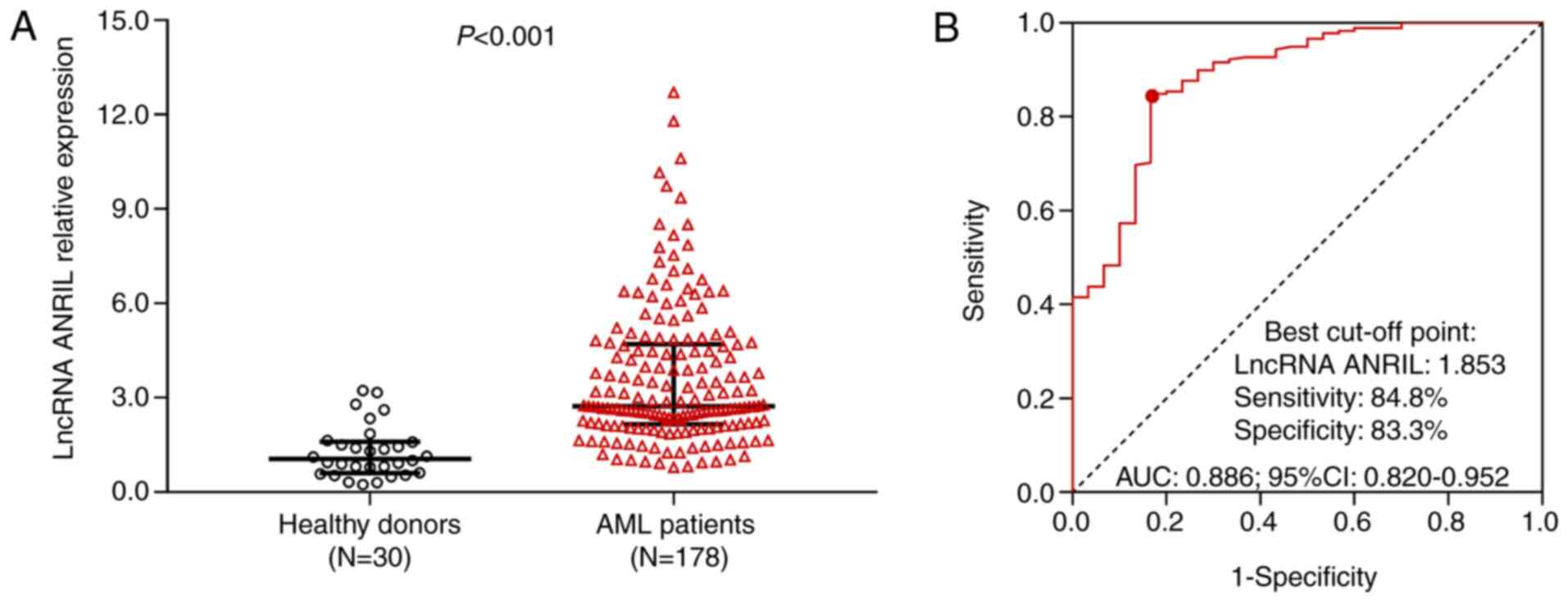
The relative expression of lncRNA ANRIL was
increased in patients with AML compared with that in healthy donors
(P<0.001; Fig. 1A). ROC curve
analysis revealed that lncRNA ANRIL was able to distinguish
patients with AML from healthy donors [area under the curve (AUC),
0.886; 95% CI, 0.820–0.952], with a sensitivity of 84.8% and a
specificity of 83.3% at the best cut-off point (where the value of
sensitivity plus specificity was the largest) (Fig. 1B).
Association of lncRNA ANRIL with FAB
classification and molecular genetics
lncRNA ANRIL was associated with elevated FLT3-ITD
mutation (P=0.046; Fig. 2; Fig. 2C, while no association of lncRNA
ANRIL with FAB classification (P=0.676; Fig. 2A), MK (P=0.578; Fig. 2B), isolated biallelic CEBPA mutation
(P=0.600; Fig. 2D) or NPMI mutation
(P=0.352; Fig. 2E) was observed in
patients with AML.
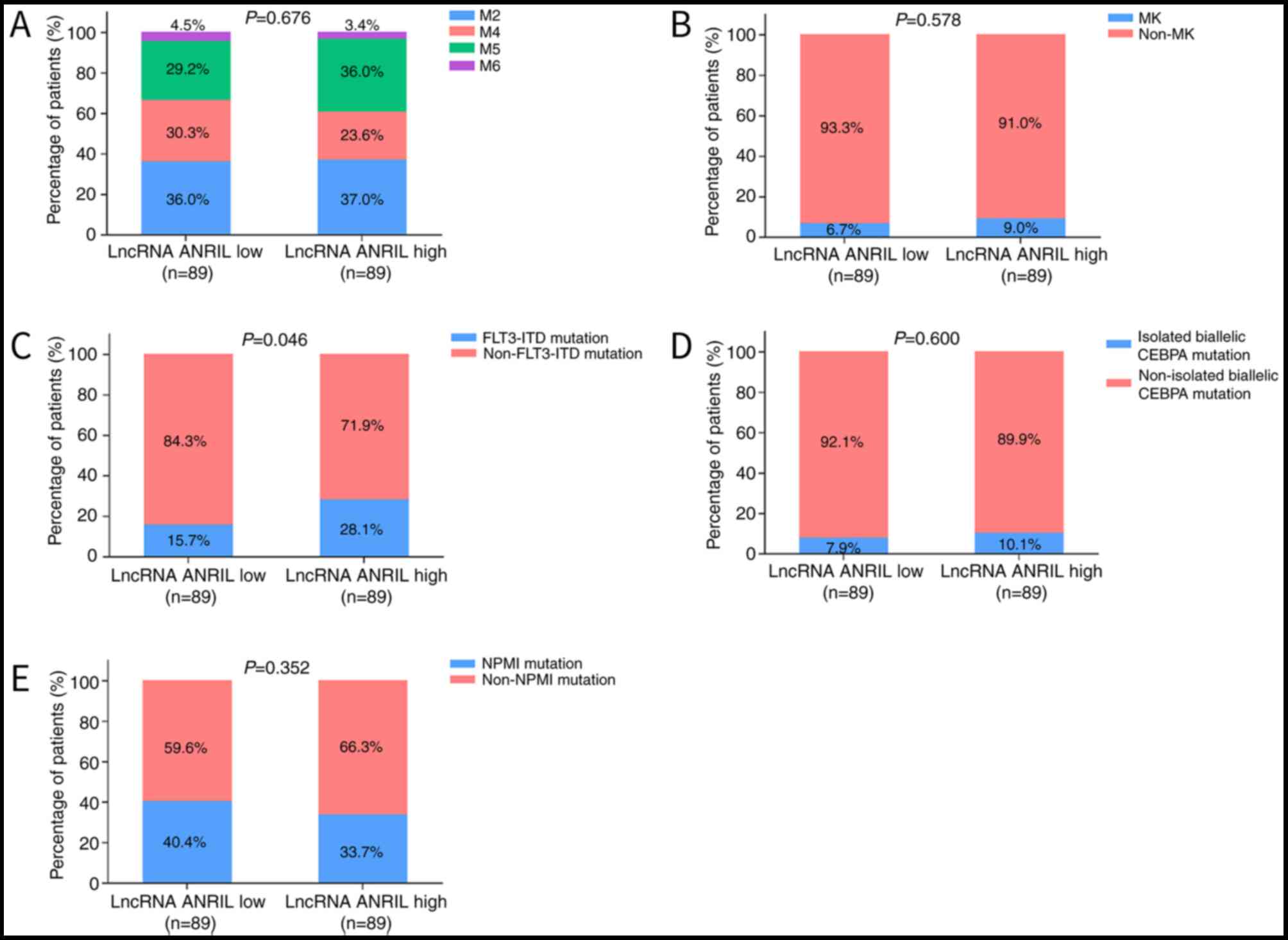 | Figure 2.Differences in FAB classification,
cytogenetics and molecular genetics between patients with acute
myeloid leukemia with high and low expression of lncRNA ANRIL.
Comparisons of (A) FAB classification, (B) MK, (C) FLT3-ITD
mutation, (D) isolated biallelic CEBPA mutation and (E) NPMI
mutation between patients with high and low expression of lncRNA
ANRIL. Comparisons were performed with χ2 tests. lncRNA,
long non-coding RNA; ANRIL, antisense noncoding RNA in the INK4
locus; FAB, French-American-Britain; MK, monosomal karyotype; NK,
normal karyotype; CK, complex karyotype; FLT3-ITD, internal tandem
duplications in the FMS-like tyrosine kinase 3; CEBPA,
CCAAT/enhancer-binding protein α; NPM1, nucleophosmin 1. |
Association of lncRNA ANRIL with
cytogenetics
lncRNA ANRIL was associated with reduced occurrence
inv(16) or t(16;16) cytogenetic type (P=0.009), while no
association of lncRNA ANRIL with NK (P=0.881), CK (P=0.202),
t(8;21) (P=1.000), −7 or 7q- (P=0.444), t(9;11) (P=1.000), +8
(P=0.368), t(9;22) (P=1.000), 11q23 (P=1.000), −5 or 5q- (P=1.000),
t(6;9) (P=1.000) or others (undefined) (P=1.000) was found in
patients with AML (Table
III).
| Table III.Comparison of cytogenetics between
lncRNA ANRIL low group and lncRNA ANRIL high group. |
Table III.
Comparison of cytogenetics between
lncRNA ANRIL low group and lncRNA ANRIL high group.
|
| lncRNA ANRIL |
|
|---|
|
|
|
|
|---|
| Item | Low | High | P-value |
|---|
| NK | 48 (53.9) | 47 (52.8) | 0.881 |
| CK | 6 (6.7) | 11 (12.4) | 0.202 |
| inv(16) or
t(16;16) | 13 (14.6) | 3 (3.4) | 0.009 |
| t(8;21) | 3 (3.4) | 4 (4.5) | 1.000 |
| −7 or 7q- | 2 (2.2) | 5 (5.6) | 0.444 |
| t(9;11) | 3 (3.4) | 3 (3.4) | 1.000 |
| +8 | 1 (1.1) | 4 (4.5) | 0.368 |
| t(9;22) | 2 (2.2) | 2 (2.2) | 1.000 |
| 11q23 | 1 (1.1) | 2 (2.2) | 1.000 |
| −5 or 5q- | 1 (1.1) | 0 (0.0) | 1.000 |
| t(6;9) | 1 (1.1) | 0 (0.0) | 1.000 |
| Others
(undefined) | 8 (9.0) | 8 (9.0) | 1.000 |
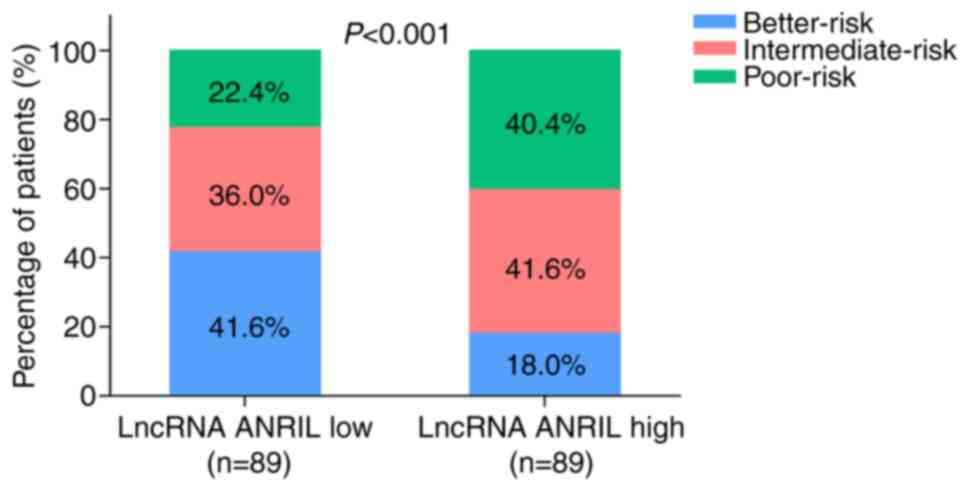
Association of lncRNA ANRIL with risk
stratification
In the lncRNA ANRIL-low expression group, 41.6, 36.0
and 22.4% of cases had a better-, intermediate- and poor-risk
stratification, respectively, while in the lncRNA ANRIL-high
expression group, 18.0, 41.6 and 40.4% of cases had a better-,
intermediate- and poor-risk stratification, respectively. Further
comparison indicated that risk stratification was poorer in lncRNA
ANRIL-high expression patients compared with that in lncRNA
ANRIL-low expression patients (P<0.001; Fig. 3).
Predictive value of lncRNA ANRIL for
treatment response
In the lncRNA ANRIL-low expression group, 84.3% of
cases achieved CR, while 15.7% did not. In the lncRNA ANRIL-high
expression group, 68.5% of cases achieved CR, while 31.5% did not.
Further analysis suggested that the CR rate was lower in the lncRNA
ANRIL-high expression group compared with that in the lncRNA
ANRIL-low expression group (P=0.013; Fig. 4).
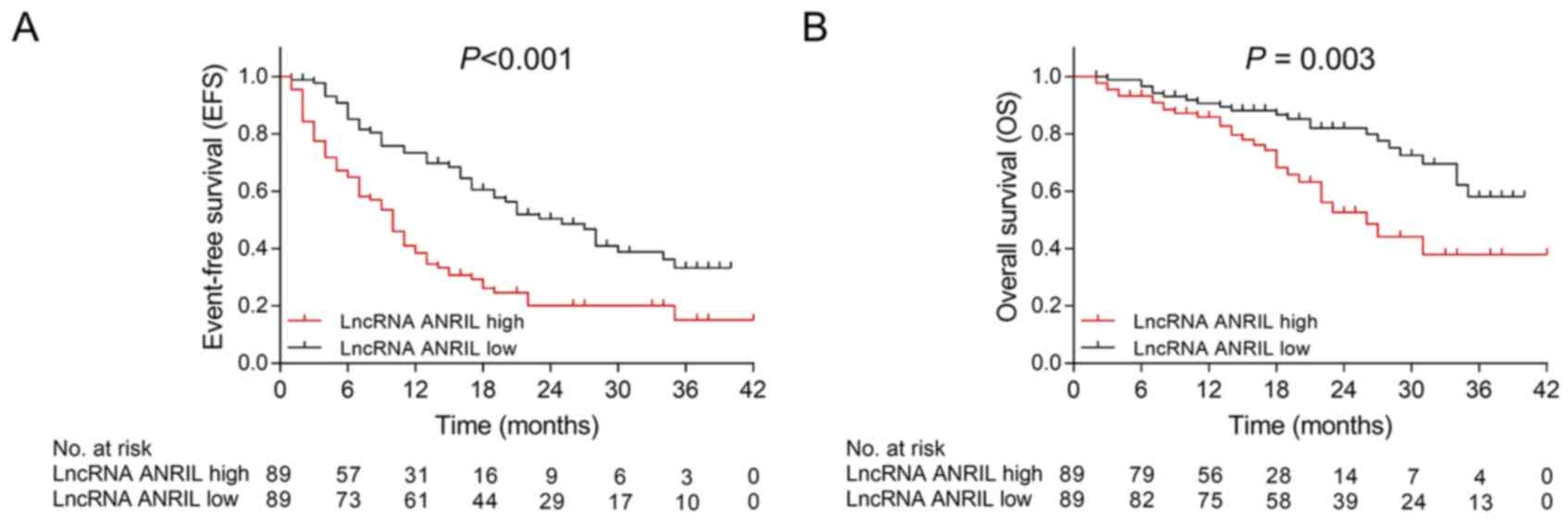
Predictive value of lncRNA ANRIL for
EFS and OS
The median EFS was shorter in the lncRNA ANRIL
high-expression group than that in the lncRNA ANRIL-low expression
group (P<0.001; Fig. 5A). The
median OS was also lower in the lncRNA ANRIL-high expression group
compared with that in the lncRNA ANRIL-low expression group
(P=0.003; Fig. 5B).
Prognostic factors for EFS
Univariate Cox regression analysis indicated that
high expression of lncRNA ANRIL [P<0.001, hazard ratio
(HR)=1.548–3.300; 95% CI=2.260], poorer risk stratification
(P<0.001, HR=1.859–3.124; 95% CI=2.410) and WBC
>10.0×10⁹ cells/l (P=0.005, HR=1.194–2.790; 95%
CI=1.825) were predictors of unfavorable EFS in patients with AML
(Table IV). Subsequent
multivariate Cox regression analysis adjusted for lncRNA ANRIL
high, age (>55 years), male, FAB classification, poorer risk
stratification and WBC (>10.0×10⁹ cells/l), which
revealed that high expression of lncRNA ANRIL (P=0.002,
HR=1.256–2.838; 95% CI=1.888), poorer risk stratification
(P<0.001, HR=1.852–3.206; 95% CI=2.436) and WBC
>10.0×10⁹ cells/l (P<0.001, HR=1.470–3.629, 95%
CI=2.309) were independent prognostic factors for poor EFS in
patients with AML.
| Table IV.Analysis of factors affecting
EFS. |
Table IV.
Analysis of factors affecting
EFS.
|
| Univariate Cox
regression | Multivariate Cox
regression |
|---|
|
|
|
|
|---|
| Factor | P-value | HR (95%CI) | P-value | HR (95%CI) |
|---|
| LncRNA ANRIL
high | <0.001 | 2.260
(1.548–3.300) | 0.002 | 1.888
(1.256–2.838) |
| Age (>55
years) | 0.856 | 0.966
(0.667–1.400) | 0.694 | 1.080
(0.736–1.584) |
| Male sex | 0.167 | 1.315
(0.892–1.938) | 0.835 | 0.955
(0.622–1.467) |
| FAB
classification |
|
|
|
|
| M2 | Reference | – | Reference | – |
| M4 | 0.275 | 0.767
(0.475–1.236) | 0.329 | 0.781
(0.475–1.283) |
| M5 | 0.501 | 1.164
(0.748–1.812) | 0.915 | 0.976
(0.623–1.529) |
| M6 | 0.737 | 0.852
(0.335–2.165) | 0.974 | 1.016
(0.394–2.623) |
| Poorer risk
stratification | <0.001 | 2.410
(1.859–3.124) | <0.001 | 2.436
(1.852–3.206) |
| WBC
(>10.0×10⁹/l) | 0.005 | 1.825
(1.194–2.790) | <0.001 | 2.309
(1.470–3.629) |
Prognostic factors for OS
Univariate Cox regression analysis revealed that
high expression of lncRNA ANRIL (P=0.004, HR=1.311–4.061, 95%
CI=2.308) and a poorer risk stratification (P<0.001,
HR=1.984–4.446, 95% CI=2.970) were predictors of poor OS in
patients with AML (Table V).
Subsequent multivariate Cox regression analysis adjusted for lncRNA
ANRIL high, age (>55 years), male, FAB classification, poorer
risk stratification and WBC (>10.0×10⁹ cells/l),
which demonstrated that high expression of lncRNA ANRIL (P=0.047,
HR=1.008–3.259, 95% CI=1.812) and poorer risk stratification
(P<0.001, HR=2.884, 95% CI=1.885–4.413) were independent
prognostic factors for shorter OS in patients with AML.
| Table V.Analysis of factors affecting OS. |
Table V.
Analysis of factors affecting OS.
|
| Univariate Cox's
regression | Multivariate Cox's
regression |
|---|
|
|
|
|
|---|
| Factor | P-value | HR (95%CI) | P-value | HR (95%CI) |
|---|
| LncRNA ANRIL
high | 0.004 | 2.308
(1.311–4.061) | 0.047 | 1.812
(1.008–3.259) |
| Age (>55
years) | 0.682 | 0.891
(0.512–1.550) | 0.917 | 0.970
(0.543–1.733) |
| Male sex | 0.823 | 0.938
(0.535–1.645) | 0.364 | 0.753
(0.409–1.388) |
| FAB
classification |
|
|
|
|
| M2 | Reference | – | Reference | – |
| M4 | 0.929 | 1.032
(0.520–2.046) | 0.612 | 0.830
(0.405–1.705) |
| M5 | 0.518 | 1.255
(0.630–2.499) | 0.770 | 0.899
(0.442–1.831) |
| M6 | 0.871 | 0.885
(0.203–3.858) | 0.845 | 0.862
(0.195–3.816) |
| Poorer risk
stratification | <0.001 | 2.970
(1.984–4.446) | <0.001 | 2.884
(1.885–4.413) |
| WBC
(>10.0×10⁹/l) | 0.705 | 0.897
(0.511–1.575) | 0.840 | 1.063
(0.590–1.915) |
LncRNA ANRIL expression in
relapsed/refractory patients with AML
The relative expression of lncRNA ANRIL was elevated
in patients with relapsed AML compared with that in patients with
de novo AML (P=0.044) and healthy donors (P<0.001;
Fig. S1). Furthermore, the
relative expression of lncRNA ANRIL was also higher in patients
with refractory AML than that in patients with de novo AML
(P=0.004) and healthy donors (P<0.001).
Discussion
The present study revealed that i) lncRNA ANRIL was
elevated in patients with AML compared with its levels in healthy
donors and was able to distinguish patients with AML from healthy
donors; ii) lncRNA ANRIL was associated with increased FLT3-ITD
mutation and poorer risk stratification in patients with AML
patients; and iii) lncRNA ANRIL was associated with a lower CR rate
after induction therapy and unfavorable survival in patients with
AML.
lncRNAs are abundant in the nucleus and cytoplasm,
and have been established as important regulators of gene
transcription, post-transcription processes, translation and
epigenetic modification (20).
Aberrant expression and dysfunction of lncRNAs are closely linked
to tumorigenesis (21). Among the
lncRNAs that have been identified thus far, lncRNA ANRIL has been
reported to be involved in the development and progression of
various cancer types (8,10,13,22,23).
In triple-negative breast cancer, lncRNA ANRIL was reported to
enhance cell proliferation and suppresses apoptosis via sponging
miR-199a (22). In gastric cancer,
lncRNA ANRIL knockdown was observed to inhibit cell viability,
migration and invasion, and facilitate cell apoptosis by
miR-99a-mediated downregulation of B-lymphoma Moloney murine
leukemiavirus insertion region 1 (23). Regarding hematological malignancies,
lncRNA ANRIL knockdown was indicated to suppress cell
proliferation, migration and invasion, and facilitate cell
apoptosis in AML (12). However,
the clinical implications of lncRNA ANRIL in patients with AML have
remained to be elucidated. In the present study, it was observed
that lncRNA ANRIL was higher in patients with AML than in healthy
donors, and it was able to differentiate patients with AML from
healthy donors (AUC, 0.886; 95% CI, 0.820–0.952). Furthermore,
lncRNA ANRIL was associated with increased FLT3-ITD mutation,
reduced inv(16) or t(16;16) cytogenetic type, and poorer
stratification in patients with AML. Several possible explanations
have been proposed: i) lncRNA ANRIL may silence the tumor
suppressor gene p15 (INK4B) by recruiting polycomb repressive
complex 2, leading to the malignant growth of myeloid precursors
(24). Thereby, lncRNA ANRIL is
associated with a higher risk of AML; and ii) lncRNA ANRIL may
repress the expression of AdipoR1 and modulate AMPK/SIRT1, which
increases glucose uptake and malignant cell survival to accelerate
AML progression (13). Thereby,
lncRNA ANRIL is associated with increased FLT3-ITD mutation and
poorer risk stratification in patients with AML.
lncRNA ANRIL is associated with dismal prognosis in
multiple solid cancer types (10,11).
In vitro, lncRNA ANRIL facilitates colorectal cancer cell
chemoresistance through the regulation of ATP-binding cassette
subfamily C member 1 by binding Let-7a (24). In the clinic, patients with head and
neck squamous cell carcinoma (HNSCC) and high lncRNA ANRIL
expression exhibit worse recurrence-free survival and OS compared
with those of patients with HNSCC and low lncRNA ANRIL expression
(10). In patients with esophageal
squamous cell carcinoma, high expression of lncRNA ANRIL was
determined to be associated with shorter disease-free survival
(DFS) and OS, and to be an independent prognostic factor for DFS
and OS according to the results of multivariate analyses (11). However, the prognostic value of
lncRNA ANRIL in AML has not been investigated to date, to the best
of our knowledge. Considering the present results that lncRNA ANRIL
was associated with FLT3-ITD mutation, intermediate-risk and
poor-risk stratification in patients with AML, it was hypothesized
that lncRNA ANRIL may also be associated with poor prognosis in
AML. Further analysis suggested that lncRNA ANRIL was associated
with a lower CR rate after induction therapy, as well as with
shorter EFS and OS in patients with AML. Furthermore, multivariate
Cox regression analyses suggested that high expression of lncRNA
ANRIL was an independent prognostic factor for shorter ESF and OS
in patients with AML. The following are possible reasons: i)
According to the present results, lncRNA ANRIL was positively
associated with FLT3-ITD mutation; this reportedly leads to
promotion of aberrant STAT5 activation and suppressed forkhead box
O3 (a member of the pro-apoptotic regulator forkhead family of
transcription factors), which in turn amplifies reactive oxygen
species production, increases the frequency of double-strand DNA
breaks and enhances AML cell survival and proliferation, resulting
in genomic instability, advanced tumor stage and poor prognosis in
patients with AML (25); ii) in the
present study, lncRNA ANRIL was positively associated with
intermediate/poor risk stratification, which was linked to poor
prognosis in patients with AML; and iii) lncRNA ANRIL may induce
the chemoresistance of tumor cells and increase their colony
formation ability via positively regulating ATP-binding cassette
subfamily C member 1, resulting in drug resistance in patients with
AML (24). Thereby, patients with
AML who exhibited high expression of lncRNA ANRIL had a lower CR
rate and shorter EFS and OS compared with those of patients with
AML who exhibited low expression of lncRNA ANRIL.
Several limitations of the present study should be
noted. First, the follow-up duration of the present study was
relatively short and should be extended to validate the present
results in future studies. Furthermore, since it was difficult to
enroll healthy donors for the present study due to the very limited
healthy volunteers for bone marrow donation, the sample size of the
healthy donor group was relatively small, which may reduce the
statistical power. Hence, further studies with a larger sample size
are required to validate the present results. Finally, only the
expression level of lncRNA ANRIL at baseline was assessed, while
changes in the expression levels of lncRNA ANRIL after treatment
were not investigated; therefore, further investigation would be
desirable in patients with AML.
To conclude, lncRNA ANRIL is associated with higher
disease risk, exacerbated clinical features and poor prognosis of
patients with AML, suggesting that lncRNA ANRIL may serve as a
biomarker for facilitating treatment decisions and improving
prognosis in patients with AML.
Supplementary Material
Supporting Data
Acknowledgements
Not applicable.
Funding
No funding was received.
Availability of data and materials
The datasets used and/or analyzed during the current
study are available from the corresponding author on reasonable
request.
Authors' contributions
ZT and ZL designed the study, KZ and YY performed
the experiments, ZT and ZL analyzed the data and all authors wrote
and revised the manuscript. All authors read and approved the final
manuscript.
Ethics approval and consent to
participate
The current study was approved by the Ethics
Committee of the Central Hospital of Xiangtan (Xiangtan, China).
All AML patients and healthy donors voluntarily participated in the
present study and provided written informed consent prior to
enrollment.
Patient consent for publication
Not applicable.
Competing interest
The authors declare that they have no competing
interests.
References
|
1
|
Döhner H, Weisdorf DJ and Bloomfield CD:
Acute myeloid leukemia. N Engl J Med. 373:1136–1152. 2015.
View Article : Google Scholar : PubMed/NCBI
|
|
2
|
Prada-Arismendy J, Arroyave JC and
Röthlisberger S: Molecular biomarkers in acute myeloid leukemia.
Blood Rev. 31:63–76. 2017. View Article : Google Scholar : PubMed/NCBI
|
|
3
|
Zimta AA, Tomuleasa C, Sahnoune I, Calin
GA and Berindan-Neagoe I: Long non-coding RNAs in myeloid
malignancies. Front Oncol. 9:10482019. View Article : Google Scholar : PubMed/NCBI
|
|
4
|
Tallman MS, Gilliland DG and Rowe JM: Drug
therapy for acute myeloid leukemia. Blood. 106:1154–1163. 2005.
View Article : Google Scholar : PubMed/NCBI
|
|
5
|
Congrains A, Kamide K, Ohishi M and Rakugi
H: ANRIL: Molecular mechanisms and implications in human health.
Int J Mol Sci. 14:1278–1292. 2013. View Article : Google Scholar : PubMed/NCBI
|
|
6
|
Pasmant E, Laurendeau I, Hèron D, Vidaud
M, Vidaud D and Bièche I: Characterization of a germ-line deletion,
including the entire INK4/ARF locus, in a melanoma-neural system
tumor family: Identification of ANRIL, an antisense noncoding RNA
whose expression coclusters with ARF. Cancer Res. 67:3963–3969.
2007. View Article : Google Scholar : PubMed/NCBI
|
|
7
|
Hao YR, Zhang DJ, Fu ZM, Guo YY and Guan
GF: Long non-coding RNA ANRIL promotes proliferation,
clonogenicity, invasion and migration of laryngeal squamous cell
carcinoma by regulating miR-181a/Snai2 axis. Regen Ther.
11:282–289. 2019. View Article : Google Scholar : PubMed/NCBI
|
|
8
|
Ma Y, Zhang H, Li G, Hu J, Liu X and Lin
L: LncRNA ANRIL promotes cell growth, migration and invasion of
hepatocellular carcinoma cells via sponging miR-144. Anticancer
Drugs. 30:1013–1021. 2019. View Article : Google Scholar : PubMed/NCBI
|
|
9
|
Deng W, Zhang Y, Cai J, Zhang J, Liu X,
Yin J, Bai Z, Yao H and Zhang Z: LncRNA-ANRIL promotes gastric
cancer progression by enhancing NF-kB signaling. Exp Biol Med
(Maywood). 244:953–959. 2019. View Article : Google Scholar : PubMed/NCBI
|
|
10
|
Zhang LM, Ju HY, Wu YT, Guo W, Mao L, Ma
HL, Xia WY, Hu JZ and Ren GX: Long non-coding RNA ANRIL promotes
tumorgenesis through regulation of FGFR1 expression by sponging
miR-125a-3p in head and neck squamous cell carcinoma. Am J Cancer
Res. 8:2296–2310. 2018.PubMed/NCBI
|
|
11
|
Cao T, Shen J, Pan W, Li C and Qiao Z:
Upregulation of long noncoding RNA ANRIL correlates with tumor
progression and poor prognosis in esophageal squamous cell
carcinoma. J BUON. 23:1862–1866. 2018.PubMed/NCBI
|
|
12
|
Wang CH, Li QY, Nie L, Ma J, Yao CJ and
Chen FP: LncRNA ANRIL promotes cell proliferation, migration and
invasion during acute myeloid leukemia pathogenesis via negatively
regulating miR-34a. Int J Biochem Cell Biol. 119:1056662020.
View Article : Google Scholar : PubMed/NCBI
|
|
13
|
Sun LY, Li XJ, Sun YM, Huang W, Fang K,
Han C, Chen ZH, Luo XQ, Chen YQ and Wang WT: LncRNA ANRIL regulates
AML development through modulating the glucose metabolism pathway
of AdipoR1/AMPK/SIRT1. Mol Cancer. 17:1272018. View Article : Google Scholar : PubMed/NCBI
|
|
14
|
Jafie ES, Harris NL, Stein H and JW W:
Tumors of haematopoietic and lymphoid tissues. IARCPress; Lyon:
2001
|
|
15
|
Bennett JM, Catovsky D, Daniel MT,
Flandrin G, Galton DA, Gralnick HR and Sultan C: Proposed revised
criteria for the classification of acute myeloid leukemia. A report
of the French-American-British cooperative group. Ann Intern Med.
103:620–625. 1985. View Article : Google Scholar : PubMed/NCBI
|
|
16
|
National Comprehensive Cancer Network AML
Guidelines Version 2. 2014.
|
|
17
|
Livak KJ and Schmittgen TD: Analysis of
relative gene expression data using real-time quantitative PCR and
the 2(-Delta Delta C(T)) method. Methods. 25:402–408. 2001.
View Article : Google Scholar : PubMed/NCBI
|
|
18
|
Tallman MS, Wang ES, Altman JK, Appelbaum
FR, Bhatt VR, Bixby D, Coutre SE, De Lima M, Fathi AT, Fiorella M,
et al: Acute myeloid leukemia, version 3.2019, NCCN clinical
practice guidelines in oncology. J Natl Compr Canc Netw.
17:721–749. 2019. View Article : Google Scholar : PubMed/NCBI
|
|
19
|
Döhner H, Estey EH, Amadori S, Appelbaum
FR, Büchner T, Burnett AK, Dombret H, Fenaux P, Grimwade D, Larson
RA, et al: Diagnosis and management of acute myeloid leukemia in
adults: Recommendations from an international expert panel, on
behalf of the European leukemianet. Blood. 115:453–474. 2010.
View Article : Google Scholar : PubMed/NCBI
|
|
20
|
Jiang MC, Ni JJ, Cui WY, Wang BY and Zhuo
W: Emerging roles of lncRNA in cancer and therapeutic
opportunities. Am J Cancer Res. 9:1354–1366. 2019.PubMed/NCBI
|
|
21
|
Bhan A, Soleimani M and Mandal SS: Long
noncoding RNA and cancer: A new paradigm. Cancer Res. 77:3965–3981.
2017. View Article : Google Scholar : PubMed/NCBI
|
|
22
|
Xu ST, Xu JH, Zheng ZR, Zhao QQ, Zeng XS,
Cheng SX, Liang YH and Hu QF: Long non-coding RNA ANRIL promotes
carcinogenesis via sponging miR-199a in triple-negative breast
cancer. Biomed Pharmacother. 96:14–21. 2017. View Article : Google Scholar : PubMed/NCBI
|
|
23
|
Liu P, Zhang M, Niu Q, Zhang F, Yang Y and
Jiang X: Knockdown of long non-coding RNA ANRIL inhibits
tumorigenesis in human gastric cancer cells via
microRNA-99a-mediated down-regulation of BMI1. Braz J Med Biol Res.
51:e68392018. View Article : Google Scholar : PubMed/NCBI
|
|
24
|
Zhang Z, Feng L, Liu P and Duan W: ANRIL
promotes chemoresistance via disturbing expression of ABCC1 by
regulating the expression of Let-7a in colorectal cancer. Biosci
Rep. 38:BSR201806202018. View Article : Google Scholar : PubMed/NCBI
|
|
25
|
Tsapogas P, Mooney CJ, Brown G and Rolink
A: The cytokine Flt3-ligand in normal and malignant hematopoiesis.
Int J Mol Sci. 18:11152017. View Article : Google Scholar
|