Introduction
Endometrial cancer is one of the most common
gynecological cancers in China and is a major threat to women's
health (1). The incidence and
mortality rates of endometrial cancer in China are 8.56 and 1.94
per 100,000 individuals, respectively (2). Furthermore, patients diagnosed with
late-stage endometrial cancer exhibit a poor prognosis (3). Thus, there is a need to elucidate the
molecular mechanisms underlying endometrial cancer to develop
reliable predictive biomarkers and effective therapeutic
agents.
Epithelial-to-mesenchymal transition (EMT) is a
phenotypic plasticity process that confers migratory and invasive
properties to epithelial cells during the pathogenesis of
endometrial cancer (4). EMT is
mediated by cell adhesion molecules (E-cadherin and N-cadherin),
transcription factors (Snail and Twist), α-smooth muscle actin
(SMA) and vimentin (5).
Additionally, various microRNAs (miRNAs/miRs) modulate the
regulatory network of EMT. Drug resistance and prognosis in
patients with various types of cancer, such as breast, colorectal,
bladder, ovarian and endometrial cancer, are directly determined by
EMT-associated factors (6,7). Previous studies have demonstrated that
EMT-associated factors, which are upregulated in cancer, mediate
cancer cell proliferation, migration and invasion. For example,
Padmanaban et al (8)
reported that E-cadherin promotes metastasis in multiple models of
breast cancer, and Li et al (9) demonstrated that Snail-induced
claudin-11 promotes tumor progression by increasing cancer cell
migration. Thus, EMT-associated factors serve vital roles in the
tumorigenesis of endometrial cancer.
Family with sequence similarity 83, member B
(FAM83B) is reported to be a novel molecule involved in the
development of various malignant tumors, including pancreatic
ductal Adenocarcinoma (10) and
gastric cancer (11). Previous
studies have demonstrated that FAM83B can directly activate
the PI3K/AKT/mTOR signaling pathway in tumors, including breast,
lung, ovarian and cervical cancer, by binding to PI3K (12,13) or
can indirectly activate the pathway by binding and hyper-activating
EGFR in lung adenocarcinoma (14).
Our previous study demonstrated that FAM83B knockdown
inhibits endometrial cancer cell proliferation and metastasis
through the inhibition of the PI3K/AKT/mTOR signaling pathway
(15). Collectively, the
aforementioned studies suggest that the pathogenesis of various
types of cancer is mediated by FAM83B.
miR-199 is a highly conserved miRNA family that
comprises miR-199a and miR-199b (16). A previous study demonstrated that
miR-199a-5p inhibits the proliferation, migration and invasion of
cancer cells (17). Additionally,
miR-199a-5p activates EMT-associated signaling pathways (18). In endometrial carcinoma, the
dysregulation of miR-199b in tissues and plasma results in the
upregulation of the mTOR kinase (19). Previous studies have reported a
one-to-one or one-to-two association of miR-199, FAM83B,
EMT, cell metastasis and endometrial cancer. However, the role of
miR-199-a/b-5p, which targets FAM83B, in endometrial cancer
has not been elucidated (11,15,20–22).
In the present study, the target genes of
miR-199-a/b-5p (including FAM83B) and the signaling pathways
in which they are enriched were predicted through data mining. The
expression levels of miR-199-a/b-5p, FAM83B, E-cadherin,
N-cadherin, Snail, α-SMA, vimentin and Twist were examined in
endometrial cancer cell lines. Additionally, the biological
functions of miR-199-a/b-5p and FAM83B in the proliferation,
migration, invasion, in vivo tumor formation and pulmonary
metastasis of endometrial cancer cells were evaluated. Furthermore,
the 3′-untranslated region (UTR) binding sites of miR-199-a/b-5p in
FAM83B was investigated.
Materials and methods
Gene Expression Omnibus (GEO) dataset
analysis
Wang et al (23) confirmed that GEO datasets can
provide a more convenient way to screen gene expression between
disease and non-disease cases, therefore GEO datasets were used to
analyze the difference in miRNA expression between endometrial
adenocarcinoma and non-cancerous tissues. Of the 48 samples in the
dataset GSE25405_RAW.tar, the microarray data of 27 samples were
downloaded from GEO of the National Center for Biotechnology
Information (NCBI) database (http://www.ncbi.nlm.nih.gov/geo). The datasets were
divided into the following groups: Seven non-cancerous endometrial
tissue datasets (GSM623853-GSM623855 and GSM623875-GSM623878) and
20 endometrioid adenocarcinoma datasets (GSM623856-GSM623874 and
GSM623881). The Agilent-019118 Human miRNA Microarray 2.0 G4470B
(GPL7731) data were analyzed using the Agilent GeneSpringGX
Software (version 11.5; Agilent Technologies, Inc.). The
differentially expressed miRNAs were identified based on the
following criteria: P<0.01 and log2 fold change (FC)
<-2. Heatmap and volcano plot were used to represent the
differentially expressed miRNAs. To determine the expression in the
microarray data, the differential gene expression profile data
(GSE35794) of the top 250 miRNAs were analyzed using NCBI GEO2R
(version R 3.2.3; http://www.ncbi.nlm.nih.gov/geo/geo2r/).
Target gene prediction
The putative miRNA targets were predicted using
TargetScan 7.2 (www.targetscan.org/vert_72). Additionally, the poorly
conserved 3′-UTR of FAM83B was predicted.
Kyoto Encyclopedia of Genes and
Genomes (KEGG) pathway enrichment analysis
The predicted target genes of miR-199-a/b-5p were
annotated using Kobas (version 3.0; kobas.cbi.pku.edu.cn/kobas3). The KEGG pathway
enrichment analysis was performed using R clusterProfiler (version
3.10.1; aipufu.com/index.html).
Cell culture and transfection
Human endometrial cancer cell lines (AN3 CA,
HEC-1-B, RL95-2, HEC-1-A, Ishikawa) were purchased from the
American Type Culture Collection, human endometrial epithelial
cells (hEECs) were purchased from Procell Life Science &
Technology Co., Ltd. (cat. no. CP-H058) and the JEC cell line was
purchased from YRGene (cat. no. CC021). The cells were cultured in
Dulbecco's modified Eagles medium (DMEM, HyClone; Cytiva)
supplemented with 10% fetal bovine serum (FBS; HyClone; Cytiva),
100 µg/ml streptomycin and 100 U/ml penicillin (Thermo Fisher
Scientific, Inc.) at 37°C and 5% CO2 conditions.
For transfection experiments, the six human
endometrial cancer cell lines (2.5×10⁵) were seeded in a
6-well plate and incubated overnight at 37°C with 5%
CO2. Subsequently, cells were transfected with 100 nM
miRNA mimics or inhibitors, 50 nM FAM83B small interfering (si)RNA
or 50 nM FAM83B overexpression plasmid for 4 h. At 24 h
post-transfection, cells were used for subsequent experiments.
Sequences of siRNA and miRNA mimics/inhibitors, including the
negative controls, were as follows: miR-199a-5p mimics,
5′-CCCAGUGUUCAGACUACCUGUUC-3′; miR-199b-5p mimics,
5′-CCCAGUGUUUAGACUAUCUGUUC-3′; miR-199a/b-5p mimics negative
control, 5′-UCACAACCUCCUAGAGGAGAGA-3′; miR-199a-5p inhibitor,
5′-GAACAGGUAGUCUGAACACUGGG-3′; miR-199b-5p inhibitor,
5′-GAACAGGTAGTCTAAACACTGGG-3′; and miR-199a/b-5p inhibitor negative
control, 5′-CAGUACUUUUGUGUAGUACAA-3′. The miR-199a/b-5p
mimics/inhibitor, si-FAM83B constructs, FAM83B overexpression
plasmid (pcDNA3.1 vector) and and their negative controls
(scramble) were synthesized by Sangon Biotech Co., Ltd. To
overexpress miR-199a/b-5p, the cells were transfected with
miR-199a-5p and miR-199b-5p mimics mixed in a ratio of 1:1. All RNA
transfections were performed using Lipofectamine® 2000
(Invitrogen; Thermo Fisher Scientific, Inc.) following the
manufacturer's instructions.
Animal experiments
A total of 48 female BALB/c-nu/nu specific
pathogen-free mice from Guangdong Medical Laboratory Animal Center
(6-weeks old; 13–15 g) were used in the present study. Mice were
housed in a temperature-controlled room (at 24±1°C) with 50±10%
humidity, 12-h light/dark cycles, and standard rodent food and
water ad libitum in polystyrene cages. All animal
experiments were approved by the Animal Care and Use Committee of
Guangzhou Medical University (Guangzhou, China; approval no.
GD2019-036) and conducted according to the National Institutes of
Health guidelines (24).
Subcutaneous xenograft animal model
and in vivo pulmonary metastasis assay
In each subgroup, mice were lively, and daily
observation revealed no abnormality in eating or any marked weight
loss. Following terminal anesthesia by intraperitoneal injection
with pentobarbital sodium (100 mg/kg body weight), mice were
euthanized by cervical dislocation and the death of the mice was
verified according to the following criteria: i) no breathing; ii)
no nerve reflexes; iii) no heartbeat; and iv) relaxed muscles. The
normal lung and tumor tissues were quickly dissected. The maximum
tumor diameter observed for a single subcutaneous tumor during the
xenograft assay was 19.7 mm.
The miR-199a/b-5p agomir/antagomir and miRNA
agomir/antagomir control (scramble) constructs were purchased from
Guangzhou RiboBio Co., Ltd. To overexpress miR-199a/b-5p, cells
were transfected with miR-199a-5p agomir and miR-199b-5p agomir
mixed in a ratio of 1:1. The sequences of the agomirs were as
follows: miR-199a-5p agomir, 5′-CCCAGUGUUCAGACUACCUGUUC-3′;
miR-199b-5p agomir, 5′-CCCAGUGUUUAGACUACCUGUUC-3′; miR-199a/b-5p
agomir negative control, 5′-GUCUCCGUCUGUCCAAUCUAGCA-3′; miR-199a-5p
antagomir, 5′-GAACAGGUAGUCUGAACACUGGG-3′; miR-199b-5p antagomir,
5′-GAACAGGTAGTCTAAACACTGGG-3′; and miR-199a/b-5p antagomir negative
control, 5′-UAUUAGGCGCACAGAGGAUCGGA-3′. Ishikawa and JEC cells were
transfected with 50 nM miR-199a/b-5p agomir or 100 nM antagomir at
37°C using Lipofectamine® 2000 (Invitrogen; Thermo
Fisher Scientific, Inc.) for 4 h. At 24 h post-transfection, cells
were used for subsequent experiments. Subequently, cells were
cultured in complete medium at 37°C with 5% CO2.
Transfected cells were harvested and resuspended in PBS. The
resuspended cells (5×10⁶) were subcutaneously injected
into the right flank of 6-week-old female BALB/c nu/nu mice. Three
weeks later, the mice were euthanized and the tumors were excised.
The tumor volume was calculated using the following equation:
Volume = (length × width2)/2. To establish an in
vivo pulmonary metastasis model, 100 µl suspension of cancer
cells (1×10⁶) was injected into the tail vein of
6-week-old female BALB/c nu/nu mice. All mice were sacrificed at
four weeks post-injection. Lung tissue was excised and fixed with
10% formalin for 48 h at 25°C, paraffin-embedded and sectioned into
4-µm-thick sections. At room temperature, sections were stained
with hematoxylin for 4 min followed by staining with eosin for 90
sec. Stained sections were observed using a light microscope
(Olympus Corporation; magnification, ×200).
Reverse transcription-quantitative PCR
(RT-qPCR)
Total RNA was extracted from Ishikawa and JEC cell
lines, as well as the lung tissues of laboratory mice using TRIzol
reagent (Invitrogen; Thermo Fisher Scientific, Inc.). The extracted
RNA was reverse transcribed into cDNA using the PrimeScript RT
reagent kit (Takara Biotechnology Co., Ltd.), following the
manufacturer's instructions. The RT-qPCR analysis was performed
using the SYBR Premix ExTaq II kit (Takara Biotechnology Co.,
Ltd.), according to the manufacturer's protocol, using a 7500
Real-Time PCR System (Applied Biosystems; Thermo Fisher Scientific,
Inc.). The PCR conditions were as follows: 95°C for 5 min, followed
by 40 cycles of 95°C for 15 sec and 60°C for 1 min. The primers
used for RT-qPCR analysis were as follows: miR-199a-5p and
miR-199b-5p forward, 5′-ACACTCCAGCTGGGCCCAGTGTTCAGACTAC-3′ and
reverse, 5′-CTCAACTGGTGTCGTGGA-3′; U6 forward,
5′-CTCGCTTCGGCAGCACA-3′ and reverse, 5′-AACGCTTCACGAATTTGCGT-3′;
FAM83B forward, 5′-AAAGCTCACCTCAGCATGGTT-3′ and reverse,
5′-AGCAAATGAACTAGGGACAC-3′; and GAPDH forward,
5′-GCTCATTTGCAGGGGGGAG-3′ and reverse, 5′-GTTGGTGGTGCAGGAGGCA-3′.
The levels of target RNA were normalized to those of U6 or GAPDH.
The relative miR-199a/b-5p and FAM83B expression levels were
calculated using the 2−ΔΔCq method (25). All PCR experiments were performed in
triplicate.
Western blotting
The harvested cells were lysed with ice-cold SDS
lysis buffer (Beyotime Institute of Biotechnology). The protein
concentration in the lysate was determined using a BCA protein
assay kit (Nanjing KeyGen Biotech Co., Ltd.), following the
manufacturer's instructions. Equal amounts of denatured proteins
(20 µg/lane) were separated via 10% SDS-PAGE and transferred to a
polyvinylidene fluoride membrane (EMD Millipore). The membrane was
blocked with 5% BSA (Beijing Solarbio Science & Technology Co.,
Ltd.) and incubated with primary antibodies at 4°C overnight. Next,
the membrane was incubated with HRP-conjugated goat anti-rabbit
(1:10,000; cat. no. ab205718; Abcam) secondary antibodies for 2 h
at 25°C. The protein bands were developed using ECL Western
Blotting Substrate (Thermo Fisher Scientific, Inc.) and examined
using the BIS 303 PC imaging system (DNR Bio-Imaging Systems Ltd.).
GAPDH was used as a loading control. Protein expression levels were
semi-quantified using ImageJ software (version 1.52n; National
Institutes of Health). Primary antibodies against FAM83B (1:500;
cat. no. PA5-28548; Invitrogen; Thermo Fisher Scientific, Inc.),
E-Cadherin (1:5,000; cat. no. ab40772; Abcam), N-Cadherin (1 µg/ml;
cat. no. ab18203; Abcam), Snail (0.3 µg/ml; cat. no. ab53519;
Abcam), α-SMA (0.5 µg/ml; cat. no. ab5694; Abcam), vimentin
(1:1,000; cat. no. ab92547; Abcam), Twist (2.5 µg/ml; cat. no.
ab49254; Abcam) and GAPDH (1:10,000; cat. no. ab181602; Abcam) were
used.
Transwell migration and invasion
assay
Cell migration and invasion assays were performed
using 24-well Transwell chambers (BD Biosciences). For the invasion
assay, the Transwell chambers were precoated with
Matrigel® (BD Biosciences) for 6 h at 37°C. The
harvested cells (1×10⁵) were placed in the top chamber
containing serum-free medium at 37°C with 5% CO2. The
bottom chamber contained complete medium supplemented with 10% FBS.
The cells were incubated at 37°C and 5% CO2 for 48 h.
The migrated and invaded cells on the reverse side of the chamber
inserts were fixed with methanol for 30 min and stained with 0.1%
crystal violet for 15 min at room temperature. The number of cells
was measured in three randomly selected high-power fields across
the center and the periphery of the membrane using a light
microscope (Olympus Corporation; magnification, ×200). All
experiments were performed in triplicates.
Dual-Luciferase reporter assay
The amplified 3′-UTR fragments of FAM83B were
cloned into the psi-CHECK-2 luciferase miRNA expression reporter
vector (Promega Corporation). The 293T cells (2×10⁴;
cat. no. CRL-11268; ATCC) were cultured in 24-well plates with DMEM
(HyClone; Cytiva) supplemented with 10% FBS. Subsequently, cells
were transfected with 50 nM miR-199a/b-5p mimics, 100 nM
miR-199a/b-5p inhibitor or mimic negative control (NC), 0.5 µg of
psi-CHECK-2 luciferase reporter vector containing the wild-type
(WT) or mutant (Mut) 3′-UTR sequences of FAM83B, or empty
vector. Transfections were performed using
Lipofectamine® 2000 (Invitrogen; Thermo Fisher
Scientific, Inc.) following the manufacturer's instructions. At 48
h post-transfection, the luciferase activity was assessed using the
Dual-Luciferase Reporter Assay System (GloMax; Promega). Firefly
luciferase activities were normalized to Renilla luciferase
activities. Three independent experiments were performed in
duplicates.
Statistical analysis
Data are presented as the mean ± standard deviation.
Multiple comparisons were performed using one-way ANOVA followed by
Dunnett's post-hoc test. All statistical analyses were performed
using SPSS (version 19.0; IBM Corp.). P<0.05 was considered to
indicate a statistically significant difference.
Results
miR-199a/b-5p expression is
downregulated in patients with endometrial cancer
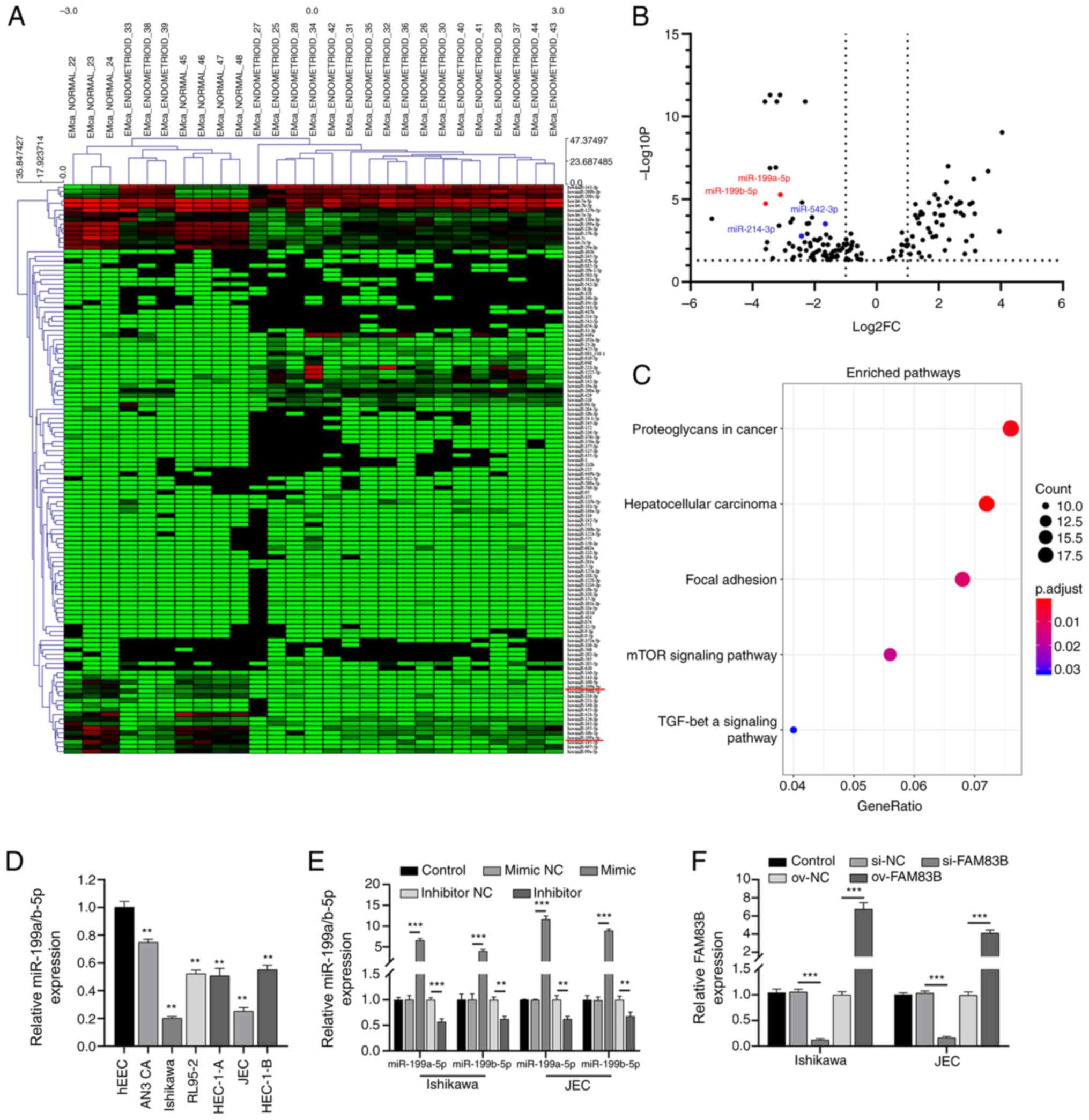
The cluster analysis of the GSE25405 dataset
revealed 150 differentially expressed miRNAs between the
non-cancerous endometrial tissues and human endometrial cancer
tissues (Fig. 1A and B). Based on
the criteria of P<0.01 and log2 FC >2, 24 miRNAs were
screened out with significantly different expression. Our previous
study confirmed that upregulated FAM38B expression in
endometrial cancer can affect the proliferation and metastasis of
cancer cells (15). Therefore,
FAM38 was chosen for further analysis. According to
TargetScan 7.2 analysis, 4/24 significantly differentially
expressed miRNAs (Fig. 1B) had
binding sites with FAM38B. Therefore, miR-199a-5p and
miR-199b-5p were chosen for further analysis. The KEGG pathway
enrichment analysis revealed that the target genes of miR-199a/b-5p
were enriched in several EMT-associated pathways, including TGF-β
and mTOR (Fig. 1C).
miR-199a/b-5p expression is
downregulated in human endometrial cancer cell lines
The expression levels of miR-199a/b-5p were examined
in different endometrial cancer cells. RT-qPCR analysis revealed
that the expression levels of miR-199a/b-5p were significantly
downregulated in all six endometrial cancer cell lines (AN3 CA,
HEC-1-B, RL95-2, HEC-1-A, Ishikawa and JEC) compared with those in
the hEECs (Fig. 1D). The JEC and
Ishikawa cell lines were chosen for further analysis as they
exhibited the lowest expression levels of miR-199a/b-5p.
Additionally, the RT-qPCR analysis revealed that compared with the
respective negative control, the expression levels of miR-199a/b-5p
in Ishikawa and JEC cells were significantly upregulated after
transfection with miR-199a-5p and miR-199b-5p mimics, and
significantly downregulated after transfection with miR-199a-5p and
miR-199b-5p inhibitors (Fig. 1E),
which indicated that the miR-199a-5p and miR-199b-5p mimics and
inhibitors were effective. Transfection with si-FAM83B knocked down
FAM83B with an efficiency of >80% in both Ishikawa and
JEC cells (Fig. 1F). Compared with
the mimic NC-transfected group, the FAM83B mimic-transfected group
exhibited significantly increased FAM83B expression
(Fig. 1F), which indicated that the
overexpression of FAM83B was effective.
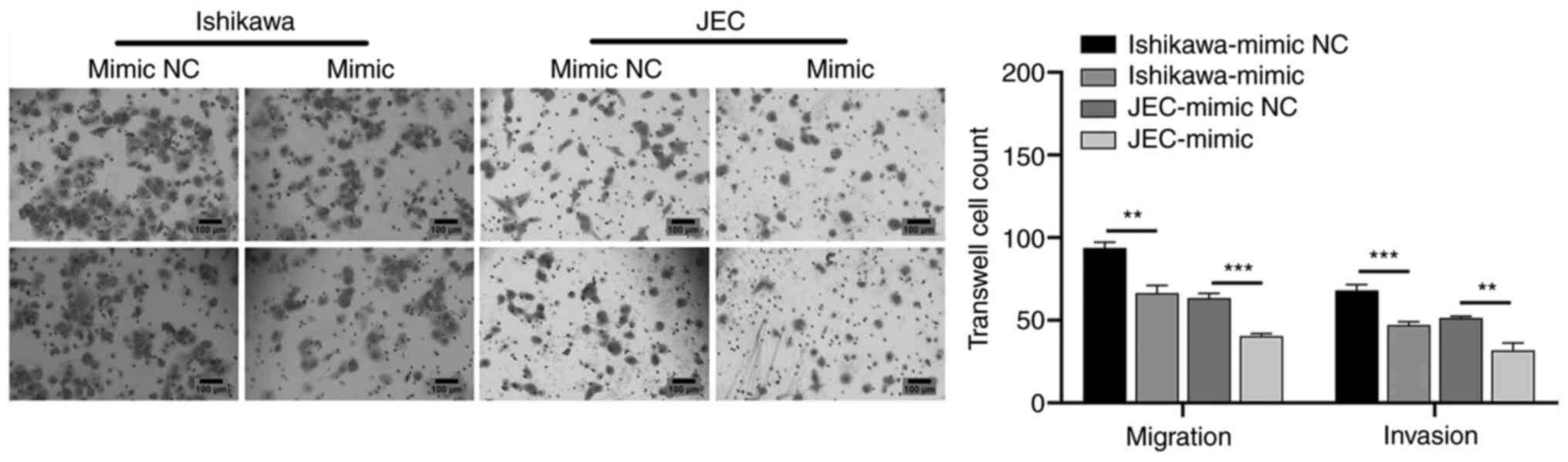
miR-199a/b-5p inhibits the migration
and invasion of JEC and Ishikawa cells
The Ishikawa and JEC cells were transfected with
miR-199a/b-5p mimics to examine the effect of miR-199a/b-5p on the
migration and invasion of cancer cells. The results of the
Transwell assay revealed that the migratory and invasive abilities
of the miR-199a/b-5p mimic-transfected cells at 48 h
post-transfection were significantly lower compared with those of
the mimic NC-transfected cells (Fig.
2).
miR-199a/b-5p inhibits the EMT
signaling pathway in Ishikawa and JEC cells
The signaling pathway through which miR-199a/b-5p
mediates cell migration and invasion in the Ishikawa and JEC cells
was investigated. Western blot analysis revealed that the protein
expression levels of FAM83B, N-cadherin, Snail, α-SMA, vimentin and
Twist in the miR-199a/b-5p mimic-transfected Ishikawa and JEC cells
were significantly lower compared with those in the mimic
NC-transfected Ishikawa and JEC cells at 48 h post-transfection. By
contrast, transfection with the miR-199a/b-5p mimics significantly
upregulated the expression levels of E-cadherin compared with those
of the mimic NC-transfected Ishikawa and JEC cells (Fig. 3A). These findings suggested that
miR-199a/b-5p inhibited the EMT signaling pathway in Ishikawa and
JEC cells.
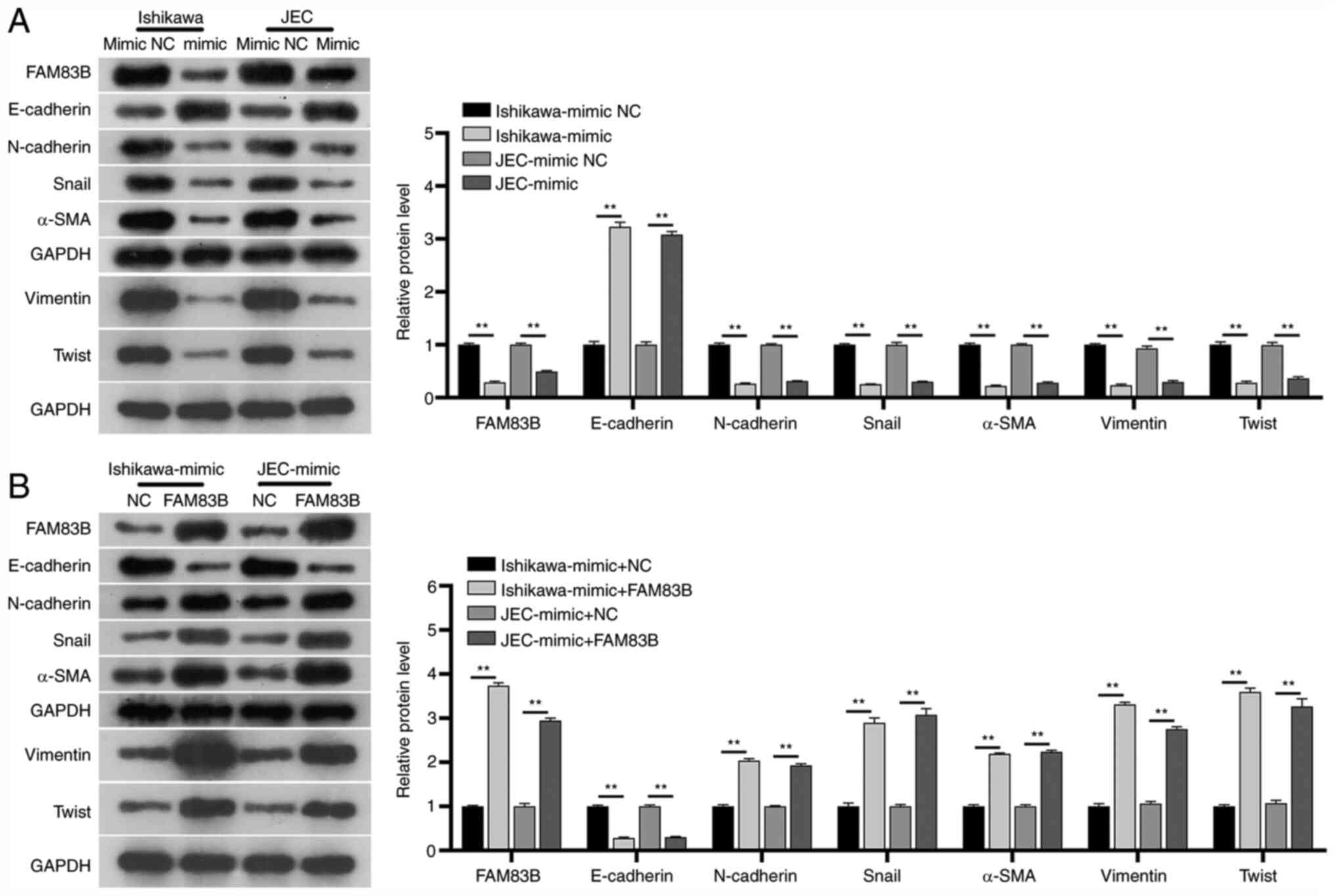 | Figure 3.Effect of miR-199a/b-5p/FAM83B
network on the EMT signaling pathway. (A) Western blot analysis
indicated that transfection with miR-199a/b-5p mimics inhibited EMT
signaling in the Ishikawa and JEC cells. The protein expression
levels of FAM83B, N-cadherin, Snail, α-SMA, vimentin and Twist were
downregulated, whereas those of E-cadherin were upregulated in the
miR-199a/b-5p mimic-transfected cells. (B) Western blot analysis
revealed that transfection with miR-199a/b-5p and FAM83B mimics
activated the EMT signaling pathway in Ishikawa and JEC cells. The
protein expression levels of FAM83B, N-cadherin, Snail, α-SMA,
vimentin and Twist were upregulated, whereas those of E-cadherin
were downregulated in the FAM83B mimic-transfected cells.
**P<0.01. EMT, epithelial-to-mesenchymal transition; miR,
microRNA; NC, negative control; α-SMA, α-smooth muscle actin;
FAM83B, family with sequence similarity 83, member B. |
FAM83B attenuates the
miR-199a/b-5p-induced EMT signaling pathway inhibition in Ishikawa
and JEC cells
The expression levels of N-cadherin, Snail, α-SMA,
vimentin and Twist were evaluated in the FAM83B mimic-transfected
Ishikawa and JEC cells at 48 h post-transfection. Transfection with
the FAM83B mimic significantly attenuated the miR-199a/b-5p
mimic-induced downregulation of the expression levels of
N-cadherin, Snail, α-SMA, vimentin and Twist in the Ishikawa and
JEC cells (Fig. 3B). Additionally,
transfection with FAM83B mimics significantly downregulated the
expression levels of E-cadherin compared with those of mimic
NC-transfected Ishikawa and JEC cells (Fig. 3B). These findings suggested that
FAM83B may activate the EMT signaling pathway in Ishikawa
and JEC cells.
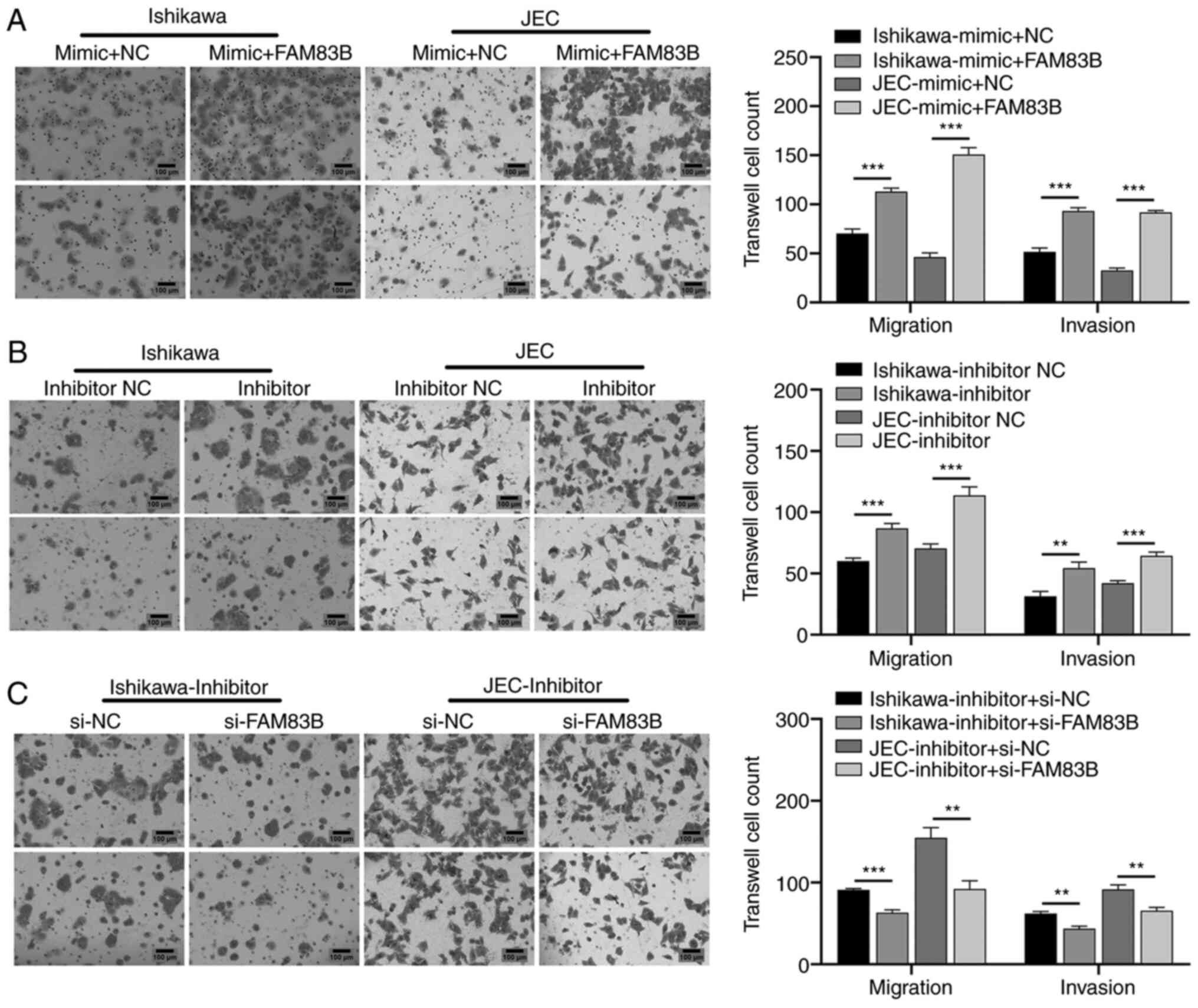
FAM83B attenuates the
miR-199a/b-5p-induced inhibition of the migratory and invasive
abilities of Ishikawa and JEC cells
To evaluate the effects of activating the EMT
signaling pathway on cancer cell migration and invasion, Ishikawa
and JEC cells were further transfected with FAM83B mimics for 48 h.
Compared with the miR-199a/b-5p mimic-transfected Ishikawa and JEC
cells, the FAM83B mimic-transfected Ishikawa and JEC cells
exhibited significantly higher migratory and invasive abilities
(Fig. 4A). Additionally, the
effects of miR-199a/b-5p inhibitors and FM83B knockdown on the
migration and invasion of Ishikawa and JEC cells were examined.
Transfection with miR-199a/b-5p inhibitors significantly enhanced
the migration and invasion of Ishikawa and JEC cells compared with
those of inhibitor NC-transfected Ishikawa and JEC cells (Fig. 4B). By contrast, transfection with
si-FAM83B significantly attenuated the miR-199a/b-5p
inhibitor-induced migration and invasion of Ishikawa and JEC cells
(Fig. 4C). These findings indicated
that FAM83B attenuated the miR-199a/b-5p-induced inhibition
of the migration and invasion of Ishikawa and JEC cells.
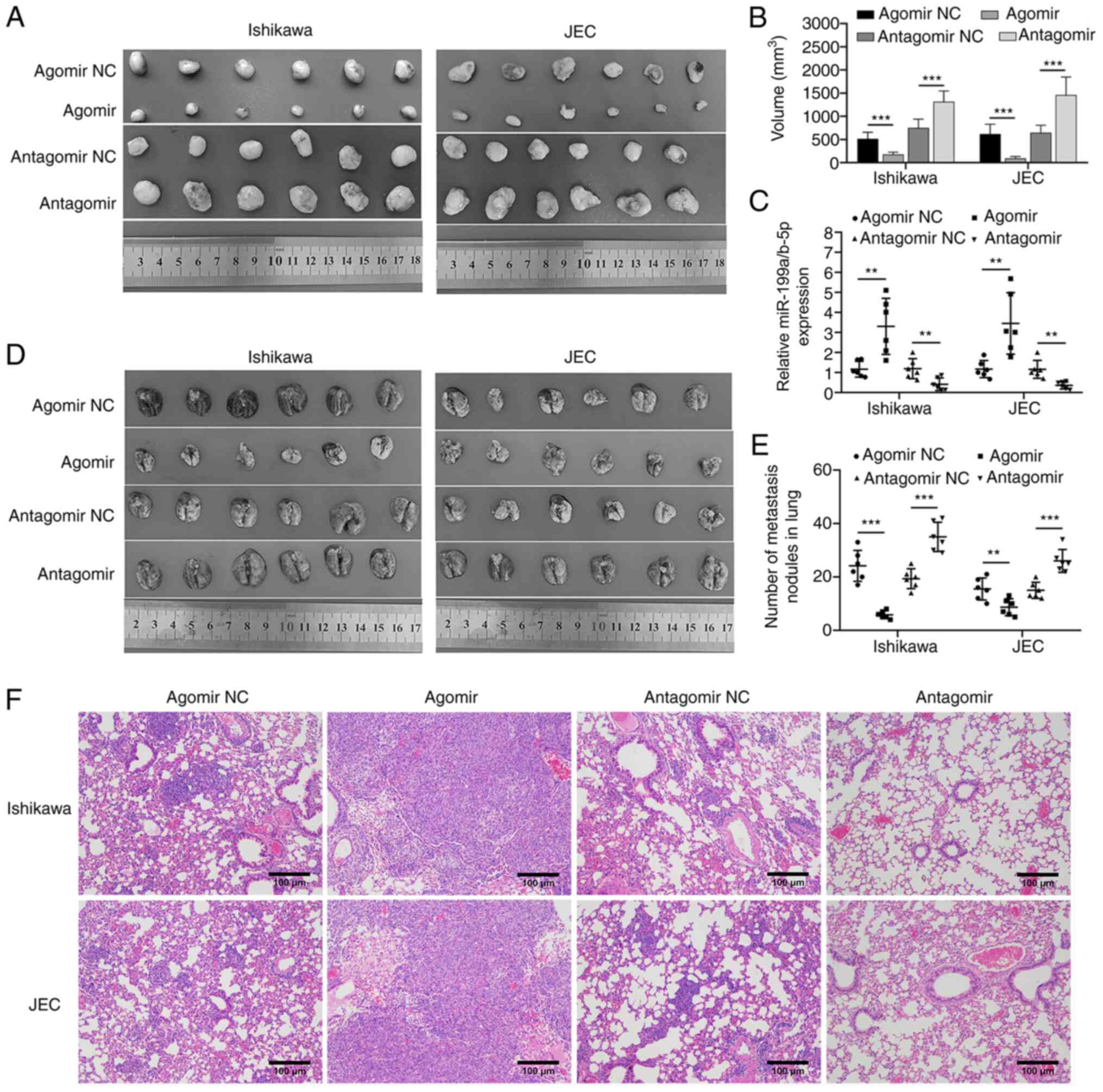
miR-199a/b-5p regulates
metastasis
The role of miR-199a/b-5p in regulating metastasis
of endometrial cancer cells was examined using the BALB/c nu/nu
mouse subcutaneous tumor and pulmonary metastasis models. The
administration of miR-199a/b-5p agomir-transfected cancer cells
significantly inhibited the growth of subcutaneous tumors in the
nude mice compared with that of the agomir NC-transfected group
(Fig. 5A and B). By contrast, the
administration of miR-199a/b-5p antagomir-transfected cancer cells
significantly enhanced the growth of subcutaneous tumors compared
with that of the antagomir NC-transfected group (Fig. 5A and B). These findings suggested
that miR-199a/b-5p inhibited the growth of endometrial cancer cells
in vivo. Meanwhile, miR-199a/b-5p expression levels in
tumors were also analyzed via RT-qPCR. The results confirmed
successful transfection of miR-199a/b-5p agomir and antagomir in
subcutaneous tumors. (Fig. 5C).
Additionally, a pulmonary metastasis model was established by
injecting the cancer cells through the tail vein. Compared with the
respective control groups, miR-199a/b-5p agomir significantly
inhibited the lung metastasis and tumor nodules, whereas
miR-199a/b-5p antagomir significantly enhanced lung metastasis and
tumor nodules. (Fig. 5D-F).
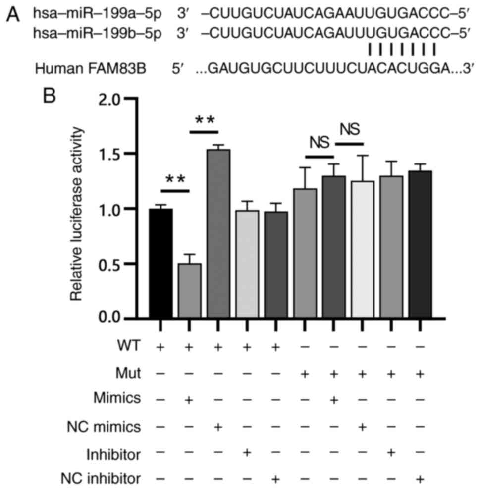
FAM83B is a direct target of
miR-199a/b-5p
The functions of miR-199a/b-5p in endometrial cancer
cell was validated using FAM83B as a target. Single putative
miR-199a/b-5p recognition site was predicted within the
FAM83B 3′-UTR sequences. FAM83B contained a
7-nucleotide site within its 3′-UTR, which matched with the seed
region of miR-199a/b-5p (Fig.
6A).
The mechanism through which miR-199a/b-5p modulates
FAM83B expression was examined by introducing the 3′-UTR of
FAM83B containing one miR-199a/b-5p binding site downstream
of a luciferase reporter. The luciferase activity of WT
FAM83B 3′-UTR construct in cells transfected with
miR-199a/b-5p mimics was significantly lower compared with that in
cells transfected with the mimic NC (Fig. 6B). A luciferase reporter containing
the Mut FAM83B 3′-UTR binding site of miR-199a/b-5p was also
constructed. The luciferase activity of the Mut FAM83B
3′-UTR construct was not suppressed upon transfection with
miR-199a/b-5p mimics or inhibitor (Fig.
6B). This indicated that miR-199a/b-5p may bind selectively to
mRNAs and that the single recognition element identified in the
3′-UTR of the FAM83B mRNA may be sufficient for
miR-199a/b-5p activity. These results indicated that miR-199a/b-5p
directly targeted FAM83B.
Discussion
Globally, the incidence and mortality rates of
endometrial cancer, which is one of the most common malignancies
among women, have increased (26).
Previous studies have suggested that various abnormally expressed
miRNAs are potential predictive biomarkers for the diagnosis or
prognosis of endometrial cancer (27,28).
However, the molecular mechanisms underlying the pathogenesis of
endometrial cancer have not been elucidated. In the present study,
miR-199a/b-5p expression was downregulated in human endometrial
cancer cells compared with in hEECs. Additionally, miR-199a/b-5p
inhibited the migration and invasion of the endometrial cancer
cells through the suppression of EMT-associated factors and EMT
signaling pathway. However, FAM83B attenuated the
miR-199a/b-5p-induced inhibition of cancer cell migration and
invasion through the activation of the EMT signaling pathway.
Additionally, miR-199a/b-5p was bound to the nucleotides 672–679 of
the FAM83B 3′-UTR.
Our previous study demonstrated that the upregulated
FAM83B expression is associated with decreased survival in
patients with endometrial cancer (15). Grant (29) demonstrated that FAM83B is a
candidate oncogene that mediates tyrosine kinase inhibitor
resistance. In contrast to FAM83B expression, the expression
levels of miR-199a/b-5p in endometrial cancer cells were
significantly downregulated compared with those in hEECs. Previous
studies on cancer, including gastric (11), lung (30) and colon (31) cancer, have focused on either
FAM83B or miR-199a/b-5p, but have not evaluated these two
markers simultaneously. Hwang et al (32) demonstrated that miR-199b-5p may be a
novel biomarker for sporadic and hereditary parathyroid tumors and
that both miR-199b-5p and FAM83B may be potential diagnosis
or prognosis markers for parathyroid tumors. Compared with healthy
individuals, patients with endometrial cancer exhibited
downregulated miR-199a/b-5p expression and upregulated
FAM83B expression (32).
Therefore, the simultaneous analysis of miR-199a/b-5p and
FAM83B may improve the accuracy and precision of cancer
diagnosis.
EMT, which is reported in various types of tumor,
including breast and lung cancer (33), can promote tumor metastasis and
invasion (34). The inhibition of
EMT is a novel therapeutic strategy for cancer (33,35,36). A
previous study demonstrated that miR-199b-5p attenuates
TGF-β1-induced EMT by binding to the 3′-UTR of N-cadherin mRNA
(37). In the present study,
miR-199a/b-5p inhibited EMT by binding to the 672–679 nucleotides
of the FAM83B 3′-UTR. Mechanistically, FAM83B
attenuates the miR-199a/b-5p-induced inhibition of cancer cell
migration and invasion through the activation of the EMT signaling
pathway. The current findings indicated that the regulatory network
of EMT, which promotes endometrial cancer metastasis, may involve
FAM83B and miRNAs, such as miR-199a/b-5p.
The present study had a number of limitations.
First, the in vitro regulatory mechanism underlying
miR-199a/b-5p-mediated FAM83B expression was not investigated in
the present study, although KEGG enrichment studies were performed.
Additionally, the effects of other hypothesized target genes that
were predicted to bind to miR-199a/b-5p in endometrial cancer cells
were not investigated. Therefore, further investigations are
acquired.
In conclusion, the present study demonstrated that
miR-199a/b-5p was downregulated in endometrial cancer and that
FAM83B promoted cancer cell metastasis and invasion by
activating the EMT signaling pathway. Additionally, miR-199a/b-5p
was demonstrated to bind to the nucleotides 672–679 of the
FAM83B 3′-UTR. The current findings may improve the
understanding of the role of FAM83B in endometrial cancer.
Furthermore, the present study indicated that FAM83B may be
a potential therapeutic target for endometrial cancer. The
elucidation of the role of miR-199a/b-5p and FAM83B in EMT
will aid in improving diagnosis and developing therapeutic agents
for endometrial cancer, as well as evaluating drug efficacy and
prognosis in patients with endometrial cancer. The role of miRNAs
and FAM83B in endometrial cancer should be evaluated using
an independent large population in future studies.
Acknowledgements
Not applicable.
Funding
The present study was supported by the Guangzhou
Medical University PhD Start Fund (grant no. 2016C22), Guangzhou
Medical University PhD Start Fund (grant no. 2016C24), Guangzhou
Medical University Affiliated Third Hospital Elite Talent Program
Initiation Fund.
Availability of data and materials
The datasets generated and/or analyzed during the
current study (GSE25405 and GSE35794) are available in the Gene
Expression Omnibus (www.ncbi.nlm.nih.gov/geo) repository. All other data
are available from the corresponding author on reasonable
request.
Authors' contributions
HX, NW and QL conceived and designed the study and
developed the methodology. HX, NW, HC, MZ and QL performed the
experiments and collected the data. HX, NW and QL analyzed and
interpreted the data. HX and NW drafted the manuscript. All authors
read and approved the final manuscript.
Ethics approval and consent to
participate
All procedures involving mice and the corresponding
experimental protocols were approved by the Animal Care and Use
Committee of Guangzhou Medical University (Guangzhou, China;
approval no. GD2019-036).
Patient consent for publication
Not applicable.
Competing interests
The authors declare that they have no competing
interests.
Glossary
Abbreviations
Abbreviations:
|
EMT
|
epithelial-to-mesenchymal
transition
|
|
UTR
|
untranslated region
|
|
KEGG
|
Kyoto Encyclopedia of Genes and
Genomes
|
|
α-SMA
|
α-smooth muscle actin
|
|
RT-qPCR
|
reverse transcription-quantitative
PCR
|
|
FC
|
fold change
|
|
miRNA/miR
|
microRNA
|
|
siRNA
|
small interfering RNA
|
|
FAM83B
|
family with sequence similarity 83,
member B
|
|
hEECs
|
human endometrial epithelial cells
|
References
|
1
|
Morice P, Leary A, Creutzberg C,
Abu-Rustum N and Darai E: Endometrial cancer. Lancet.
387:1094–1108. 2016. View Article : Google Scholar : PubMed/NCBI
|
|
2
|
Geng YH, Wang ZF, Jia YM, Zheng LY, Chen
L, Liu DG, Li XH, Tian XX and Fang WG: Genetic polymorphisms in
CDH1 are associated with endometrial carcinoma susceptibility among
Chinese Han women. Oncol Lett. 16:6868–6878. 2018.PubMed/NCBI
|
|
3
|
Rodriguez AM, Schmeler KM and Kuo YF: Lack
of improvement in survival rates for women under 50 with
endometrial cancer, 2000–2011. J Cancer Res Clin Oncol.
142:783–793. 2016. View Article : Google Scholar : PubMed/NCBI
|
|
4
|
Zhang HH, Li R, Li YJ, Yu XX, Sun QN, Li
AY and Kong Y: eIF4Erelated miR320a and miR3405p inhibit
endometrial carcinoma cell metastatic capability by preventing
TGF-β-1induced epithelial-mesenchymal transition. Oncol Rep.
43:447–460. 2020.PubMed/NCBI
|
|
5
|
Su J, Morgani SM, David CJ, Wang Q, Er EE,
Huang YH, Basnet H, Zou Y, Shu W, Soni RK, et al: TGF-beta
orchestrates fibrogenic and developmental EMTs via the RAS effector
RREB1. Nature. 577:566–571. 2020. View Article : Google Scholar : PubMed/NCBI
|
|
6
|
Qureshi R, Arora H and Rizvi MA: EMT in
cervical cancer: Its role in tumour progression and response to
therapy. Cancer Lett. 356:321–331. 2015. View Article : Google Scholar : PubMed/NCBI
|
|
7
|
Mutlu M, Raza U, Saatci O, Eyupoglu E,
Yurdusev E and Sahin O: miR-200c: A versatile watchdog in cancer
progression, EMT, and drug resistance. J Mol Med (Berl).
94:629–644. 2016. View Article : Google Scholar : PubMed/NCBI
|
|
8
|
Padmanaban V, Krol I, Suhail Y, Szczerba
BM, Aceto N, Bader JS and Ewald AJ: E-cadherin is required for
metastasis in multiple models of breast cancer. Nature.
573:439–444. 2019. View Article : Google Scholar : PubMed/NCBI
|
|
9
|
Li CF, Chen JY, Ho YH, Hsu WH, Wu LC, Lan
HY, Hsu DS, Tai SK, Chang YC and Yang MH: Snail-induced claudin-11
prompts collective migration for tumour progression. Nat Cell Biol.
21:251–262. 2019. View Article : Google Scholar : PubMed/NCBI
|
|
10
|
Shen CQ, Yan TT, Liu W, Zhu XQ, Tian XL,
Fu XL, Hua R, Zhang JF, Huo YM, Liu DJ, et al: High expression of
FAM83B predicts poor prognosis in patients with pancreatic ductal
adenocarcinoma and correlates with cell cycle and cell
proliferation. J Cancer. 8:3154–3165. 2017. View Article : Google Scholar : PubMed/NCBI
|
|
11
|
Zou Z, Ma T, He X, Zhou J, Ma H, Xie M,
Liu Y, Lu D, Di S and Zhang Z: Long intergenic non-coding RNA 00324
promotes gastric cancer cell proliferation via binding with HuR and
stabilizing FAM83B expression. Cell Death Dis. 9:7172018.
View Article : Google Scholar : PubMed/NCBI
|
|
12
|
Cipriano R, Graham J, Miskimen KL, Bryson
BL, Bruntz RC, Scott SA, Brown HA, Stark GR and Jackson MW: FAM83B
mediates EGFR- and RAS-driven oncogenic transformation. J Clin
Invest. 122:3197–3210. 2012. View Article : Google Scholar : PubMed/NCBI
|
|
13
|
Cipriano R, Miskimen KL, Bryson BL, Foy
CR, Bartel CA and Jackson MW: FAM83B-mediated activation of
PI3K/AKT and MAPK signaling cooperates to promote epithelial cell
transformation and resistance to targeted therapies. Oncotarget.
4:729–738. 2013. View Article : Google Scholar : PubMed/NCBI
|
|
14
|
Yamaura T, Ezaki J, Okabe N, Takagi H,
Ozaki Y, Inoue T, Watanabe Y, Fukuhara M, Muto S, Matsumura Y, et
al: Family with sequence similarity 83, member B is a predictor of
poor prognosis and a potential therapeutic target for lung
adenocarcinoma expressing wild-type epidermal growth factor
receptor. Oncol Lett. 15:1549–1558. 2018.PubMed/NCBI
|
|
15
|
Lin Q, Chen H, Zhang M, Xiong H and Jiang
Q: Knocking down FAM83B inhibits endometrial cancer cell
proliferation and metastasis by silencing the PI3K/AKT/mTOR
pathway. Biomed Pharmacother. 115:1089392019. View Article : Google Scholar : PubMed/NCBI
|
|
16
|
Wang Q, Ye B, Wang P, Yao F, Zhang C and
Yu G: Overview of microRNA-199a Regulation in Cancer. Cancer Manag
Res. 11:10327–10335. 2019. View Article : Google Scholar : PubMed/NCBI
|
|
17
|
Ye H, Pang L, Wu Q, Zhu Y, Guo C, Deng Y
and Zheng X: A critical role of mir-199a in the cell biological
behaviors of colorectal cancer. Diagn Pathol. 10:652015. View Article : Google Scholar : PubMed/NCBI
|
|
18
|
Hu Y, Liu J, Jiang B, Chen J, Fu Z, Bai F,
Jiang J and Tang Z: MiR-199a-5p loss up-regulated DDR1 aggravated
colorectal cancer by activating epithelial-to-mesenchymal
transition related signaling. Dig Dis Sci. 59:2163–2172. 2014.
View Article : Google Scholar : PubMed/NCBI
|
|
19
|
Torres A, Torres K, Pesci A, Ceccaroni M,
Paszkowski T, Cassandrini P, Zamboni G and Maciejewski R:
Deregulation of miR-100, miR-99a and miR-199b in tissues and plasma
coexists with increased expression of mTOR kinase in endometrioid
endometrial carcinoma. BMC Cancer. 12:3692012. View Article : Google Scholar : PubMed/NCBI
|
|
20
|
Zhang HY, Li CH, Wang XC, Luo YQ, Cao XD
and Chen JJ: MiR-199 inhibits EMT and invasion of hepatoma cells
through inhibition of Snail expression. Eur Rev Med Pharmacol Sci.
23:7884–7891. 2019.PubMed/NCBI
|
|
21
|
Chu Y, Wang Y, Peng W, Xu L, Liu M, Li J,
Hu X, Li Y, Zuo J and Ye Y: STAT3 activation by IL-6 from
adipose-derived stem cells promotes endometrial carcinoma
proliferation and metastasis. Biochem Biophys Res Commun.
500:626–631. 2018. View Article : Google Scholar : PubMed/NCBI
|
|
22
|
Cai J, Zhang Y, Huang S, Yan M, Li J, Jin
T and Bao S: MiR-100-5p, miR-199a-3p and miR-199b-5p induce
autophagic death of endometrial carcinoma cell through targeting
mTOR. Int J Clin Exp Pathol. 10:9262–9272. 2017.PubMed/NCBI
|
|
23
|
Wang Z, Monteiro CD, Jagodnik KM,
Fernandez NF, Gundersen GW, Rouillard AD, Jenkins SL, Feldmann AS,
Hu KS, McDermott MG, et al: Extraction and analysis of signatures
from the Gene Expression Omnibus by the crowd. Nat Commun.
7:128462016. View Article : Google Scholar : PubMed/NCBI
|
|
24
|
Wang Y, Yu D, Liu Z, Zhou F, Dai J, Wu B,
Zhou J, Heng BC, Zou XH, Ouyang H, et al: Exosomes from embryonic
mesenchymal stem cells alleviate osteoarthritis through balancing
synthesis and degradation of cartilage extracellular matrix. Stem
Cell Res Ther. 8:1892017. View Article : Google Scholar : PubMed/NCBI
|
|
25
|
Livak KJ and Schmittgen TD: Analysis of
relative gene expression data using real-time quantitative PCR and
the 2(-Delta Delta C(T)) method. Methods. 25:402–408. 2001.
View Article : Google Scholar : PubMed/NCBI
|
|
26
|
Sorosky JI: Endometrial cancer. Obstet
Gynecol. 120:383–397. 2012. View Article : Google Scholar : PubMed/NCBI
|
|
27
|
Siegel RL, Miller KD and Jemal A: Cancer
statistics, 2020. CA Cancer J Clin. 70:7–30. 2020. View Article : Google Scholar : PubMed/NCBI
|
|
28
|
Zheng Y and Yang X, Wang C, Zhang S, Wang
Z, Li M, Wang Y, Wang X and Yang X: HDAC6, modulated by miR-206,
promotes endometrial cancer progression through the PTEN/AKT/mTOR
pathway. Sci Rep. 10:35762020. View Article : Google Scholar : PubMed/NCBI
|
|
29
|
Grant S: FAM83A and FAM83B: Candidate
oncogenes and TKI resistance mediators. J Clin Invest.
122:3048–3051. 2012. View Article : Google Scholar : PubMed/NCBI
|
|
30
|
Richtmann S, Wilkens D, Warth A,
Lasitschka F, Winter H, Christopoulos P, Herth FJF, Muley T,
Meister M and Schneider MA: FAM83A and FAM83B as prognostic
biomarkers and potential new therapeutic targets in NSCLC. Cancers
(Basel). 11:6522019. View Article : Google Scholar
|
|
31
|
Chao CC, Wu PH, Huang HC, Chung HY, Chou
YC, Cai BH and Kannagi R: Downregulation of miR-199a/b-5p is
associated with GCNT2 induction upon epithelial-mesenchymal
transition in colon cancer. FEBS Lett. 591:1902–1917. 2017.
View Article : Google Scholar : PubMed/NCBI
|
|
32
|
Hwang S, Jeong JJ, Kim SH, Chung YJ, Song
SY, Lee YJ and Rhee Y: Differential expression of miRNA199b-5p as a
novel biomarker for sporadic and hereditary parathyroid tumors. Sci
Rep. 8:120162018. View Article : Google Scholar : PubMed/NCBI
|
|
33
|
Pattabiraman DR, Bierie B, Kober KI, Thiru
P, Krall JA, Zill C, Reinhardt F, Tam WL and Weinberg RA:
Activation of PKA leads to mesenchymal-to-epithelial transition and
loss of tumor-initiating ability. Science. 351:aad36802016.
View Article : Google Scholar : PubMed/NCBI
|
|
34
|
Hamilton G and Rath B:
Mesenchymal-epithelial transition and circulating tumor cells in
small cell lung cancer. Adv Exp Med Biol. 994:229–245. 2017.
View Article : Google Scholar : PubMed/NCBI
|
|
35
|
Dongre A and Weinberg RA: New insights
into the mechanisms of epithelial-mesenchymal transition and
implications for cancer. Nat Rev Mol Cell Biol. 20:69–84. 2019.
View Article : Google Scholar : PubMed/NCBI
|
|
36
|
Wang ZC, Gao Q, Shi JY, Guo WJ, Yang LX,
Liu XY, Liu LZ, Ma LJ, Duan M, Zhao YJ, et al: Protein tyrosine
phosphatase receptor S acts as a metastatic suppressor in
hepatocellular carcinoma by control of epithermal growth factor
receptor-induced epithelial-mesenchymal transition. Hepatology.
62:1201–1214. 2015. View Article : Google Scholar : PubMed/NCBI
|
|
37
|
Zhou SJ, Liu FY, Zhang AH, Liang HF, Wang
Y, Ma R, Jiang YH and Sun NF: MicroRNA-199b-5p attenuates
TGF-beta1-induced epithelial-mesenchymal transition in
hepatocellular carcinoma. Br J Cancer. 117:233–244. 2017.
View Article : Google Scholar : PubMed/NCBI
|