Introduction
Breast cancer is the most common cancer in women and
is one of the three most common malignancies worldwide (1). In 2018, an estimated 2,088,849 new
cases of breast cancer were diagnosed worldwide, and it was
predicted that 626,679 patients would die from the disease in the
same year (2). The incidence of
breast cancer has increased in the majority of countries over the
past few decades (3). Although
multidisciplinary synthetic compound therapies have used for the
treatment of breast cancer, the morbidity and mortality rates
remain high in developing countries and the prognosis of breast
cancer remains poor, particularly in patients with triple-negative
breast cancer (4,5). Early diagnosis and correct prognostic
judgment can aid medical experts to implement individualized
treatment strategies for patients, resulting in a significant
improvement to their quality of life and disease prognosis
(6).
MicroRNAs (miRNAs/miRs) are short non-coding RNAs,
~22-nucleotides in length, that form an indispensable and important
aspect of gene regulation (7,8).
Circulating miRNAs have received increasing attention in the field
of tumor biomarkers over the past two decades (9). Serum miRNAs are ideal biomarkers for
tumor diagnosis and prognosis due to their low cost, easy
application, high stability and minimal invasiveness (8,10,11).
Previous studies have reported the potential of serum miRNAs as
biomarkers for early cancer diagnosis and prognosis, including
breast cancer (12–14). Moreover, miRNAs are associated with
cell migration and invasion in several different types of cancer,
including breast cancer (15,16).
Transducer of ERBB2, 1 (TOB1) acts as a tumor
suppressor in certain types of tumors. Previous findings have
indicated that TOB1 is downregulated in breast, pancreatic,
thyroid and stomach cancer (17).
Moreover, it has been reported that TOB1 inhibits cancer
cell proliferation, migration and invasion, and induces cancer cell
apoptosis. In lung and gastric cancer cells, TOB1 knockdown
promoted tumor cell migration and invasion (18,19),
and in breast cancer cells, TOB1 overexpression induced cell
apoptosis (20). miR-25-3p, which
is one of the most extensively studied miRNAs, belongs to the
miR-25 family and is a member of the 93 and 106b cluster (21). Increasing evidence has demonstrated
that miR-25-3p can be used as a prognosis biomarker and that it can
regulate cell functions in numerous types of cancer (22), including epithelial ovarian cancer
(23), as well as hepatocellular
and esophageal squamous cell carcinoma (24,25).
One study analyzed tumor tissue miRNA expression and patient
survival collecting data from The Cancer Genome Atlas, which used
miRNA sequencing and overall survival datasets of 759 breast cancer
patients to demonstrate that increased expression of miR-25
predicts improved breast cancer survival (26). A previous study demonstrated that
miR-25-3p functions as an oncogene and promotes proliferation via
BTG anti-proliferation factor 2 induction in breast cancer
(27). Analysis of RNA in serum of
patients with breast cancer also showed that miR-25-3p is highly
overexpressed (28). However, the
diagnostic and prognostic roles of miR-25-3p, as well as the
mechanisms underlying miR-25-3p during breast cancer are not
completely understood. Therefore, the present study aimed to
investigate the diagnostic and prognostic role of miR-25-3p, as
well as its molecular mechanism in breast cancer progression.
Materials and methods
Tissue specimens and serum
samples
Tumor tissue samples and corresponding non-tumor
breast tissue samples (>3 cm from tumor) were obtained from 25
female patients with breast cancer (median age, 51 years; age
range, 37–68 years) in Tianjin Medical University Cancer Institute
and Hospital from January 2017 to January 2018. Tissue specimens
were verified and classified by an experienced pathologist with
regard to their origin and type. Tissue specimens were immediately
immersed in RNAlater reagent (Qiagen GmbH) for 30 min at room
temperature and stored in liquid nitrogen until further
analysis.
Blood samples were collected from 88 female patients
with breast cancer prior to surgery, the aforementioned 25 female
patients with breast cancer on day 1 and at 1 month-post surgery
and 40 cancer-free blood donor female volunteers. All the blood
samples were collected from Tianjin Medical University Cancer
Institute and Hospital from January 2017 to January 2018. The blood
samples were processed within 3 h of collection. To obtain serum
samples, the blood samples were centrifuged at 1,200 × g for 10 min
at 4°C. A second centrifugation was performed at 10,000 × g for 10
min at 4°C to remove residual cellular debris. Subsequently, serum
samples were stored in liquid nitrogen until further analysis.
The present study was approved by the Ethics
Committee of Tianjin Medical University Cancer Institute and
Hospital. Written informed consent was obtained from each
participant. The clinical characteristics of the patients with
breast cancer are listed in Table
I.
 | Table I.Association between serum miR-25-3p
expression and clinicopathological characteristics of breast
cancer. |
Table I.
Association between serum miR-25-3p
expression and clinicopathological characteristics of breast
cancer.
| Clinical
characteristics | n | Serum miR-25-3p
expression | P-value |
|---|
| Age (years) |
|
| 0.882 |
|
≤50 | 45 | 0.397±0.065 |
|
|
>50 | 43 | 0.401±0.062 |
|
| Clinical stage |
|
| 0.112 |
| I,
II | 60 | 0.391±0.066 |
|
| III,
IV | 28 | 0.417±0.053 |
|
| Lymph node
metastasis |
|
| 0.002 |
|
Positive | 31 | 0.424±0.053 |
|
|
Negative | 57 | 0.385±0.064 |
|
| ER |
|
| 0.563 |
|
Positive | 36 | 0.404±0.059 |
|
|
Negative | 52 | 0.395±0.066 |
|
| PR |
|
| 0.912 |
|
Positive | 47 | 0.400±0.067 |
|
|
Negative | 41 | 0.398±0.059 |
|
| HER2 |
|
| 0.523 |
|
Positive | 51 | 0.394±0.060 |
|
|
Negative | 37 | 0.405±0.068 |
|
RNA extraction
Total RNA was extracted from tissue samples and cell
lines (SUM-159PT and MCF-7 cells) using TRIzol® reagent
(Invitrogen; Thermo Fisher Scientific, Inc.), according to the
manufacturer's protocol. Total RNA was extracted from serum samples
(2.5 ml) using the Qiagen miRNeasy Mini Kit (Qiagen GmbH),
according to the manufacturer's protocol. RNA quantity and purity
were determined using the NanoDrop 2000c spectrophotometer (Thermo
Fisher Scientific, Inc.). RNA samples with an optical density ratio
(260/280) of 1.8–2.0 were used for subsequent experiments. RNA
samples were stored at −80°C until further analysis.
Reverse transcription-quantitative PCR
(RT-qPCR)
Total RNA (0.1 µg) was reverse transcribed into cDNA
using the miScript Reverse Transcription kit II (Qiagen GmbH) with
the following thermocycling conditions: 37°C for 60 min and 95°C
for 5 min. Subsequently, qPCR was performed using the miScript
SYBR® Green PCR kit (Qiagen GmbH) and the Roche
Lightcycler 480 Real-Time PCR system (Roche Diagnostics). The
following thermocycling conditions were used for qPCR: 95°C for 15
min; followed by 40 cycles of 94°C for 15 sec, 55°C for 30 sec and
70°C for 30 sec. The following primer pairs were used for qPCR:
miR-25-3p forward, 5′-CATTGCACTTGTCTCGGTCTGA-3′ and reverse,
5′-GCTGTCAACGATACGCTACGTAACG-3′. The miR-25-3p expression levels
were quantified using the 2−ΔΔCq method (29) and normalized to the internal
reference genes cel-miR-39 for blood samples (forward,
5′-UAAGGUGCAUCUAGUGCAGAUAG-3′ and reverse,
5′-AUCUGCACUAGAUGCACCUUAUU-3′) or U6 for tissue samples (forward,
5′-CTCGCTTCGGCAGCACA-3′ and reverse,
5′-ACGCTTCACGAATTTGCGT-3′).
Cell culture and transfection
The normal breast cell line MCF-10A and the breast
cancer cell lines SUM-159PT and MCF-7 used in the present study
were purchased from American Type Culture Collection. Cells were
cultured in complete medium consisting of DMEM (Gibco; Thermo
Fisher Scientific), FBS (Invitrogen; Thermo Fisher Scientific), 100
µl/ml penicillin, 100 mg/ml streptomycin sulfates and glutamine.
Cells were incubated at 37°C with 5% CO2.
SUM-159PT and MCF-7 cells (4×10⁶) were
transfected with miR-25-3p inhibitor (100 pmol;
5′-CUAUCAGACUAGAUCGACCUUA-3′; Shanghai GenePharma Co., Ltd.) or the
negative control (NC; 5′-CAGUACUUUUGUGUAGUACAA-3′; Shanghai
GenePharma Co., Ltd.) using Lipofectamine® 3000 reagent
(Invitrogen; Thermo Fisher Scientific) according to the
manufacturer's protocol. To confirm transfection efficiency,
RT-qPCR was performed 24 h after transfection.
Cell proliferation assay
The Cell Counting Kit-8 (CCK-8) assay (US Everbright
Inc.) was performed to assess SUM-159PT and MCF-7 cell
proliferation. Transfected cells were seeded (3×10³
cells/well) into 96-well plates and cultured for 24, 48 or 72 h.
Subsequently, 10 µl CCK-8 solution was added to each well and
incubated for 1 h at 36.5°C. The optical density of each well at a
wavelength of 450 nm was measured every 24 h for 72 h using an
ELISA microplate reader (model 680; Bio Rad Laboratories,
Inc.).
Cell invasion assay
The Transwell assay was conducted to assess breast
cancer cell migration. Transwell membranes were precoated with
Matrigel® at 36.5°C for 12 h. Transfected cells with
serum-free DMEM (3×10³) were seeded into the upper
chambers of Transwell plates. DMEM (450 µl) supplemented with 50 µl
FBS was plated into the lower chambers of Transwell plates to act
as a chemoattractant. Following incubation for 36 h, cells on the
upper surface of the Transwell membrane were washed with sterile
water and removed. Invading cells were stained using 1% crystal
violet at 36.5°C for 1 h. Stained cells were observed in three
random fields of view using a light microscope (×200 magnification;
Olympus Corporation).
Bioinformatic analysis
The potential target gene regulated by miR-25-3p was
predicted using three miRNA-binding site prediction databases:
TargetScan (www.targetscan.org/vert_72), miRWalk (zmf.umm.uni-heidelberg.de/apps/zmf/mirwalk2) and
starBase (starbase.sysu.edu.cn). TOB1
expression levels were explored using the University of Alabama
Cancer Database (UALCAN; ualcan.path.uab.edu). The correlation between
TOB1 and miR-25-3p was explored using LinkedOmics
(www.linkedomics.org).
Dual-luciferase reporter assay
The dual-luciferase reporter assay was performed to
measure the translation coupling efficiency of recoding mechanisms
and to evaluate gene expression regulation. The full-length
wild-type (WT) TOB1−3′-untranslated region (UTR) containing
the predicted binding site of miR-25-3p and its mutant (MT)
fragment were cloned into the luciferase reporter vector (Syngene).
Cells were co-transfected with WT or MT luciferase reporter vector
and miR-25-3p mimic or NC using Lipofectamine® 3000.
Luciferase activities were detected using the Dual-Luciferase
Reporter assay system (Promega Corporation). Firefly luciferase
activities were normalized to Renilla luciferase activities
(30,31).
Western blotting
Transfected cells were washed using PBS.
Subsequently, total protein was extracted from the cells using
ice-cold RIPA buffer. Following homogenization, centrifugation
(3,000 × g, 4°C, 60 min) was performed and the supernatant was
collected. Protein determination was performed via BCA assay. Total
protein (50 µg) was separated via 12% SDS-PAGE and transferred to
PVDF membranes, which were blocked with 5% skimmed milk (room
temperature, 60 min). Subsequently, the membranes were incubated at
4°C for 24 h with the following primary antibodies:
Anti-TOB1 (1:1,000; ProteinTech Group, Inc.) and an
anti-GAPDH (1:2,000; ProteinTech Group, Inc.). Following primary
incubation, the membranes were incubated with a horseradish
peroxidase-conjugated goat anti-rabbit secondary antibody (1:1,000;
Sigma-Aldrich; Merck KGaA) for 2 h at room temperature. Protein
bands were visualized using enhanced chemiluminescence substrates
(EMD Millipore). Protein expression was quantified using Bio-Rad
Gel Doc XR+ system (Bio-Rad Laboratories, Inc.).
Statistical analysis
Statistical analysis was performed using SPSS
(version 21.0; IBM Corp.) or GraphPad Prism (version 7; GraphPad
Software, Inc.) software. Data are presented as the mean ± SD or
SEM (n≥3). Differences among multiple groups were analyzed using
one-way ANOVA followed by Tukey's post hoc test. The correlation
between miR-25-3p expression and clinicopathological variables was
assessed using the χ2 test. Receiver-operating
characteristics (ROC) and area under the curve (AUC) calculations
were performed to assess the diagnostic value of serum miRNAs for
the discrimination between normal control subjects and patients
with breast cancer. The overall survival curve was drawn using the
Kaplan-Meier method. Multivariate Cox regression analyses were
performed to evaluate the potential prognostic factors for breast
cancer. P<0.05 was considered to indicate a statistically
significant difference.
Results
miR-25-3p expression in tissue and
serum samples
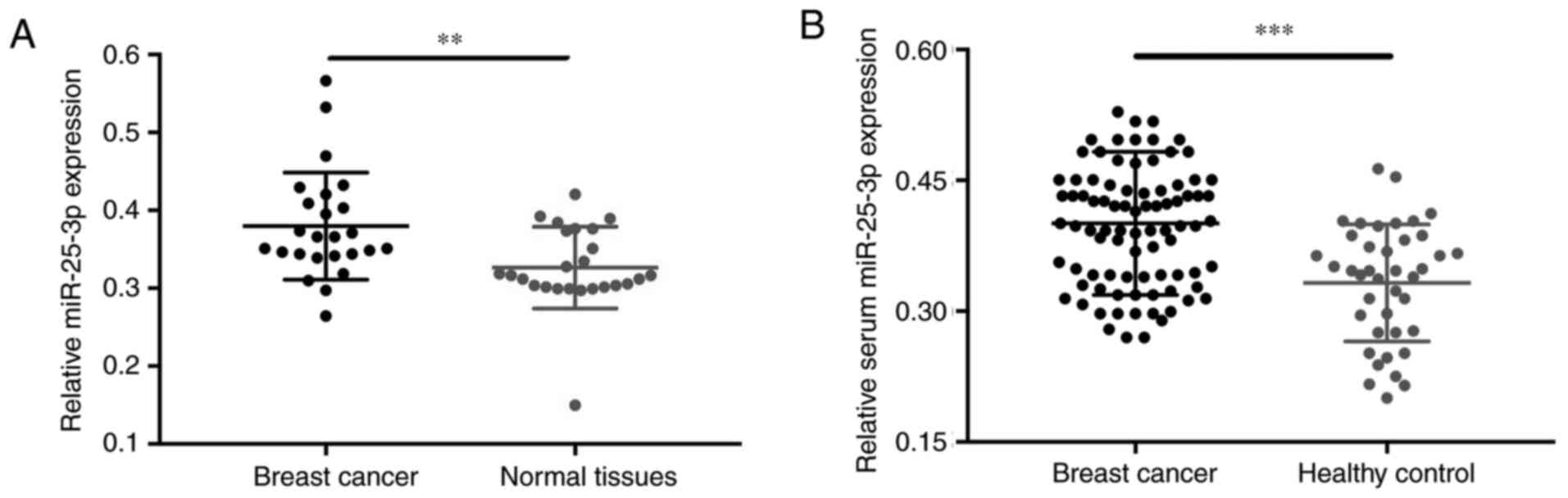
miR-25-3p expression levels were assessed in 50
tissue samples (25 breast cancer and 25 normal tissues) and 176
serum samples (86 samples obtained from patients with breast
cancer, 25 paired samples obtained from patients with breast cancer
1 day and 1 month post-surgery, and 40 healthy control samples).
The association between age and breast cancer was not significant
(Table I). The RT-qPCR results
demonstrated that miR-25-3p expression(± SD) was upregulated in
breast cancer tissues compared with corresponding non-tumor tissues
(Fig. 1A; P=0.005). Moreover,
miR-25-3p expression levels were significantly increased in serum
samples of patients with breast cancer compared with healthy
control subjects (Fig. 1B;
P<0.001).
Association between serum miR-25-3p
expression and clinicopathological characteristics of patients with
breast cancer
Subsequently, the association between the expression
levels of serum miR-25-3p and the clinicopathological
characteristics of patients with breast cancer was analyzed.
Patients with breast cancer (n=88) were split into two groups and
the median expression level of serum miR-25-3p (44 low serum
miR-25-3p expression and 44 high serum miR-25-3p expression) was
used as the cut-off point. Serum miR-25-3p expression levels were
not significantly associated with age, clinical stage, estrogen
receptor, progesterone receptor or human epidermal growth factor
receptor 2 levels (Table I). By
contrast, serum miR-25-3p expression levels were significantly
associated with lymph node metastasis (Table I; P=0.002). The results suggested
that patients with breast cancer with high serum miR-25-3p
expression levels were more likely to display lymph node
metastasis.
Serum miR-25-3p may serve as a
promising diagnostic biomarker for breast cancer
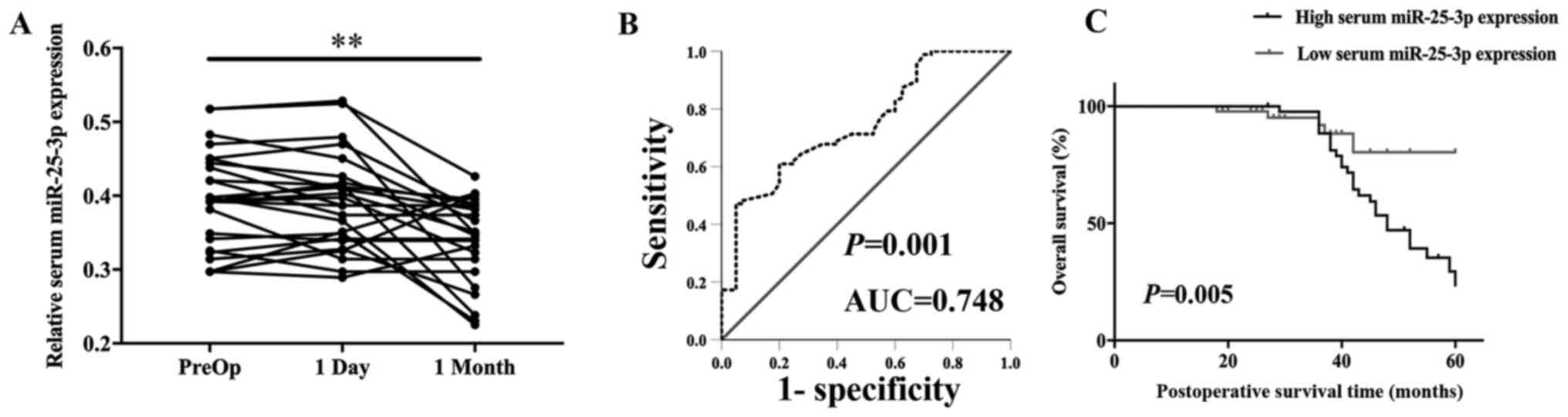
Serum miR-25-3p expression levels of 25
postoperative patients with breast cancer were detected. Serum
miR-25-3p expression levels were not significantly different in
samples obtained on day 1 post-surgery (P>0.05). However,
miR-25-3p expression levels were significantly decreased in samples
obtained at 1month post-surgery (P=0.002) compared with
preoperative samples (Fig. 2A).
Moreover, ROC curve analysis was performed to evaluate the
diagnostic value of miR-25-3p in breast cancer (88 patients with
breast cancer and 40 cancer-free blood donor volunteers). The AUC
for miR-25-3p was 0.748, with 57.1% sensitivity and 95.0%
specificity (Fig. 2B; P=0.001). The
ROC curve corresponded to the diagnostic value of miR-25-3p for
breast cancer.
Serum miR-25-3p may serve as a
prognostic biomarker in breast cancer
The association between serum miR-25-3p expression
levels and breast cancer prognosis was analyzed. The multivariate
Cox proportional hazard regression analysis suggested that clinical
stage [hazard ratio (HR)=2.235; 95% confidence interval
(CI)=1.094–5.853; P=0.047], lymph node metastasis (HR=4.974; 95%
CI=1.786–9.843; P=0.011) and serum miR-25-3p expression levels
(HR=6.683; 95% CI=3.343–9.789; P=0.007) were potential factors
associated with the overall survival of patients with breast cancer
(Table II). The Kaplan-Meier
survival curves indicated that patients with breast cancer with low
expression of serum miR-25-3p displayed a higher overall survival
rate compared with patients with breast cancer with high expression
of serum miR-25-3p (P=0.005; Fig.
2C). The results demonstrated that serum miR-25-3p may serve as
a promising prognostic biomarker for breast cancer.
| Table II.Univariate and multivariate survival
analysis of 88 patients with breast cancer. |
Table II.
Univariate and multivariate survival
analysis of 88 patients with breast cancer.
|
| Univariate
analysis | Multivariate
analysis |
|---|
|
|
|
|
|---|
| Variable | HR | 95% CI | P-value | HR | 95% CI | P-value |
|---|
| Age (≤50 years vs.
>50 years) | 0.844 | 0.417–1.709 | 0.638 | – | – | – |
| Clinical stage (I,
II vs. III, IV) | 5.423 | 2.741–8.658 | 0.012 | 2.235 | 1.094–5.853 | 0.047 |
| Lymph node
metastasis (positive vs. negative) | 10.356 | 4.404–24.353 | 0.001 | 4.974 | 1.786–9.843 | 0.011 |
| ER (positive vs.
negative) | 1.172 | 0.578–2.378 | 0.660 | – | – | – |
| PR (positive vs.
negative) | 0.828 | 0.409–1.678 | 0.601 | – | – | – |
| HER2 (positive vs.
negative) | 0.799 | 0.393–1.622 | 0.534 | – | – | – |
| Serum miR-25-3p
expression | 8.333 | 2.900–23.944 | 0.001 | 6.683 | 3.343–9.789 | 0.007 |
miR-25-3p expression levels in breast
cancer and normal cell lines
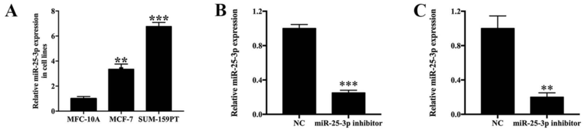
The expression levels of miR-25-3p in MCF-7 and
SUM-159PT cells were measured by RT-qPCR. In addition, alterations
to the expression levels of miR-25-3p following transfection with
miR-25-3p inhibitor, miR-25-3p mimic or NC were also monitored.
miR-25-3p expression levels were 3.45- and 6.67-fold higher in
MCF-7 (P<0.01) and SUM-159PT (P<0.001) cells compared with
MCF-10A cells (Fig. 3A). Moreover,
miR-25-3p inhibitor-transfected SUM-159PT cells displayed a
0.249-fold decrease in miR-25-3p expression levels compared with
the NC group (P<0.001; Fig. 3B).
miR-25-3p inhibitor-transfected MCF-7 cells displayed a 0.201-fold
decrease in miR-25-3p expression levels compared with the NC group
(P<0.01; Fig. 3C). By contrast,
miR-25-3p mimic-transfected SUM-159PT and MCF-7 cells displayed a
12.49- (Fig. S1A; P<0.001) and
13.21-fold (Fig. S1B; P<0.001)
increase in miR-25-3p expression levels compared with the mimic NC
group, respectively. The results indicated that the transfections
were successful.
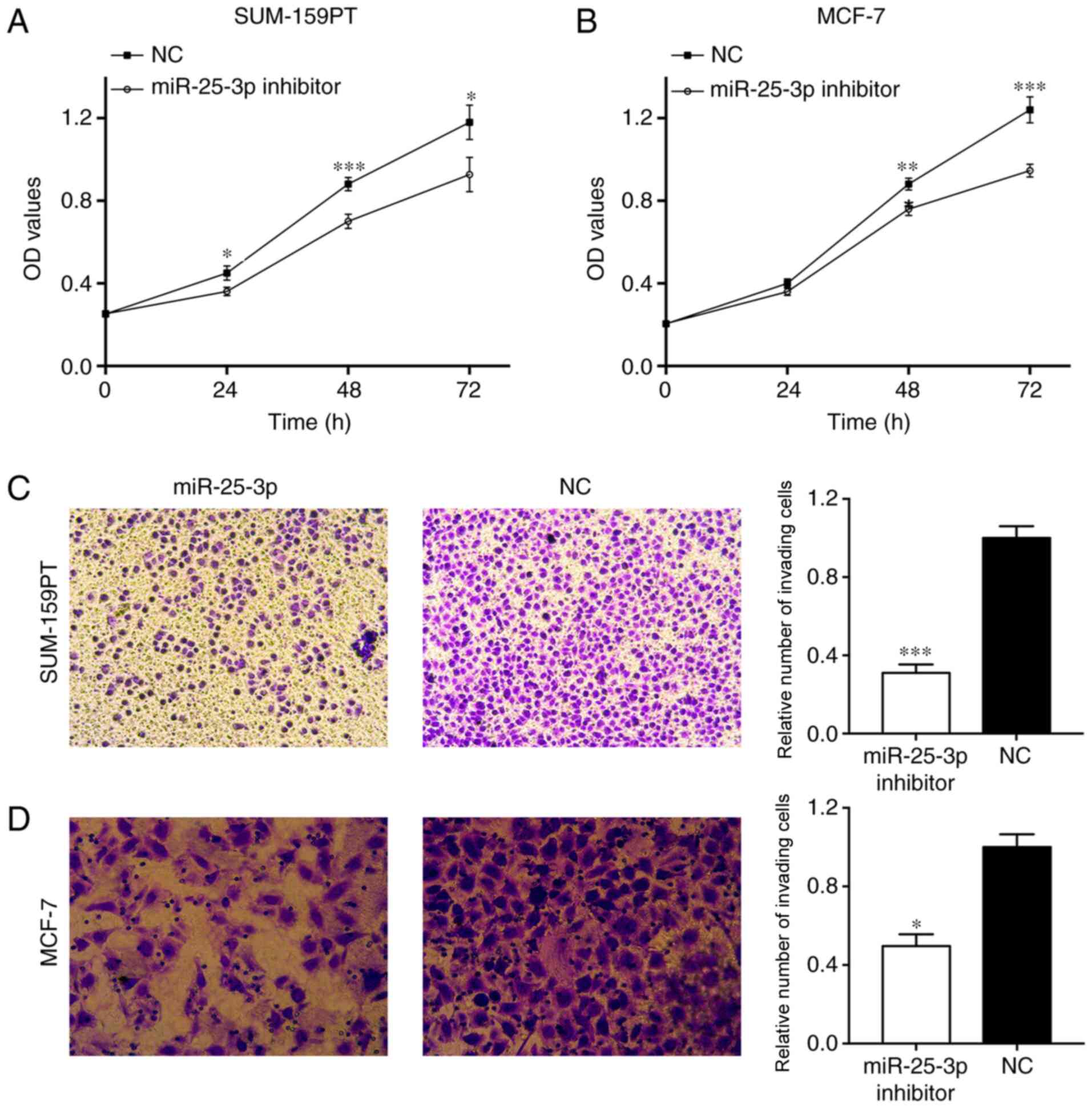
miR-25-3p knockdown suppresses cell
proliferation and invasion
The CCK-8 assay indicated that SUM-159PT cell
proliferation was significantly inhibited following miR-25-43p
knockdown compared with the NC group (Fig. 4A). A similar result was observed in
MCF-7 cells (Fig. 4B). In addition,
the Transwell assay indicated that SUM-159PT cell invasion was
significantly inhibited by miR-25-3p knockdown compared with the NC
group (P<0.001; Fig. 4C).
Similarly, MCF-7 cell invasion was also significantly decreased by
miR-25-3p knockdown compared with the NC group (P<0.05; Fig. 4D). The results indicated that
miR-25-3p knockdown suppressed cell proliferation and invasion.
TOB1 is a target gene regulated by
miR-25-3p
In the present study, three miRNA-binding site
prediction databases (TargetScan, miRWalk and starBase) were
utilized to predict the potential target gene regulated by
miR-25-3p. TOB1 was identified as a target gene. The binding
sequence between miR-25-3p and TOB1 is presented in Fig. 5A. TOB1 expression levels were
0.35- (P<0.01) and 0.44- (P<0.05) fold lower in MCF-7 and
SUM-159PT cells compared with MCF-10A cells (Fig. 5B). In addition, the UALCAN analysis
revealed that TOB1 expression levels were significantly
decreased in breast cancer tissues compared with normal tissues
(P=8.85×10⁻⁹; Fig.
S2A). Furthermore, a negative association between TOB1
and miR-25-3p expression levels was identified
(P=9.38×10⁻⁸; Fig.
S2B). The results suggested that TOB1 is a potential
target gene regulated by miR-25-3p. Subsequently, a dual-luciferase
reporter assay was performed to verify the binding of miR-25-3p to
TOB1. The luciferase activity of the TOB1-UTR-WT
group in 293T cells was significantly inhibited by miR-25-3p
overexpression compared with the NC group (P<0.05). By contrast,
the luciferase activity of the TOB1-UTR-MT group was not
altered by miR-25-3p expression (Fig.
5C). To confirm the specificity of TOB1 in breast cancer, a
dual-luciferase reporter assay was also performed in breast cancer
cell lines, and similar results were obtained in SUM159-PT
(P<0.05; Fig. 5D) and MCF-7
cells (P<0.05; Fig. 5E). Western
blotting was performed for target gene validation. In SUM-159PT
cells, TOB1 protein expression levels in the miR-25-3p inhibitor
group were significantly increased compared with the inhibitor NC
group (P<0.001; Fig. 5F).
Moreover, TOB1 protein expression levels in the miR-25-3p inhibitor
group were significantly increased compared with the inhibitor NC
group (P<0.01; Fig. 5G).
Collectively, the results demonstrated that TOB1 was a
potential target gene regulated by miR-25-3p in breast cancer.
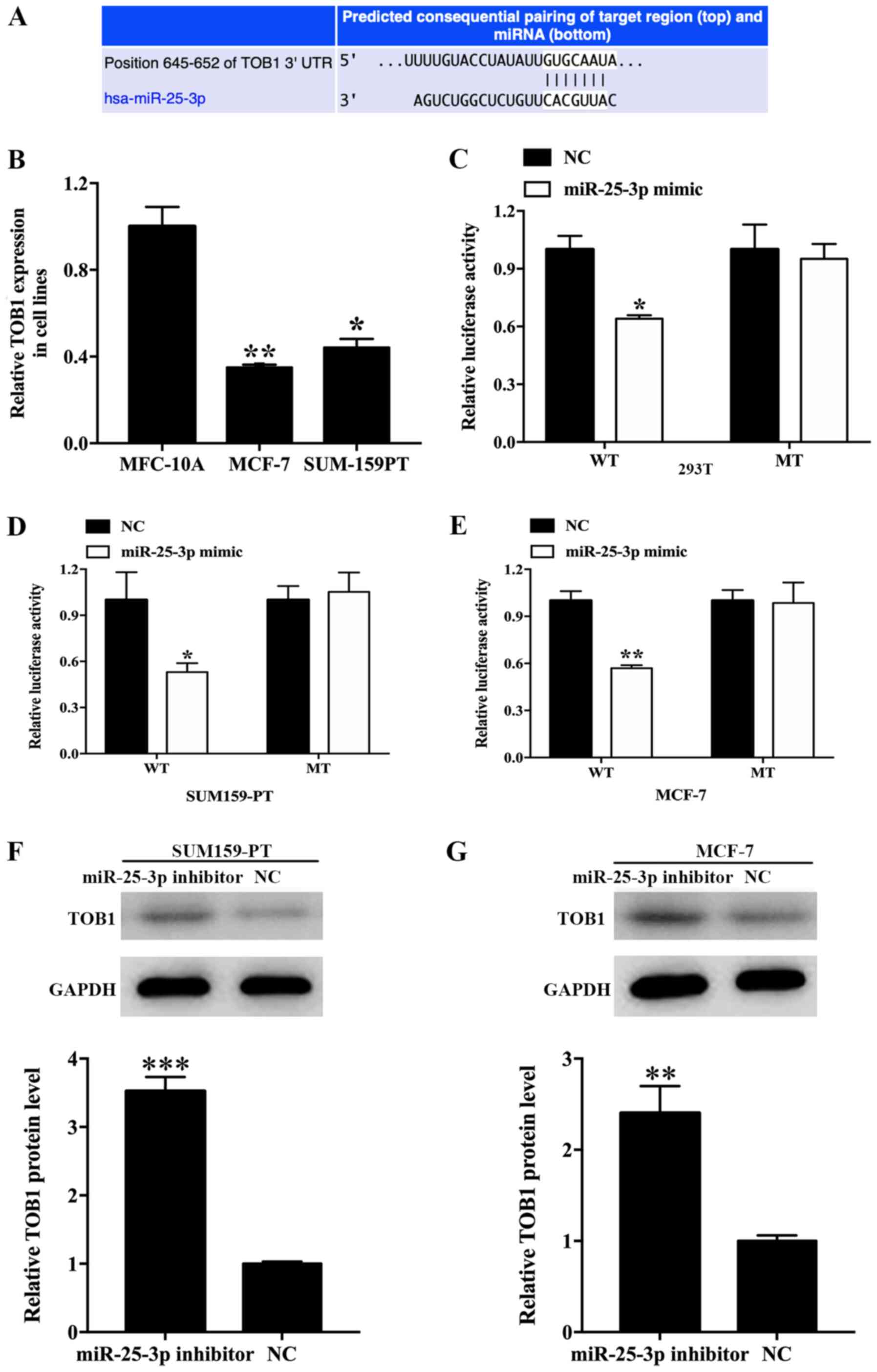 | Figure 5.Validation of TOB1 as a direct target
of miR-25-3p. (A) The binding sites between miR-25-3p and TOB1
3′-UTR. (B) Relative expression of TOB1 in cell lines. *P<0.05,
**P<0.01 vs. MFC-10A. (C) Luciferase activities in the WT and MT
groups treated with miR-25-3p mimic or NC in (C) 293T, (D)
SUM159-PT and (E) MCF-7 cells. TOB1 protein expression levels
following miR-25-3p knockdown in (D) SUM-159-PT and (E) MCF-7
cells. Relative TOB1 protein expression in (F) SUM159-PT and (G)
MCF7 cells after treatment with miR-25-3p compared with NC group.
*P<0.05, **P<0.01 and ***P<0.001 vs. NC. TOB1, transducer
of ERBB2, 1; miR, microRNA; 3′-UTR, 3′-untranslated region; WT,
wild-type; MT, mutant; NC, negative control. |
Discussion
miRNAs are a distinct class of small non-coding RNAs
that display various effects in post-transcriptional gene silencing
of target mRNAs (32). miRNAs are
involved in numerous biological processes, including cell
proliferation, invasion, migration, apoptosis, cell cycle control,
differentiation, metabolism, immunity, neuronal patterning and
stress responses (33–37). Previous findings have revealed that
miRNAs exist in the 12 body fluids, which includes serum, urine,
saliva, peritoneal fluid, pleural fluid, seminal fluid, tears,
amniotic fluid, breast milk, bronchial lavage, cerebrospinal fluid
and colostrum (38). Mitchell et
al (39) initially described
serum miRNAs and demonstrated that they remain in a stable form
that is not degraded by endogenous RNase enzymes. It has also been
suggested that serum miRNAs may serve as predictive prognostic
biomarkers in various malignancies, including breast cancer
(39–41).
miR-25-3p is a member of the of miR-25-93-106b
cluster, which commonly act as oncogenes, is involved in
carcinogenesis and is often upregulated in various malignancies
(42). Wang et al (43) reported that miR-25 expression was
significantly upregulated in hepatocellular carcinoma tissues,
where it promoted cancer cell growth, migration and invasion via
Rho GDP dissociation inhibitor-1 (43). miR-25 upregulation was also observed
in esophageal squamous cell carcinoma cells and miR-25 knockdown
significantly inhibited cell migration and invasion (44). Moreover, it has been reported that
miR-25-3p may serve as a biomarker for numerous types of cancer,
such as osteosarcoma and pancreatic cancer (45,46).
Previous findings demonstrated that miR-25-3p expression was
upregulated in breast cancer and that the abnormal expression
levels promoted breast cancer cell proliferation (27). Therefore, the present study aimed to
investigate the diagnostic and prognostic role of miR-25-3p in
breast cancer.
In the present study, miR-25-3p expression levels
were increased in breast cancer tissue and serum samples compared
with normal breast tissue and serum samples. Moreover, serum
miR-25-3p expression levels were not significantly different on day
1 post-surgery compared with presurgery; however, at 1 month
post-surgery, miR-25-3p expression levels were significantly
decreased compared with prior to surgery, which indicated that
miR-25-3p downregulation was associated with the removal of the
tumor and not due to surgical stress. Furthermore, the results
indicated that serum miR-25-3p expression levels could successfully
discriminate patients with breast cancer from healthy subjects. The
AUC, sensitivity and specificity of serum miR-25-3p for the
diagnosis of breast cancer were 0.748, 57.1 and 95.0%,
respectively. The Kaplan-Meier survival curves demonstrated that
patients with breast cancer with low serum miR-25-3p expression
showed a higher overall survival rate compared with patients with
breast cancer with high serum miR-25-3p expression. Therefore, the
results demonstrated that serum miR-25-3p may serve as an
alternative biomarker for the diagnosis and prognosis of breast
cancer.
Previous studies revealed that serum miRNAs could be
used as diagnostic biomarkers in breast cancer. For example, serum
miR-103 expression levels were significantly increased in patients
with breast cancer compared with healthy control subjects.
Additionally, for the diagnosis of breast cancer, serum miR-103
displayed 84% sensitivity and 70% specificity, suggesting that
serum miR-103 was a promising diagnostic marker for breast cancer
detection (47). Serum miR-195
downregulation was observed in patients with breast cancer, and
serum miR-195 displayed 69.0% sensitivity and 89.2% specificity for
breast cancer detection. Furthermore, serum miR-195 was used to
distinguish patients with breast cancer from healthy subjects,
indicating that serum miR-195 was a potential tumor biomarker for
breast cancer diagnosis (48). A
meta-analysis was performed with 438 patients and 228 healthy
subjects, and the results suggested that serum miR-21 displayed
0.79% sensitivity, 0.85% specificity and 0.89 AUC in the diagnosis
of breast cancer; therefore, the study indicated that miR-21 was a
novel biomarker for early detection of breast cancer (49).
Similarly, previous studies have indicated that
serum miRNAs have the potential to be used as prognostic biomarkers
in breast cancer. A meta-analysis of 1,629 cases demonstrated that
patients with breast cancer with elevated miR-21 expression
displayed a poor overall survival. miR-21 expression levels were
significantly associated with lymph node metastasis, suggesting
that circulating miR-21 could be used as a prognostic biomarker in
patients with breast cancer (50).
Hsieh et al (51) reported
that serum miR-125a-5p expression was negatively associated with
tumor grade, lymph-node status and tumor size. Moreover, low
miR-125a-5p expression was an independent biomarker for the
prediction of poor prognosis in patients with breast cancer.
Another study indicated that serum miR-99a expression levels were
significantly decreased in patients with breast cancer compared
with healthy subjects. Furthermore, it was reported that patients
with breast cancer with lower miR-99a expression levels displayed a
poor overall survival, and that serum miR-99a expression is an
independent risk factor for breast cancer, indicating that serum
miR-99a was a tumor suppressor and a prognostic biomarker for
breast cancer (52).
TOB1 is a transducer of ErbB-2 that is
ubiquitously expressed in human adult tissues (53). The TOB1 gene is located on
chromosome 17q21 and codes for a 45 kDa protein (54). Previous studies have revealed that
TOB1 is associated with tumor cell proliferation and invasion
(54,55). In the present study, TOB1 was
downregulated in breast cancer cells compared with normal cells,
and miR-25-3p knockdown suppressed breast cancer cell proliferation
and invasion by regulating TOB1 expression.
The main limitation of the present study was that
the sample size was small; therefore, a future study employing a
larger sample size should be performed to verify the results of the
present study. Moreover, the relevant signaling pathways and
targets of miR-25-3p in breast cancer, as well as the function of
TOB1 in breast cancer were not clarified in the present study.
In conclusion, the present study demonstrated that
miR-25-3p, which regulated breast cancer cellular functions via
TOB1, may serve as a breast cancer biomarker.
Supplementary Material
Supporting Data
Acknowledgements
Not applicable.
Funding
The present study was supported by the Natural
Science Foundation of Tianjin (grant no. 18JCYBJC94100) and Key
Task Project of Tianjin Health and Family Planning Commission
(grant no. 2015KZ089).
Availability of data and materials
The datasets used and/or analyzed during the current
study are available from the corresponding author on reasonable
request.
Authors' contributions
TZ, XP and LH designed the experiments and analyzed
the data. TZ, WM, LG, YC and XY performed the experiments. SS
recruited and examined participants. TZ and WM wrote the
manuscript. All authors discussed the results. All authors confirm
the authenticity of all the raw data and read and approved the
final manuscript.
Ethics approval and consent to
participate
The present study was approved by the Ethics
Committee of Tianjin Medical University Cancer Institute and
Hospital. Written informed consent was obtained from all
participants.
Patient consent for publication
Not applicable.
Competing interests
The authors declare that they have no competing
interests.
References
|
1
|
Harbeck N and Gnant M: Breast cancer.
Lancet. 389:1134–1150. 2017. View Article : Google Scholar : PubMed/NCBI
|
|
2
|
Bray F, Ferlay J, Soerjomataram I, Siegel
RL, Torre LA and Jemal A: Global cancer statistics 2018: GLOBOCAN
estimates of incidence and mortality worldwide for 36 cancers in
185 countries. CA Cancer J Clin. 68:394–424. 2018. View Article : Google Scholar : PubMed/NCBI
|
|
3
|
Bray F, McCarron P and Parkin DM: The
changing global patterns of female breast cancer incidence and
mortality. Breast Cancer Res. 6:229–239. 2004. View Article : Google Scholar : PubMed/NCBI
|
|
4
|
Jemal A, Center MM, DeSantis C and Ward
EM: Global patterns of cancer incidence and mortality rates and
trends. Cancer Epidemiol Biomarkers Prev. 19:1893–1907. 2010.
View Article : Google Scholar : PubMed/NCBI
|
|
5
|
Pogoda K, Niwińska A, Murawska M and
Pieńkowski T: Analysis of pattern, time and risk factors
influencing recurrence in triple-negative breast cancer patients.
Med Oncol. 30:3882013. View Article : Google Scholar : PubMed/NCBI
|
|
6
|
Liang F, Liang H, Li Z and Huang P: JAK3
is a potential biomarker and associated with immune infiltration in
kidney renal clear cell carcinoma. Int Immunopharmacol.
86:1067062020. View Article : Google Scholar : PubMed/NCBI
|
|
7
|
Halvorsen AR, Helland Å, Gromov P,
Wielenga VT, Talman MM, Brunner N, Sandhu V, Børresen-Dale AL,
Gromova I and Haakensen VD: Profiling of microRNAs in tumor
interstitial fluid of breast tumors-a novel resource to identify
biomarkers for prognostic classification and detection of cancer.
Mol Oncol. 11:220–234. 2017. View Article : Google Scholar : PubMed/NCBI
|
|
8
|
Madhavan D, Cuk K, Burwinkel B and Yang R:
Cancer diagnosis and prognosis decoded by blood-based circulating
microRNA signatures. Front Genet. 4:1162013. View Article : Google Scholar : PubMed/NCBI
|
|
9
|
Christou N, Meyer J, Popeskou S, David V,
Toso C, Buchs N, Liot E, Robert J, Ris F and Mathonnet M:
Circulating tumour cells, circulating tumour DNA and circulating
tumour miRNA in blood assays in the different steps of colorectal
cancer management, a review of the evidence in 2019. Biomed Res
Int. 2019:59530362019. View Article : Google Scholar : PubMed/NCBI
|
|
10
|
Zhang K, Wu X, Wang J, Lopez J, Zhou W,
Yang L, Wang SE, Raz DJ and Kim JY: Circulating miRNA profile in
esophageal adenocarcinoma. Am J Cancer Res. 6:2713–2721.
2016.PubMed/NCBI
|
|
11
|
Liu X, Zhang H, Qin S, Wang Q, Yang X and
Wang K: Optical fiber amplifier for quantitative and sensitive
point-of-care testing of myoglobin and miRNA-141. Biosens
Bioelectron. 129:87–92. 2019. View Article : Google Scholar : PubMed/NCBI
|
|
12
|
Imaoka H, Toiyama Y, Fujikawa H, Hiro J,
Saigusa S, Tanaka K, Inoue Y, Mohri Y, Mori T, Kato T, et al:
Circulating microRNA-1290 as a novel diagnostic and prognostic
biomarker in human colorectal cancer. Ann Oncol. 27:1879–1886.
2016. View Article : Google Scholar : PubMed/NCBI
|
|
13
|
Toiyama Y, Takahashi M, Hur K, Nagasaka T,
Tanaka K, Inoue Y, Kusunoki M, Boland CR and Goel A: Serum miR-21
as a diagnostic and prognostic biomarker in colorectal cancer. J
Natl Cancer Inst. 105:849–859. 2013. View Article : Google Scholar : PubMed/NCBI
|
|
14
|
Bhagirath D, Yang TL, Bucay N, Sekhon K,
Majid S, Shahryari V, Dahiya R, Tanaka Y and Saini S: microRNA-1246
Is an exosomal biomarker for aggressive prostate cancer. Cancer
Res. 78:1833–1844. 2018. View Article : Google Scholar : PubMed/NCBI
|
|
15
|
Li Z, Zhou L, Lin C, Pan X, Xie J, Zhao L,
Quan J, Xu J, Guan X, Xu W, et al: miR-302b regulates cell
functions and acts as a potential biomarker to predict recurrence
in bladder cancer. Life Sci. 209:15–23. 2018. View Article : Google Scholar : PubMed/NCBI
|
|
16
|
Zhao C, Ling X, Li X, Hou X and Zhao D:
MicroRNA-138-5p inhibits cell migration, invasion and EMT in breast
cancer by directly targeting RHBDD1. Breast Cancer. 26:817–825.
2019. View Article : Google Scholar : PubMed/NCBI
|
|
17
|
Lee HS, Kundu J, Kim RN and Shin YK:
Transducer of ERBB2.1 (TOB1) as a tumor suppressor: A mechanistic
perspective. Int J Mol Sci. 16:29815–29828. 2015. View Article : Google Scholar : PubMed/NCBI
|
|
18
|
Kundu J, Wahab SM, Kundu JK, Choi YL,
Erkin OC, Lee HS, Park SG and Shin YK: Tob1 induces apoptosis and
inhibits proliferation, migration and invasion of gastric cancer
cells by activating Smad4 and inhibiting β-catenin signaling. Int J
Oncol. 41:839–848. 2012. View Article : Google Scholar : PubMed/NCBI
|
|
19
|
Jiao Y, Sun KK, Zhao L, Xu JY, Wang LL and
Fan SJ: Suppression of human lung cancer cell proliferation and
metastasis in vitro by the transducer of ErbB-2.1 (TOB1). Acta
Pharmacol Sin. 33:250–260. 2012. View Article : Google Scholar : PubMed/NCBI
|
|
20
|
Wu D, Zhou W, Wang S, Zhou Z, Wang S and
Chen L: Tob1 enhances radiosensitivity of breast cancer cells
involving the JNK and p38 pathways. Cell Biol Int. 39:1425–1430.
2015. View Article : Google Scholar : PubMed/NCBI
|
|
21
|
Cioffi M, Trabulo SM, Vallespinos M, Raj
D, Kheir TB, Lin ML, Begum J, Baker AM, Amgheib A, Saif J, et al:
The miR-25-93-106b cluster regulates tumor metastasis and immune
evasion via modulation of CXCL12 and PD-L1. Oncotarget.
8:21609–21625. 2017. View Article : Google Scholar : PubMed/NCBI
|
|
22
|
Sárközy M, Kahán Z and Csont T: A myriad
of roles of miR-25 in health and disease. Oncotarget.
9:21580–21612. 2018. View Article : Google Scholar : PubMed/NCBI
|
|
23
|
Wang X, Meng X, Li H, Liu W, Shen S and
Gao Z: MicroRNA-25 expression level is an independent prognostic
factor in epithelial ovarian cancer. Clin Transl Oncol. 16:954–958.
2014. View Article : Google Scholar : PubMed/NCBI
|
|
24
|
Wen Y, Han J, Chen J, Dong J, Xia Y, Liu
J, Jiang Y, Dai J, Lu J, Jin G, et al: Plasma miRNAs as early
biomarkers for detecting hepatocellular carcinoma. Int J Cancer.
137:1679–1690. 2015. View Article : Google Scholar : PubMed/NCBI
|
|
25
|
Jia Y, Lu H, Wang C, Wang J and Zhang C,
Wang F and Zhang C: miR-25 is upregulated before the occurrence of
esophageal squamous cell carcinoma. Am J Transl Res. 9:4458–4469.
2017.PubMed/NCBI
|
|
26
|
Chang JT, Wang F, Chapin W and Huang RS:
Identification of MicroRNAs as breast cancer prognosis markers
through the cancer genome atlas. PLoS One. 11:e01682842016.
View Article : Google Scholar : PubMed/NCBI
|
|
27
|
Chen H, Pan H, Qian Y, Zhou W and Liu X:
miR-25-3p promotes the proliferation of triple negative breast
cancer by targeting BTG2. Mol Cancer. 17:42018. View Article : Google Scholar : PubMed/NCBI
|
|
28
|
Zou X, Xia T, Li M, Wang T, Liu P, Zhou X,
Huang Z and Zhu W: MicroRNA profiling in serum: Potential
signatures for breast cancer diagnosis. Cancer Biomark. 30:41–53.
2021. View Article : Google Scholar : PubMed/NCBI
|
|
29
|
Livak KJ and Schmittgen TD: Analysis of
relative gene expression data using real-time quantitative PCR and
the 2(-Delta Delta C(T)) method. Methods. 25:402–408. 2001.
View Article : Google Scholar : PubMed/NCBI
|
|
30
|
Dyer BW, Ferrer FA, Klinedinst DK and
Rodriguez R: A noncommercial dual luciferase enzyme assay system
for reporter gene analysis. Anal Biochem. 282:158–161. 2000.
View Article : Google Scholar : PubMed/NCBI
|
|
31
|
Grentzmann G, Ingram JA, Kelly PJ,
Gesteland RF and Atkins JF: A dual-luciferase reporter system for
studying recoding signals. RNA. 4:479–486. 1998.PubMed/NCBI
|
|
32
|
Cortez MA, Welsh JW and Calin GA:
Circulating microRNAs as noninvasive biomarkers in breast cancer.
Recent Results Cancer Res. 195:151–161. 2012. View Article : Google Scholar : PubMed/NCBI
|
|
33
|
Mens MMJ and Ghanbari M: Cell cycle
regulation of stem cells by MicroRNAs. Stem Cell Rev Rep.
14:309–322. 2018. View Article : Google Scholar : PubMed/NCBI
|
|
34
|
Qi Y, Wang X, Kong X, Zhai J, Fang Y, Guan
X and Wang J: Expression signatures and roles of microRNAs in
inflammatory breast cancer. Cancer Cell Int. 19:232019. View Article : Google Scholar : PubMed/NCBI
|
|
35
|
Li Z, Lin C, Zhao L, Zhou L, Pan X, Quan
J, Peng X, Li W, Li H, Xu J, et al: Oncogene miR-187-5p is
associated with cellular proliferation, migration, invasion,
apoptosis and an increased risk of recurrence in bladder cancer.
Biomed Pharmacother. 105:461–469. 2018. View Article : Google Scholar : PubMed/NCBI
|
|
36
|
Olejniczak M, Kotowska-Zimmer A and
Krzyzosiak W: Stress-induced changes in miRNA biogenesis and
functioning. Cell Mol Life Sci. 75:177–191. 2018. View Article : Google Scholar : PubMed/NCBI
|
|
37
|
Singh SK, Pal Bhadra M, Girschick HJ and
Bhadra U: MicroRNAs-micro in size but macro in function. FEBS J.
275:4929–4944. 2008. View Article : Google Scholar : PubMed/NCBI
|
|
38
|
Armand-Labit V and Pradines A: Circulating
cell-free microRNAs as clinical cancer biomarkers. Biomol Concepts.
8:61–81. 2017. View Article : Google Scholar : PubMed/NCBI
|
|
39
|
Mitchell PS, Parkin RK, Kroh EM, Fritz BR,
Wyman SK, Pogosova-Agadjanyan EL, Peterson A, Noteboom J, O'Briant
KC, Allen A, et al: Circulating microRNAs as stable blood-based
markers for cancer detection. Proc Natl Acad Sci USA.
10:10513–10518. 2008. View Article : Google Scholar
|
|
40
|
Li Q, Liu M, Ma F, Luo Y, Cai R, Wang L,
Xu N and Xu B: Circulating miR-19a and miR-205 in serum may predict
the sensitivity of luminal A subtype of breast cancer patients to
neoadjuvant chemotherapy with epirubicin plus paclitaxel. PLoS One.
9:e1048702014. View Article : Google Scholar : PubMed/NCBI
|
|
41
|
Shen L, Wan Z, Ma Y, Wu L, Liu F, Zang H
and Xin S: The clinical utility of microRNA-21 as novel biomarker
for diagnosing human cancers. Tumour Biol. 36:1993–2005. 2015.
View Article : Google Scholar : PubMed/NCBI
|
|
42
|
Gruszka R and Zakrzewska M: The oncogenic
relevance of miR-17-92 cluster and its paralogous miR-106b-25 and
miR-106a-363 clusters in brain tumors. Int J Mol Sci. 19:8792018.
View Article : Google Scholar
|
|
43
|
Wang C, Wang X, Su Z, Fei H, Liu X and Pan
Q: miR-25 promotes hepatocellular carcinoma cell growth, migration
and invasion by inhibiting RhoGDI1. Oncotarget. 6:36231–36244.
2015. View Article : Google Scholar : PubMed/NCBI
|
|
44
|
Hua Y, Zhao K, Tao G, Dai C and Su Y:
miR-25 promotes metastasis via targeting FBXW7 in esophageal
squamous cell carcinoma. Oncol Rep. 38:3030–3038. 2017. View Article : Google Scholar : PubMed/NCBI
|
|
45
|
Fujiwara T, Uotani K, Yoshida A, Morita T,
Nezu Y, Kobayashi E, Yoshida A, Uehara T, Omori T, Sugiu K, et al:
Clinical significance of circulating miR-25-3p as a novel
diagnostic and prognostic biomarker in osteosarcoma. Oncotarget.
8:33375–33392. 2017. View Article : Google Scholar : PubMed/NCBI
|
|
46
|
Deng T, Yuan Y, Zhang C, Zhang C, Yao W,
Wang C, Liu R and Ba Y: Identification of circulating miR-25 as a
potential biomarker for pancreatic cancer diagnosis. Cell Physiol
Biochem. 39:1716–1722. 2016. View Article : Google Scholar : PubMed/NCBI
|
|
47
|
Wang X, Wu X, Yan L and Shao J: Serum
miR-103 as a potential diagnostic biomarker for breast cancer. Nan
Fang Yi Ke Da Xue Xue Bao. 32:631–634. 2012.(In Chinese).
PubMed/NCBI
|
|
48
|
Zhao FL, Dou YC, Wang XF, Han DC, Lv ZG,
Ge SL and Zhang YK: Serum microRNA-195 is down-regulated in breast
cancer: A potential marker for the diagnosis of breast cancer. Mol
Biol Rep. 41:5913–5922. 2014. View Article : Google Scholar : PubMed/NCBI
|
|
49
|
Li S, Yang X, Yang J, Zhen J and Zhang D:
Serum microRNA-21 as a potential diagnostic biomarker for breast
cancer: A systematic review and meta-analysis. Clin Exp Med.
16:29–35. 2016. View Article : Google Scholar : PubMed/NCBI
|
|
50
|
Jinling W, Sijing S, Jie Z and Guinian W:
Prognostic value of circulating microRNA-21 for breast cancer: A
systematic review and meta-analysis. Artif Cells Nanomed
Biotechnol. 45:1–6. 2017. View Article : Google Scholar : PubMed/NCBI
|
|
51
|
Hsieh TH, Hsu CY, Tsai CF, Long CY, Chai
CY, Hou MF, Lee JN, Wu DC, Wang SC and Tsai EM: miR-125a-5p is a
prognostic biomarker that targets HDAC4 to suppress breast
tumorigenesis. Oncotarget. 6:494–509. 2015. View Article : Google Scholar : PubMed/NCBI
|
|
52
|
Li J, Song ZJ, Wang YY, Yin Y, Liu Y and
Nan X: Low levels of serum miR-99a is a predictor of poor prognosis
in breast cancer. Genet Mol Res. 152016.doi:
10.4238/gmr.15038338.
|
|
53
|
Lin S, Zhu Q, Xu Y, Liu H, Zhang J, Xu J,
Wang H, Sang Q, Xing Q and Fan J: The role of the TOB1 gene in
growth suppression of hepatocellular carcinoma. Oncol Lett.
4:981–987. 2012. View Article : Google Scholar : PubMed/NCBI
|
|
54
|
O'Malley S, Su H, Zhang T, Ng C, Ge H and
Tang CK: TOB suppresses breast cancer tumorigenesis. Int J Cancer.
125:1805–1813. 2009. View Article : Google Scholar : PubMed/NCBI
|
|
55
|
Guo H, Ji F, Zhao X, Yang X, He J, Huang L
and Zhang Y: MicroRNA-371a-3p promotes progression of gastric
cancer by targeting TOB1. Cancer Lett. 443:179–188. 2019.
View Article : Google Scholar : PubMed/NCBI
|