Introduction
In recent years, there has been a consensus that
physical exercise is effective in maintaining or improving
cardiovascular and musculoskeletal health, both of which are
fundamental for the preservation of physiological functioning and
independence (1). Previous studies
have reported that some physical exercise modalities are effective
in reducing falls (2–4), and in improving balance (2) and symptoms of anxiety and depression
(5,6) in healthy individuals.
There has been increasing evidence of the benefits
of physical exercise in improving mobility (7–10),
postural stability (7–10), gait (11,12),
quality of life (13,14), cognitive function (13–15)
and severity of symptoms in individuals with Parkinson's disease
(PD) (10). PD has a lifetime risk
of 2% and is considered the second most common neurodegenerative
disorder (16). Functional
disability caused by PD leads to an increased inability to perform
activities of daily living, loss of independence, decreased quality
of life, and socioeconomic and occupational losses (17). Motor deficiencies in individuals
with PD, caused by bradykinesia, rigidity, tremor and postural
instability, accelerate the declines in functional capacity,
especially when associated with decreasing physical activity and a
sedentary lifestyle (18,19).
Recent advances in understanding the molecular
mechanisms of PD have revealed the involvement of small non-coding
RNA molecules, known as microRNAs (miRNAs/miRs), in the
derangements and perturbation of processes associated with the
regulation of the expression levels of genes involved in PD
development (20–22). Moreover, a systematic review of the
literature identified ~91 different miRNAs associated with PD, with
the expression levels of 39 of these miRNAs differing significantly
between individuals with PD and healthy controls and/or between
treated and untreated patients with PD (23), such as miR-30b, miR-30c, miR-26a,
miR-450b-3p, miR-148b, miR-1, miR-22*, miR-29a, miR-103a-3p,
miR-30b-5p, miR-29a-3p, miR-1249, miR-20a, miR-18b, miR-378c,
miR-4293, miR-652, miR-15a*, miR-29c, miR-376c, miR-143, and
miR-19b (downregulated miRNAs) and miR-1826, miR-626, miR-505,
miR-16-2a*, miR-26a2*, miR-30a, miR-7, miR-9-3-p, miR-9-5p,
miR-129, miR-132, miR-423, miR-365, miR-486, miR-1260, miR-218 and
miR-331-5p (upregulated miRNAs).
A systematic review of experimental and
quasi-experimental studies published between 2007 and 2017
(24) revealed the acute and/or
chronic effects of physical exercise (aerobic and resistance) on
the expression of several miRNAs in healthy individuals (25–36),
athletes (37–40), young and older adults (41–50),
as well as in patients with chronic heart failure (CHF) (51,52),
chronic kidney disease (CKD) (53),
type 2 diabetes mellitus (DM2) associated with morbid obesity
(54), prediabetes (55) and intermittent claudication
(56,57). However, to the best of our
knowledge, there have been no studies investigating the effects of
physical exercise on the expression levels of miRNAs in individuals
with PD. Therefore, additional studies should be performed to
examine the effect of physical exercise on circulating miRNAs in
individuals with pathological conditions, such as neurological
diseases, as physical exercise serves an important role in
mitigating or delaying the emergence of symptoms, which ultimately
results in a certain degree of independence and improved quality of
life, indicating its consideration as an auxiliary method in
traditional therapies (24).
Based on these premises, the present study aimed to
investigate the effects of an interval training program using a
cycle ergometer on the expression levels of miRs associated with
PD, including miR-106a-5p, miR-103a-3p and miR-29a-3p, in serum
samples from men with PD.
Materials and methods
Ethics approval and consent
This quasi-experimental study with pre- and
post-tests, and with a non-equivalent group design was approved by
the Ethics Committee for Research involving Human Beings of the
State University of Santa Catarina in 2015 (certificate of
presentation for ethical appreciation no. 50032415.1.0000.0118).
The objectives of this study and the procedures adopted throughout
the research were explained to all participants, who voluntarily
agreed to continue participation by signing an informed consent
document.
Study design
The evaluations were performed at the beginning of
the study (week 0) and after 8 weeks of the intervention program
(week 9) at the Center for Health and Sports Sciences (CEFID;
Florianópolis, Brazil) from March to June 2018.
Identification, selection and
allocation of participants
A total of eight men with PD participated in this
study. The participants fulfilled the following inclusion criteria:
i) Age ≥50 years old and ≤80 years old; ii) classification in
stages I, II and III of the Hoehn & Yahr scale; iii) a score
≥24 in the Mini-Mental State Examination (MMSE); iv) use of stable
medication during the last 3 months; and v) physical ability to
walk 300 m or more during the 6-min walk test (6MWT). The decision
to include only men in this study was made given the need to
minimize possible sex-specific confounding effects, such as those
arising from typical hormonal implications of the menstrual cycle
in women, which may cause changes in circulating miRNA expression
levels (58,59).
During the recruitment phase, some individuals
indicated that they would meet the inclusion criteria. After the
initial assessments, these potential participants were excluded as
they may i) have a deep brain stimulator implant; ii) present with
other neurological disorders, musculoskeletal diseases and
uncontrolled hypertension; iii) show changes in cognitive functions
that would impair cooperation and comprehension of the proposed
activities; and iv) have visual deficits.
The participants were selected based on the
eligibility criteria and classified into two groups (both n=8):
Experimental group (EG, interval training program) and control
group (CG). The medications prescribed [levodopa (L-dopa) or
Prolopa] for the participants were not changed by the researchers
during the present study. All evaluations and training sessions
were performed at the same time of day to minimize the effects of
scheduled medication on motor function.
Demographic, economic and clinical
assessments
Demographic and economic information was collected
using a clinical report form specifically developed for this study
in March 2018. Clinical characteristics, namely time of diagnosis,
disease severity and motor function were respectively assessed
using a clinical report form, the modified Hoehn & Yahr Scale
(60), with scores ranging from 0
(no signs of disease) to 5 (wheelchair bound or bedridden unless
aided), and the Unified Parkinson's Rating Scale-section III
(UPDRS) (61), where the score of
each of the 14 items ranges from 0–4 and higher values indicate
greater impairment.
Overall cognition was assessed through the MMSE
(62), with a score ranging from
0–30 points and lower scores indicating a possible cognitive
deficit, and the Montreal Cognitive Assessment (MoCA) (63), with a total score of 30 points and
scores ≥26 indicating absence of cognitive impairment.
Cardiorespiratory performance was assessed in field
tests, and all participants were submitted to the 6MWT following
the Brazilian guidelines for the application of the test (64). The 6MWT is useful to evaluate
clinical data and to help design rehabilitation therapies for
individuals with PD (65).
Blood samples
Blood was collected following the recommendations of
the Brazilian Society of Clinical Pathology/Laboratory Medicine for
venous blood collection (66). All
participants had their blood samples collected in the afternoon
(week 0 and 9). Peripheral blood samples (5 ml) were collected in
Vacutainer tubes without anticoagulant (BD Biosciences). The
samples were processed for serum isolation within 2 h of
collection. Serum samples were centrifuged at 3,000 × g for 10 min
at 4°C (Omega® centrifuge). The serum was divided into
aliquots and stored in a freezer at −80°C for subsequent analyses,
which were performed by the Group of Studies on Micro-Macromolecule
Interactions of the Federal University of Santa Catarina. The
aliquots were transported by land on dry ice following the
Brazilian Health Regulatory Agency guidelines for blood transport
and components (67). Serum samples
with hemolysis were not included in the study.
Extraction of total RNA and reverse
transcription-quantitative PCR
Total RNA was extracted using the mirVana™ PARIS™
RNA kit (Thermo Fisher Scientific, Inc.) according to the
manufacturer's instructions. During RNA extraction, 25 fmol
spike-in cel-miR-238-3p from Caenorhabditis elegans was added to
each serum sample after denaturation. To evaluate the amount and
quality of total RNA, the NanoVue Plus spectrophotometer (GE
Healthcare Life Sciences) was used. Finally, the total RNA obtained
was stored in a freezer at −80°C.
For qPCR, polyadenylation, RT and qPCR were
performed using the miRNA RT-qPCR Master Mix Detection kit (Agilent
Technologies, Inc.) according to the manufacturer's instructions.
For the qPCR experiment, the specific primers of each miRNA were
designed following the recommendations of the miRNA RT-qPCR Master
Mix Detection kit protocol (Agilent Technologies, Inc.). Primers
were designed for evaluation of the expression levels of the
following miRNAs: Cel-miR-238-3p, hsa-miR-106a-5p, hsa-miR-103a-3p
and hsa-miR-29a-5p. The sequences of the primers are presented in
Table SI. The efficiency of the
primers was determined using LinRegPCR version 2018.0 (68).
The qPCR data were analyzed on 96-well plates using
the StepOnePlus™ Real-Time PCR System (Applied Biosystems; Thermo
Fisher Scientific, Inc.). The qPCR reactions were performed in a
final volume of 25 µl according to the manufacturer's instructions.
The qPCR experimental conditions were as follows: Initial
denaturation at 95°C for 10 min, followed by 40 cycles at 95°C for
10 sec, 53°C for 15 sec and 72°C for 20 sec. Finally, the
specificity of the reactions was evaluated via analysis of
dissociation curves (95°C for 1 min, 53°C for 30 sec, annealing
temperature up to 95°C with ramp of 0.3°C per second and 95°C for 1
min). The miRNA with Cq value >35 was considered undetected. The
reactions were performed in triplicate, assuming the geometric mean
between the Cq values found for each sample to calculate relative
gene expression. For the calculation of relative gene expression,
the 2−ΔΔCq method described by Livak and Schmittgen was
used (69).
Prediction of target genes
Prediction analyses of target genes of miR-106a-5p,
−103a-3p and −29a-3p were performed using two databases (TargetScan
version 7.2: http://www.targetscan.org/vert_72/ and miRDB January
2019 version: http://www.mirdb.org/) (70,71).
The final list of target genes was obtained through the
intersection of the results from the two databases. The analysis of
the main biological processes (P<0.05) involved in the target
genes list was performed using the Gene Ontology (GO) database
(Released 2018-12-01; http://geneontology.org/) (72). The most relevant biological
processes were visualized using REViGO (version 2017;
revigo.irb.hr/) (73).
Interval training program
The interval training program was performed on a
cycle ergometer (Embreex® stationary bicycle; Embreex)
for 30 min, three times a week during an 8-week period. The program
was supervised by a physiotherapist specialized in Exercise
Physiology and Customized Training for Special Groups. All
activities were performed at the CEFID. The exercise sessions were
standardized and divided into five parts: i) Initial part (prior to
training): Blood pressure (BP), heart rate (HR) and rate of
perceived exertion (RPE) were measured; ii) Preparation/warm-up (5
min): Participants were instructed to pedal at a comfortable rate;
iii) Training (20 min): Participants were asked to pedal for 15 sec
at an intensity corresponding to 80% HR6MWT (74). Due to cardiovascular adjustments in
the first sec of the training, perceived exertion was controlled
during the stimulus phase according to the RPE scale 6–7 (74). In the recovery phase, the
participants were recommended to pedal for 45 sec at an intensity
of 55–60% HR6MWT, which corresponds to the RPE scale
2–3. Thus, the participants performed the interval training
composed of 20 stimuli of 15 sec and recovery of 45 sec (74). The HR was monitored throughout the
training using an FT1 Polar® heart rate monitor
(74). Sound signals indicated the
moment when the participant should change the pedaling
intensity.
iv) Cooling-off/calm down (5 min): Participants were
instructed to pedal at a comfortable rate; and v) Final part
(similar to the initial part): BP, HR and RPE were measured.
Each participant was monitored on a daily basis
using a control chart including the participant's name, date of
activity, BP, HR, initial and final perceived exertion rates. In
addition, HR and perceived exertion were recorded at 15 and 45 sec
of each min for 20 min.
Statistical analysis
The data were initially entered into a Microsoft
Excel® spreadsheet (2010; Microsoft Corporation) and
were subsequently exported to the IBM® SPSS program
(version 20.0; IBM Corp.) and to the GraphPad Prism 6 statistics
software (GraphPad Software, Inc.). Data are presented as the mean
± SD of two independent experimental repeats. The normality of the
data was analyzed using the Shapiro-Wilk test. The t-test for
independent samples was used to compare the groups (EG and CG).
Comparisons among the participants before (week 0) and after (week
9) the intervention were conducted using the t-test for paired
samples with GraphPad Prism 6 statistics software. The correlation
between the variables UPDRS, MMSE, MoCA, 6MWT and the relative
expression levels of the miRNAs (before and after training) were
evaluated using the Spearman's rank correlation coefficient.
P<0.05 was considered to indicate a statistically significant
difference.
Results
Participant characteristics
After the research was advertised on social media,
nine assessed individuals met the eligibility criteria to
participate in the study. However, between the assessment phase and
the beginning of the exercise program, one individual decided to
withdraw participation due to personal reasons. Demographic and
economic data, clinical characteristics, global cognition and
cardiorespiratory performance at baseline are summarized in
Table I.
 | Table I.Characteristics of the study
participants at baseline (n=8). |
Table I.
Characteristics of the study
participants at baseline (n=8).
|
| Groups |
|
|---|
|
|
|
|
|---|
|
Characteristics | EG (n=4) | CG (n=4) | P-value |
|---|
| Demographic and
economic data |
|
|
|
| Age,
years | 66.25±12.97 | 63.50±9.60 | 0.745 |
| Years
of schooling | 11.00±2.16 | 10.25±6.94 | 0.843 |
| ABEP,
points | 26.50±7.41 | 29.25±14.86 | 0.752 |
| Clinical
characteristics |
|
|
|
| Time of
diagnosis, years | 5.75±3.68 | 9.25±5.73 | 0.344 |
| Hoehn & Yahr
scale |
|
|
|
| Stage
I | 3 (75) | 3 (75) | – |
| Stage
II | 1 (25) | 1 (25) | – |
| Stage
III | 0 | 0 | – |
| UPDRS- section
III | 12.50±2.38 | 8.50±2.51 | 0.069 |
| Global
cognition |
|
|
|
|
MMSE | 27.50±1.73 | 26.75±2.63 | 0.651 |
|
MoCA | 25.50±4.65 | 23±5.47 | 0.513 |
| Cardiorespiratory
performance |
|
|
|
|
Distance in 6MWT, m | 422.00±84.36 | 546.79±74.99 | 0.060 |
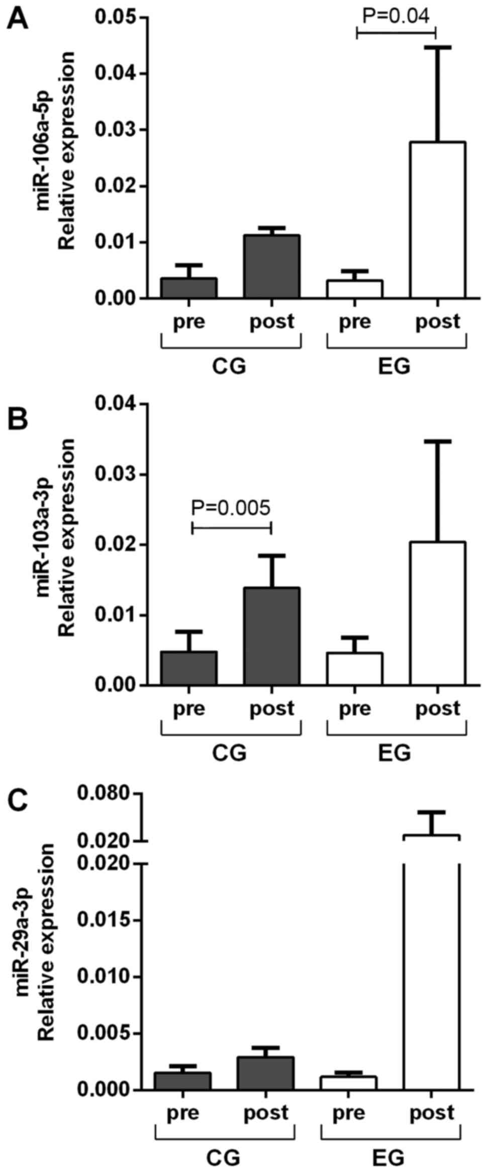
Relative expression of miRNAs before
and after cycling training program
There was no difference in the relative expression
levels of the miRNAs analyzed between the EG and CG before and
after the cycling training program. Regarding the effect of the
interval training on a cycle ergometer, there was an increase in
the relative expression levels of miR-106a-5p in the EG (P=0.04)
and no difference in the CG (P=0.07) compared with the levels
before training (Fig. 1A). There
was an increase in the relative expression levels of miR-103a-3p in
the CG (P=0.005), but no significant difference was observed in the
EG (P=0.09) after training (Fig.
1B). The expression levels of miR-29a-3p were upregulated in
the CG (P=0.06) (Fig. 1C), while no
statistical difference was noted in the EG (P=0.16).
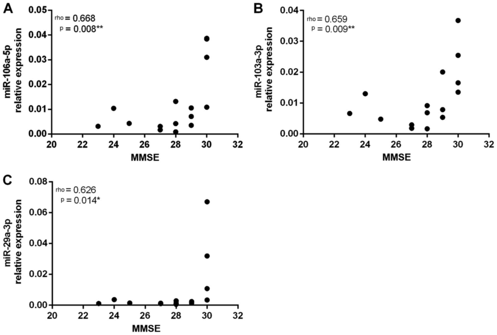
Correlation between relative
expression levels of miRNAs, MMSE, UPDRS, MoCA and 6MWT
Spearman's rank correlation coefficient was used to
identify the correlation between the analyzed variables. The
cognitive profile assessed using the MMSE was moderately,
positively correlated with the relative expression levels of
miR-106a-5p, miR-103a-3p and miR-29a-3p (Fig. 2).
The variables UPDRS, MoCA and 6MWT were not
correlated with the relative expression levels of the assessed
miRNAs (Table II).
| Table II.Correlation between the variables
UPDRS, MoCA, 6MWT and the relative expression levels of
miR-106a-5p, miR-103a-3p and miR-29a-3p. |
Table II.
Correlation between the variables
UPDRS, MoCA, 6MWT and the relative expression levels of
miR-106a-5p, miR-103a-3p and miR-29a-3p.
|
| miR-106a-5p | miR-103a-3p | miR-29a-3p |
|---|
|
|
|
|
|
|---|
| Variables | ρ | P-value | ρ | P-value | ρ | P-value |
|---|
| MoCA | 0.450 | 0.093 | 0.340 | 0.214 | 0.457 | 0.088 |
| UPDRS | −0.234 | 0.365 | −0.248 | 0.337 | −0.304 | 0.241 |
| 6MWT | 0.085 | 0.760 | 0.237 | 0.390 | 0.143 | 0.608 |
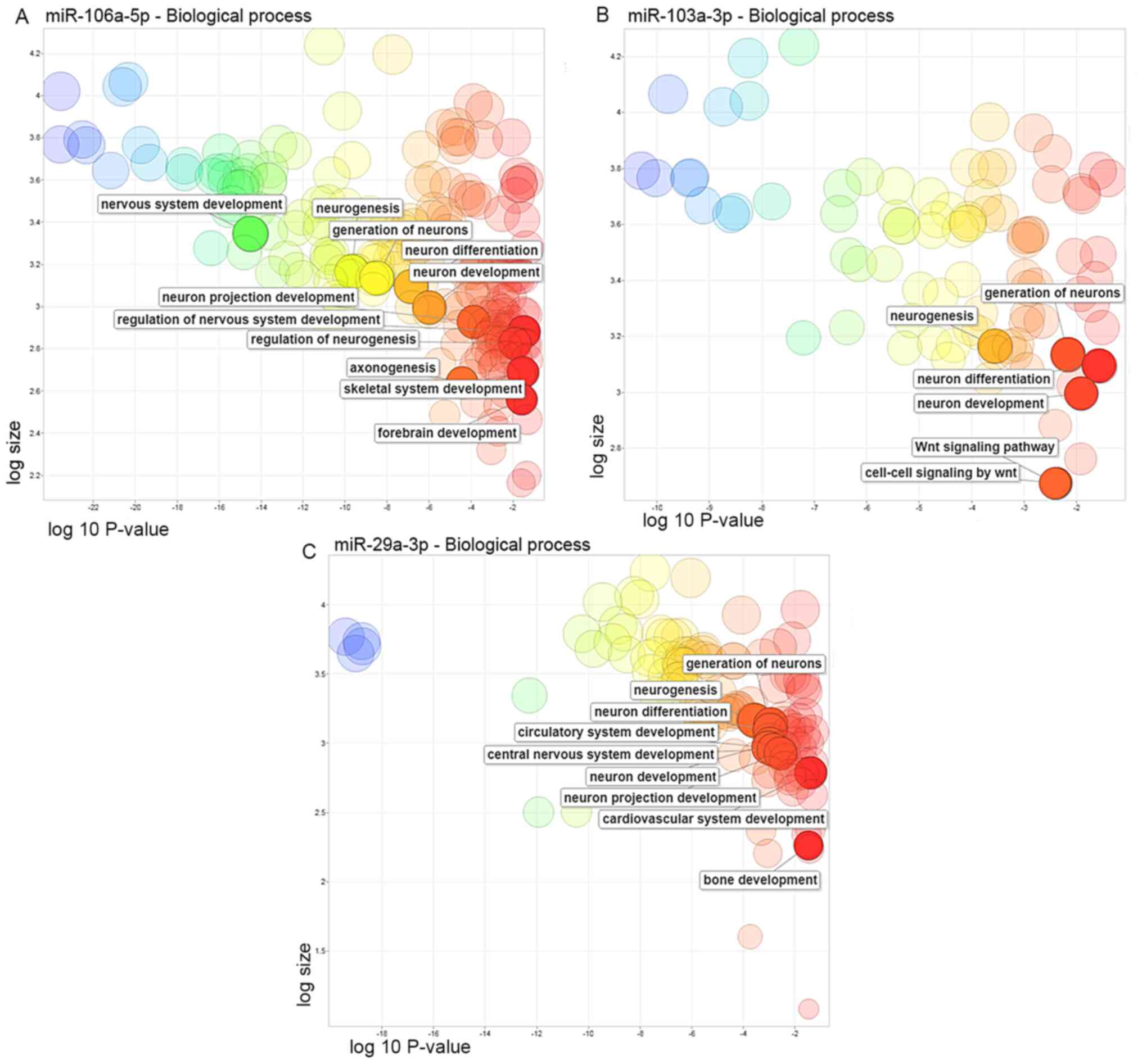
Target genes
The target genes for the three miRNAs were
identified using the TargetScan and miRDB databases. Among these
genes, 943 genes were predicted for miR-106a-5p, 438 genes for
miR-103a-3p and 771 genes for miR-29a-3p in both databases
(Fig. S1 and Data S1). The analysis of the main
biological processes involved with the target genes identified an
important relation with the ‘Wnt signaling pathway’, as well as
with the development of the nervous system, bones and muscles
(Fig. 3 and Table SII).
Discussion
PD is the most prevalent movement disorder of the
central nervous system, characterized by the progressive loss of
dopaminergic neurons in the substantia nigra pars compacta and the
accumulation of α-synuclein (10,16,20).
The accumulation of the α-synuclein protein leads to neuronal
dysfunction. miRNAs can modulate the accumulation of this factor
and other proteins by regulating the genes that encode these toxic
proteins and transcription factors, or by altering the expression
of proteins that regulate survival of neuronal cells (75). In this context, miRNAs are involved
directly and indirectly in the pathogenesis of PD (75–79),
and therefore, miRNAs that target genes involved in
neurodegeneration are of potential therapeutic value (80).
In the present study, it was possible to observe an
increase in relative expression levels of miR-106a-5p, miR-103a-3p
and miR-29a-3p in serum of individuals who participated in a
supervised cycling training program of 30 min, three times a week
for a period of 8 weeks. Notably, the program promoted a
significant increase in the expression of miR-106a-5p.
In healthy individuals, previous studies have
reported changes in miRNA expression levels after aerobic exercise.
For instance, Denham et al (34) observed a reduction in miR-210 and an
increase in miR-21 after 12 weeks of physical exercise on the
treadmill. In four studies, changes were identified in 38 miRNAs in
neutrophils (25), 33 miRNAs in
peripheral blood mononuclear cells (26), 22 miRNAs in natural killer cells
(27) and 18 in monocytes (28) in healthy men after exercise using a
cycle ergometry. Another study revealed the acute and chronic
effects of stationary bike exercise in healthy young individuals on
the expression levels of miRNAs (46). In a previous study, miR-106a,
miR-221, miR-30b, miR-151-5p, let-7i, miR-146a, miR-652 and
miR-151-3p expression levels were decreased, while miR-338-3p,
miR-330-3p, miR-223, miR-139-5p, miR-143, miR-145 and miR-424
expression levels were increased after 1 h of training. After 12
weeks, there was a decrease in miR-342-3p, let-7d, miR-766, miR-25,
miR-148a, miR-185 and miR-21 expression levels and an increase in
miR-103 and miR-107 expression levels (46). These results suggested the potential
value of miRNAs as a possible physiological mediator of
exercise-induced adaptation in healthy individuals and in
individuals with neurological diseases, such as PD.
In the present study, positive correlations were
observed between the MMSE values and the relative expression levels
of miR-106a-5p, miR-103a-3p and miR-29a-3p, indicating that the
increase in the expression level of these miRNAs was associated
with decreased cognitive deficit. Other studies also reported a
correlation between miR-29a expression and MMSE in patients with
vascular dementia (81), and
between miR-103a expression and MMSE in Japanese subjects without a
clinical diagnosis of dementia who attended a health examination in
Yakumo, Hokkaido (82). A study
involving an ovariectomized mouse model of cognitive impairment
suggested that miR-106a may be a marker of onset or a potential
therapeutic target for cognitive disturbances (83). At present, to the best of our
knowledge, only Serafin et al (79) demonstrated dysregulation of
miR-103a-3p and miR-29a-3p in PD; additionally, overexpression in
peripheral blood mononuclear cells of patients treated with L-dopa
was observed.
Prediction analyses of target genes in the present
study revealed that these miRNAs are associated with important
biological processes involved in motor function and in the
development of the central nervous system. Some of these target
genes have been validated in functional experiments, where it has
been reported that miR-106a-5p regulates the expression of the
autophagy-related gene 7 (84), and
suppression of this gene is associated with the death of
dopaminergic neurons and accumulation of α-synuclein (85). Moreover, miR-106a-5p has been
reported to regulate the expression of the hypoxia-induced factor-1
gene (86), which is associated
with cell death in various neurodegenerative diseases. In addition
to regulating processes related to neurogenesis, miR-106a-5p serves
a role in myogenesis. Recently, miR-106a-5p has been identified in
animal models as a repressor of myogenesis as it inhibits
differentiation and promotes atrophy by blocking the PI3K/AKT
signaling pathway via PIK3R1 (87).
A previous study also observed that miR-106a-5p was downregulated
immediately after acute exercise in young healthy men (88); however, the impact of exercises in
older men is yet to be elucidated.
Prediction analyses of target genes of miR-103a-3p
have demonstrated a relationship with the Wnt signaling pathway
(89,90). This pathway is associated with the
survival of dopaminergic neurons and their dysregulation may be a
potential cause for PD (89,90),
suggesting that miR-103a-3p can regulate the expression of the
extracellular Dickkopf-1 protein (DKK1) (88). A recent study confirmed that
miR-103a-3p can regulate DKK1 expression (88). Furthermore, this protein can block
the canonical Wnt pathway, contributing to neurotoxicity in PD
(91,92). miR-103a-3p is also involved in the
regulation of several other PD-related processes, such as the
regulation of myocyte enhancer factor-2 (MEF2D) transcription
factor, and the regulation of MEF2D is associated with loss of
dopaminergic neurons (93,94). miR-103a-3p and miR-29a-3p are
related to insulin resistance, a process that may be linked to
neurodegeneration in DP (95–100).
In the present study, although miR-29a-3p did not
show a statistically significant difference between the pre- and
post-exercise groups, a correlation with cognitive profile assessed
using the MMSE was noted. One of the targets of miR-29a-3p is G
protein-coupled receptor 37 (GPR37) (99). The intracellular accumulation of
GPR37 is neurotoxic and is associated with PD (99). Inactivation of GPR37 produces motor
and non-motor phenotypes relevant to PD (99). Thus, poor regulation of GPR37 due to
overexpression of miR-29a-3p may be associated with PD (79). CDC42, a candidate gene for PD
involved in neuronal death (100),
was also identified as a potential target for miR-29a-3p.
An emerging paradigm of post-transcriptional
molecular regulation due to physical exercises involves the action
of miRNAs, which regulate mRNA translation (38). Several studies have examined the
acute and/or chronic effects of physical exercises s(aerobic and
resistance) on various miRNAs in healthy individuals (25–36),
athletes (37–40) and in young and older adults
(41–50), as well as in patients with CHF
(51,52), CKD (53), DM2 associated with morbid obesity
(54), prediabetes (55) and intermittent claudication
(56,57). In these studies, most of the
investigated subjects were men. Moreover, to the best of our
knowledge, the effects of physical exercises on the expression of
miRNAs in individuals with PD have not been investigated. Thus, the
present study was the first to investigate the effects of an
interval cycling training program on the expression levels of
miRNAs in serum samples from men with PD.
In the present study, the interval cycling training
program increased the expression levels of miR-106a-5p, miR-103a-3p
and miR-29a-3p, with a statistically significant increase observed
in the expression of miR-106a-5p. However, there is limited
information regarding this miRNA in PD. The current results
indicated that these miRNAs may participate in the response to
exercise; however, further studies with a larger sample size are
required to confirm these results. The present results suggested
that miR-106a-5p, miR-103a-3p and miR-29a-3p may partially reflect
exercise-induced responses or time regulation controlled by miRNAs
during exercise.
Limitations of the present work are that the
selected participants were only men, and that the dosage and
treatment were not investigated. Therefore, future research should
be performed to investigate whether this is a sex-specific issue,
as well as to examine the relationship between patients were
treated with L-dopa or Prolopa.
In conclusion, in men with PD, serum expression
levels of miR-106a-5p, miR-103a-3p and miR-29a-3p were increased in
response to a supervised interval training program on a cycle
ergometer for 30 min, three times a week for a period of 8 weeks.
It was found that the program promoted a significant increase in
the expression of miR-106a-5p. Additionally, the expression levels
of miR106a-5p, miR-103a-3p and miR-29a-3p were positively
correlated with MMSE values. Further studies are required to
clarify the potential use of these circulating miRNAs as markers of
adaptation to physical exercise. The present results indicated that
these three miRNAs were associated with the exercise response and
cognitive improvement in men with PD.
Supplementary Material
Supporting Data
Supporting Data
Supporting Data
Acknowledgements
Not applicable.
Funding
No funding was received.
Availability of data and materials
The datasets used and/or analyzed during the current
study are available from the corresponding author upon reasonable
request.
Authors' contributions
FCDS, MPR and RDS designed the study. FCDS, MPR,
GGV, RDRI and TBCP performed the experiments. FCDS, MPR and ASM
analyzed the data. FCDS and MPR prepared the manuscript. FCDS, MPR,
GGV and RDRI confirmed the authenticity of all the raw data. All
authors read and approved the final version of the manuscript.
Ethics approval and consent to
participate
This study was approved by the Ethics Committee for
Research involving Human Beings of the State University of Santa
Catarina, with the Certificate of Presentation for Ethical
Appreciation no. 50032415.1.0000.0118, in 2015. The objectives of
this study and the procedures to be adopted throughout the research
were explained to all participants, who voluntarily agreed to
continue participation by signing an informed consent document.
Patient consent for publication
Not applicable.
Competing interests
The authors declare that they have no competing
interests.
References
|
1
|
The World Health Organization 2017.
Physical activity and adults. http://www.who.int/dietphysicalactivity/factsheet_adults/en/January
8–2019
|
|
2
|
Barnett A, Smith B, Lord SR, Williams M
and Baumand A: Community-based group exercise improves balance and
reduces falls in at-risk older people: A randomised controlled
trial. Age Ageing. 32:407–414. 2003. View Article : Google Scholar : PubMed/NCBI
|
|
3
|
Gillespie LD, Robertson MC, Gillespie WJ,
Sherrington C, Gates S, Clemson LM and Lamb SE: Interventions for
preventing falls in older people living in the community. Cochrane
Database Syst Rev. CD0071462012.PubMed/NCBI
|
|
4
|
Sherrington C, Whitney JC, Lord SR,
Herbert RD, Cumming RG and Close JC: Effective exercise for the
prevention of falls: A systematic review and meta-analysis. J Am
Geriatr Soc. 56:2234–2243. 2008. View Article : Google Scholar : PubMed/NCBI
|
|
5
|
Fox KR: The influence of physical activity
on mental well-being. Public Health Nutr. 2:411–418. 1999.
View Article : Google Scholar : PubMed/NCBI
|
|
6
|
Cooney GM, Dwan K, Greig CA, Lawlor DA,
Rimer J, Waugh FR, McMurdo M and Mead GE: Exercise for depression.
Cochrane Database Syst Rev. CD0043662013.PubMed/NCBI
|
|
7
|
Latt MD, Lord SR, Morris JG and Fung VS:
Clinical and physiological assessments for elucidating falls risk
in Parkinson's disease. Mov Disord. 24:1280–1289. 2009. View Article : Google Scholar : PubMed/NCBI
|
|
8
|
Allen NE, Canning CG, Sherrington C, Lord
SR, Latt MD, Close JC, O'Rourke SD, Murray SM and Fung VS: The
effects of an exercise program on fall risk factors in people with
Parkinson's disease: A randomized controlled trial. Mov Disord.
25:1217–1225. 2010. View Article : Google Scholar : PubMed/NCBI
|
|
9
|
Lun V, Pullan N, Labelle N, Adams C and
Suchowersky O: Comparison of the effects of a self-supervised home
exercise program with a physiotherapist-supervised exercise program
on the motor symptoms of Parkinson's disease. Mov Disord.
20:971–975. 2005. View Article : Google Scholar : PubMed/NCBI
|
|
10
|
Tomlinson CL, Patel S, Meek C, Clarke CE,
Stowe R, Shah L, Sackley CM, Deane KHO, Herd CP, Wheatley K and
Ives N: Physiotherapy versus placebo or no intervention in
Parkinson's disease. Cochrane Database Syst Rev. CD0028172012.
|
|
11
|
Goodwin VA, Richards SH, Taylor RS, Taylor
AH and Campbell JL: The effectiveness of exercise interventions for
people with Parkinson's disease: A systematic review and
meta-analysis. Mov Disord. 23:631–640. 2008. View Article : Google Scholar : PubMed/NCBI
|
|
12
|
Rochester L, Nieuwboer A and Lord S:
Physiotherapy for Parkinson's disease: Defining evidence within a
framework for intervention. Neurodegenerative Disease Management.
1:57–65. 2011. View
Article : Google Scholar
|
|
13
|
da Silva FC, Iop RDR, de Oliveira LC, Boll
AM, de Alvarenga JGS, Gutierres Filho PJB, de Melo LMAB, Xavier AJ
and da Silva R: Effects of physical exercise programs on cognitive
function in Parkinson's disease patients: A systematic review of
randomized controlled trials of the last 10 years. PLoS One.
13:e01931132018. View Article : Google Scholar : PubMed/NCBI
|
|
14
|
Oguh O, Eisenstein A, Kwasny M and Simuni
T: Back to the basics: Regular exercise matters in parkinson's
disease: Results from the national Parkinson foundation QII
registry study. Parkinsonism Relat Disord. 20:1221–1225. 2014.
View Article : Google Scholar : PubMed/NCBI
|
|
15
|
Murray DK, Sacheli MA, Eng JJ and Stoessl
AJ: The effects of exercise on cognition in Parkinson's disease: A
systematic review. Transl Neurodegener. 3:52014. View Article : Google Scholar : PubMed/NCBI
|
|
16
|
Schapira AH: Neurobiology and treatment of
Parkinson's disease. Trends Pharmacol Sci. 30:41–47. 2009.
View Article : Google Scholar : PubMed/NCBI
|
|
17
|
Morris ME, Iansek R and Kirkwood B: A
randomized controlled trial of movement strategies compared with
exercise for people with Parkinson's disease. Mov Disord. 24:64–71.
2009. View Article : Google Scholar : PubMed/NCBI
|
|
18
|
van Nimwegen M, Speelman AD, Hofman-van
Rossum EJ, Overeem S, Deeg DJ, Borm GF, van der Horst MH, Bloem BR
and Munneke M: Physical inactivity in Parkinson's disease. J
Neurol. 258:2214–2221. 2011. View Article : Google Scholar : PubMed/NCBI
|
|
19
|
van Hilten JJ, Hoogland G, van der Velde
EA, Middelkoop HA, Kerkhof GA and Roos RA: Diurnal effects of motor
activity and fatigue in Parkinson's disease. J Neurol Neurosurg
Psychiatry. 56:874–877. 1993. View Article : Google Scholar : PubMed/NCBI
|
|
20
|
Harraz MM, Dawson TM and Dawson VL:
MicroRNAs in Parkinson's disease. J Chem Neuroanat. 4:127–130.
2011. View Article : Google Scholar : PubMed/NCBI
|
|
21
|
Mouradian MM: MicroRNAs in Parkinson's
disease. Neurobiol Dis. 46:279–284. 2012. View Article : Google Scholar : PubMed/NCBI
|
|
22
|
Filatova EV, Alieva AKh, Shadrina MI and
Slominsky PA: MicroRNAs: Possible role in pathogenesis of
Parkinson's disease. Biochemistry (Mosc). 77:813–819. 2012.
View Article : Google Scholar : PubMed/NCBI
|
|
23
|
da Silva FC, Iop RD, Vietta GG, Kair DA,
Gutierres Filho PJ, de Alvarenga JG and da Silva R: microRNAs
involved in Parkinson's disease: A systematic review. Mol Med Rep.
14:4015–4022. 2016. View Article : Google Scholar : PubMed/NCBI
|
|
24
|
Silva FCD, Iop RDR, Andrade A, Costa VP,
Gutierres Filho PJB and Silva RD: Effects of physical exercise on
the expression of MicroRNAs: A Systematic review. J Strength Cond
Res. 34:270–280. 2020. View Article : Google Scholar : PubMed/NCBI
|
|
25
|
Radom-Aizik S, Zaldivar F, Oliver S,
Galassetti P and Cooper DM: Effects of exercise on miRNA expression
levels in human peripheral blood mononuclear cells (PBMCs). FASEB
J. 24:626.42010. View Article : Google Scholar
|
|
26
|
Radom-Aizik S, Zaldivar F Jr, Leu SY,
Adams GR, Oliver S and Cooper DM: Effects of exercise on microRNA
expression in young males peripheral blood mononuclear cells. Clin
Transl Sci. 5:32–38. 2012. View Article : Google Scholar : PubMed/NCBI
|
|
27
|
Radom-Aizik S, Zaldivar F, Haddad F and
Cooper DM: Impact of brief exercise on peripheral blood NK cell
gene and microRNA expression in young adults. J Appl Physiol
(1985). 114:628–636. 2013. View Article : Google Scholar : PubMed/NCBI
|
|
28
|
Radom-Aizik S, Zaldivar FP Jr, Haddad F
and Cooper DM: Impact of brief exercise on circulating monocyte
gene and microRNA expression: Implications for atherosclerotic
vascular disease. Brain Behav Immun. 39:121–129. 2014. View Article : Google Scholar : PubMed/NCBI
|
|
29
|
Guescini M, Canonico B, Lucertini F,
Maggio S, Annibalini G, Barbieri E, Luchetti F, Papa S and Stocchi
V: Muscle releases alpha-sarcoglycan positive extracellular
vesicles carrying miRNAs in the bloodstream. PLoS One.
10:e01250942015. View Article : Google Scholar : PubMed/NCBI
|
|
30
|
Chilton WL, Marques FZ, West J,
Kannourakis G, Berzins SP, O'Brien BJ and Charchar FJ: Acute
exercise leads to regulation of telomere-associated genes and
microRNA expression in immune cells. PLoS One. 9:e920882014.
View Article : Google Scholar : PubMed/NCBI
|
|
31
|
Uhlemann M, Möbius-Winkler S, Fikenzer S,
Adam J, Redlich M, Möhlenkamp S, Hilberg T, Schuler GC and Adams V:
Circulating microRNA-126 increases after different forms of
endurance exercise in healthy adults. Eur J Prev Cardiol.
21:484–491. 2014. View Article : Google Scholar : PubMed/NCBI
|
|
32
|
McLean CS, Mielke C, Cordova JM, Langlais
PR, Bowen B, Miranda D, Coletta DK and Mandarino LJ: Gene and
MicroRNA expression responses to exercise; relationship with
insulin sensitivity. PLoS One. 10:e01270892015. View Article : Google Scholar : PubMed/NCBI
|
|
33
|
Fyfe JJ, Bishop DJ, Zacharewicz E, Russell
AP and Stepto NK: Concurrent exercise incorporating high-intensity
interval or continuous training modulates mTORC1 signaling and
microRNA expression in human skeletal muscle. Am J Physiol Regul
Integr Comp Physiol. 310:R1297–R1311. 2016. View Article : Google Scholar : PubMed/NCBI
|
|
34
|
Denham J, O'Brien BJ, Marques FZ and
Charchar FJ: Changes in the leukocyte methylome and its effect on
cardiovascular-related genes after exercise. J Appl Physiol (1985).
118:475–488. 2015. View Article : Google Scholar : PubMed/NCBI
|
|
35
|
Dias RG, Silva MS, Duarte NE, Bolani W,
Alves CR, Lemos JR, da Silva JL, de Oliveira PA, Alves GB, de
Oliveira EM, et al: PBMCs express a transcriptome signature
predictor of oxygen uptake responsiveness to endurance exercise
training in men. Physiol Genomics. 7:13–23. 2015. View Article : Google Scholar : PubMed/NCBI
|
|
36
|
Nielsen S, Scheele C, Yfanti C, Åkerström
T, Nielsen AR, Pedersen BK and Laye MJ: Muscle specific microRNAs
are regulated by endurance exercise in human skeletal muscle. J
Physiol. 588:4029–4037. 2010. View Article : Google Scholar : PubMed/NCBI
|
|
37
|
Backes C, Leidinger P, Keller A, Hart M,
Meyer T, Meese E and Hecksteden A: Blood born miRNAs signatures
that can serve as disease specific biomarkers are not significantly
affected by overall fitness and exercise. PLoS One. 9:e1021832014.
View Article : Google Scholar : PubMed/NCBI
|
|
38
|
Tonevitsky AG, Maltseva DV, Abbasi A,
Samatov TR, Sakharov DA, Shkurnikov MU, Lebedev AE, Galatenko VV,
Grigoriev AI and Northoff H: Dynamically regulated miRNA-mRNA
networks revealed by exercise. BMC Physiol. 13:92013. View Article : Google Scholar : PubMed/NCBI
|
|
39
|
Mooren FC, Viereck J, Kruger K and Thum T:
Circulating microRNAs as potential biomarkers of aerobic exercise
capacity. Am J Physiol Heart Circ Physiol. 4:H557–H563. 2014.
View Article : Google Scholar : PubMed/NCBI
|
|
40
|
Baggish AL, Hale A, Weiner RB, Lewis GD,
Systrom D, Wang F, Wang TJ and Chan SY: Dynamic regulation of
circulating MicroRNA during acute exhaustive exercise and sustained
aerobic exercise training. J Physiol. 589:3983–3994. 2011.
View Article : Google Scholar : PubMed/NCBI
|
|
41
|
Cui SF, Li W, Niu J, Zhang CY, Chen X and
Ma JZ: Acute responses of circulating microRNAs to low-volume
sprint interval cycling. Front Physiol. 6:3112015. View Article : Google Scholar : PubMed/NCBI
|
|
42
|
Cui SF, Wang C, Yin X, Tian D, Lu QJ,
Zhang CY, Chen X and Ma JZ: Similar responses of circulating
microRNAs to acute high-intensity interval exercise and
vigorous-intensity continuous exercise. Front Physiol. 7:1022016.
View Article : Google Scholar : PubMed/NCBI
|
|
43
|
Zacharewicz E, Della Gatta P, Reynolds J,
Garnham A, Crowley T, Russell AP and Lamon S: Identification of
microRNAs linked to regulators of muscle protein synthesis and
regeneration in young and old skeletal muscle. PLoS One.
9:e1140092014. View Article : Google Scholar : PubMed/NCBI
|
|
44
|
Mueller M, Breil FA, Lurman G, Klossner S,
Fluck M, Billeter R, Dapp C and Hoppeler H: Different molecular and
structural adaptations with eccentric and conventional strength
training in elderly men and women. Gerontology. 57:528–538. 2011.
View Article : Google Scholar : PubMed/NCBI
|
|
45
|
Drummond MJ, McCarthy JJ, Fry CS, Esser KA
and Rasmussen BB: Aging differentially affects human skeletal
muscle microRNA expression at rest and after an anabolic stimulus
of resistance exercise and essential amino acids. Am J Physiol
Endocrinol Metab. 295:E1333–E1340. 2008. View Article : Google Scholar : PubMed/NCBI
|
|
46
|
Davidsen PK, Gallagher IJ, Hartman JW,
Tarnopolsky MA, Dela F, Helge JW, Timmons JA and Phillips SM: High
responders to resistance exercise training demonstrate differential
regulation of skeletal muscle microRNA expression. J Appl Physio
(1985). 110:309–317. 2011. View Article : Google Scholar : PubMed/NCBI
|
|
47
|
Nielsen S, Hvid T, Kelly M, Lindegaard B,
Dethlefsen C, Winding K, Mathur N, Scheele C, Pedersen BK and Laye
MJ: Muscle specific miRNAs are induced by testosterone and
independently upregulated by age. Front Physiol. 4:3942014.
View Article : Google Scholar : PubMed/NCBI
|
|
48
|
Zhang T, Birbrair A, Wang ZM, Messi ML,
Marsh AP, Leng I, Nicklas BJ and Delbono O: Improved knee extensor
strength with resistance training associates with muscle specific
miRNAs in older adults. Exp Gerontol. 62:7–13. 2015. View Article : Google Scholar : PubMed/NCBI
|
|
49
|
Aoi W, Ichikawa H, Mune K, Tanimura Y,
Mizushima K, Naito Y and Yoshikawa T: Muscle-enriched microRNA
miR-486 decreases in circulation in response to exercise in young
men. Front Physiol. 4:802013. View Article : Google Scholar : PubMed/NCBI
|
|
50
|
Margolis LM, Lessard SJ, Ezzyat Y,
Fielding RA and Rivas DA: Circulating MicroRNA are predictive of
aging and acute adaptive response to resistance exercise in men. J
Gerontol A Biol Sci Med Sci. 72:1319–1326. 2017.PubMed/NCBI
|
|
51
|
Xu T, Liu Q, Yao J, Dai Y, Wang H and Xiao
J: Circulating microRNAs in response to exercise. Scand J Med Sci
Sports. 25:e149–e154. 2015. View Article : Google Scholar : PubMed/NCBI
|
|
52
|
Riedel S, Radzanowski S, Bowen TS, Werner
S, Erbs S, Schuler G and Adams V: Exercise training improves
high-density lipoprotein-mediated transcription of proangiogenic
microRNA in endothelial cells. Eur J Prev Cardiol. 22:899–903.
2015. View Article : Google Scholar : PubMed/NCBI
|
|
53
|
Van Craenenbroeck AH, Ledeganck KJ, Van
Ackeren K, Jürgens A, Hoymans VY, Fransen E, Adams V, De Winter BY,
Verpooten GA, Vrints CJ, et al: Plasma levels of microRNA in
chronic kidney disease: Patterns in acute and chronic exercise. Am
J Physiol Heart Circ Physiol. 309:H2008–H2016. 2015. View Article : Google Scholar : PubMed/NCBI
|
|
54
|
Rowlands DS, Page RA, Sukala WR, Giri M,
Ghimbovschi SD, Hayat I, Cheema BS, Lys I, Leikis M, Sheard PW, et
al: Multi-omic integrated networks connect DNA methylation and
miRNA with skeletal muscle plasticity to chronic exercise in Type 2
diabetic obesity. Physiol Genomics. 46:747–765. 2014. View Article : Google Scholar : PubMed/NCBI
|
|
55
|
Parrizas M, Brugnara L, Esteban Y,
Gonzalez-Franquesa A, Canivell S, Murillo S, Gordillo-Bastidas E,
Cusso R, Cadefau JA, Garcia-Roves PM, et al: Circulating miR-192
and miR-193b are markers of prediabetes and are modulated by an
exercise intervention. J Clin Endocrinol Metab. 100:E407–E415.
2015. View Article : Google Scholar : PubMed/NCBI
|
|
56
|
da Silva ND Jr, Roseguini BT, Chehuen M,
Fernandes T, Mota GF, Martin PK, Han SW, Forjaz CL, Wolosker N and
de Oliveira EM: Effects of oral N-acetylcysteine on walking
capacity, leg reactive hyperemia, and inflammatory and angiogenic
mediators in patients with intermittent claudication. Am J Physiol
Heart Circ Physiol. 309:H897–H905. 2015. View Article : Google Scholar : PubMed/NCBI
|
|
57
|
Nowak WN, Mika P, Nowobilski R, Kusinska
K, Bukowska-Strakova K, Nizankowski R, Józkowicz A, Szczeklik A and
Dulak J: Exercise training in intermittent claudication: Effects on
antioxidant genes, inflammatory mediators and proangiogenic
progenitor cells. Thromb Haemost. 108:824–831. 2012. View Article : Google Scholar : PubMed/NCBI
|
|
58
|
Shen JJ, Wang YF and Yang W:
Sex-interacting mRNA- and miRNA-eQTLs and their implications in
gene expression regulation and disease. Front Genet. 10:3132019.
View Article : Google Scholar : PubMed/NCBI
|
|
59
|
Sharma S and Eghbali M: Influence of sex
differences on microRNA gene regulation in disease. Biol Sex
Differ. 5:32014. View Article : Google Scholar : PubMed/NCBI
|
|
60
|
Hoehn MM and Yahr MD: Parkinsonism: Onset,
progression and mortality. Neurology. 17:427–442. 1967. View Article : Google Scholar : PubMed/NCBI
|
|
61
|
Fahn S and Elton R: Members of the UPDRS
Development Committee. Recent Developments in Parkinson's Disease.
2. Fahn S, Marsden CD, Calne DB and Goldstein M: Macmillan Health
Care Information; Florham park, NJ: pp. 153–163, 293-304. 1987
|
|
62
|
Folstein MF, Folstein SE and McHugh PR:
‘Mini-mental state’. A practical method for grading the cognitive
state of patients for the clinician. J Psychiatr Res. 12:189–198.
1975. View Article : Google Scholar : PubMed/NCBI
|
|
63
|
Nasreddine ZS, Phillips NA, Bédirian V,
Charbonneau S, Whitehead V, Collin I, Cummings JL and Chertkow H:
The montreal cognitive assessment, MoCA: A brief screening tool for
mild cognitive impairment. J Am Geriatr Soc. 53:695–699. 2005.
View Article : Google Scholar : PubMed/NCBI
|
|
64
|
Britto RR and Sousa LAP: Teste de
caminhada de seis minutos-uma normatização brasileira. Fisioter
Mov. 19:49–54. 2006.
|
|
65
|
Kobayashi E, Himuro N and Takahashi M:
Clinical utility of the 6-min walk test for patients with moderate
Parkinson's disease. Int J Rehabil Res. 40:66–70. 2017. View Article : Google Scholar : PubMed/NCBI
|
|
66
|
Sociedade Brasileira de Patologia
Clínica/Medicina Laboratorial. Recomendações da Sociedade
Brasileira de Patologia Clínica/Medicina Laboratorial para coleta
de sangue venoso. 2nd edition. Minha Editora; Barueri, SP: 2010
|
|
67
|
ANVISA, . Agência Nacional de Vigilância
Sanitária. Guia para transportes de sangue e componentes. 2013.
|
|
68
|
Ramakers C, Ruijter JM, Deprez RH and
Moorman AF: Assumption-free analysis of quantitative real-time
polymerase chain reaction (PCR) data. Neurosci Lett. 339:62–66.
2003. View Article : Google Scholar : PubMed/NCBI
|
|
69
|
Livak KJ and Schmittgen TD: Analysis of
relative gene expression data using real-time quantitative PCR and
the 2(-Delta Delta C(T)) method. Methods. 25:402–408. 2001.
View Article : Google Scholar : PubMed/NCBI
|
|
70
|
Agarwal V, Bell GW, Nam JW and Bartel DP:
Predicting effective microRNA target sites in mammalian mRNAs.
Elife. 4:e050052015. View Article : Google Scholar : PubMed/NCBI
|
|
71
|
Wong N and Wang X: miRDB: An online
resource for microRNA target prediction and functional annotations.
Nucleic Acids Res. 43((Database Issue)): D146–D152. 2015.
View Article : Google Scholar : PubMed/NCBI
|
|
72
|
Ashburner M, Ball CA, Blake JA, Botstein
D, Butler H, Cherry JM, Davis AP, Dolinski K, Dwight SS, Eppig JT,
et al: Gene ontology: Tool for the unification of biology. The gene
ontology consortium. Nat Genet. 25:25–29. 2000. View Article : Google Scholar : PubMed/NCBI
|
|
73
|
Supek F, Bošnjak M, Škunca N and Šmuc T:
REVIGO summarizes and visualizes long lists of gene ontology terms.
PLoS One. 6:e218002011. View Article : Google Scholar : PubMed/NCBI
|
|
74
|
Uygur M, Bellumori M and Knight CA:
Effects of a low-resistance, interval bicycling intervention in
Parkinson's disease. Physiother Theory Pract. 33:897–904. 2017.
View Article : Google Scholar : PubMed/NCBI
|
|
75
|
Eacker SM, Dawson TM and Dawson VL:
Understanding microRNAs in neurodegeneration. Nat Rev Neurosci.
10:837–841. 2009. View Article : Google Scholar : PubMed/NCBI
|
|
76
|
Margis R, Margis R and Rieder CR:
Identification of blood microRNAs associated to Parkinsonis
disease. J Biotechnol. 152:96–101. 2011. View Article : Google Scholar : PubMed/NCBI
|
|
77
|
Martins M, Rosa A, Guedes LC, Fonseca BV,
Gotovac K, Violante S, Mestre T, Coelho M, Rosa MM, Martin ER, et
al: Convergence of miRNA expression profiling, α-synuclein
interacton and GWAS in Parkinson's disease. PLoS One. 6:e254432011.
View Article : Google Scholar : PubMed/NCBI
|
|
78
|
Alvarez-Erviti L, Seow Y, Schapira AH,
Rodriguez-Oroz MC, Obeso JA and Cooper JM: Influence of microRNA
deregulation on chaperone-mediated autophagy and α-synuclein
pathology in Parkinson's disease. Cell Death Dis. 4:e5452013.
View Article : Google Scholar : PubMed/NCBI
|
|
79
|
Serafin A, Foco L, Zanigni S, Blankenburg
H, Picard A, Zanon A, Giannini G, Pichler I, Facheris MF, Cortelli
P, et al: Overexpression of blood microRNAs 103a, 30b, and 29a in
L-dopa-treated patients with PD. Neurology. 84:645–653. 2015.
View Article : Google Scholar : PubMed/NCBI
|
|
80
|
Roshan R, Ghosh T, Scaria V and Pillai B:
MicroRNAs: Novel therapeutic targets in neurodegenerative diseases.
Drug Discov Today. 14:1123–1129. 2009. View Article : Google Scholar : PubMed/NCBI
|
|
81
|
Ragusa M, Bosco P, Tamburello L,
Barbagallo C, Condorelli AG, Tornitore M, Spada RS, Barbagallo D,
Scalia M, Elia M, et al: miRNAs plasma profiles in vascular
dementia: Biomolecular data and biomedical implications. Front Cell
Neurosci. 10:512016. View Article : Google Scholar : PubMed/NCBI
|
|
82
|
Kondo M, Yamada H, Munetsuna E, Yamazaki
M, Hatta T, Iwahara A, Ohashi K, Ishikawa H, Tsuboi Y, Inoue T, et
al: Associations of serum microRNA-20a, −27a, and −103a with
cognitive function in a Japanese population: The Yakumo study. Arch
Gerontol Geriatr. 82:155–160. 2019. View Article : Google Scholar : PubMed/NCBI
|
|
83
|
Zhang M, Ye Y, Cong J, Pu D, Liu J, Hu G
and Wu J: Regulation of STAT3 by miR-106a is linked to cognitive
impairment in ovariectomized mice. Brain Res. 1503:43–52. 2013.
View Article : Google Scholar : PubMed/NCBI
|
|
84
|
Hao H, Xia G, Wang C, Zhong F, Liu L and
Zhang D: miR-106a suppresses tumor cells death in colorectal cancer
through targeting ATG7. Med Mol Morphol. 50:76–85. 2017. View Article : Google Scholar : PubMed/NCBI
|
|
85
|
Ahmed I, Liang Y, Schools S, Dawson VL,
Dawson TM and Savitt JM: Development and characterization of a new
Parkinson's disease model resulting from impaired autophagy. J
Neurosci. 32:16503–16509. 2012. View Article : Google Scholar : PubMed/NCBI
|
|
86
|
Liu Y, Zhang J, Sun X and Li M: EMMPRIN
Down-regulating miR-106a/b modifies breast cancer stem-like cell
properties via interaction with fibroblasts through STAT3 and
HIF-1α. Sci Rep. 6:283292016. View Article : Google Scholar : PubMed/NCBI
|
|
87
|
Li X, Zhu Y, Zhang H, Ma G, Wu G, Xiang A,
Shi X, Yang GS and Sun S: MicroRNA-106a-5p Inhibited C2C12
myogenesis via targeting PIK3R1 and modulating the PI3K/AKT
signaling. Genes (Basel). 9:3332018. View Article : Google Scholar : PubMed/NCBI
|
|
88
|
Nielsen S, Åkerström T, Rinnov A, Yfanti
C, Scheele C, Pedersen BK and Laye MJ: The miRNA plasma signature
in response to acute aerobic exercise and endurance training. PLoS
One. 9:e873082014. View Article : Google Scholar : PubMed/NCBI
|
|
89
|
Berwick DC and Harvey K: The importance of
Wnt signalling for neurodegeneration in Parkinson's disease.
Biochem Soc Trans. 40:1123–1128. 2012. View Article : Google Scholar : PubMed/NCBI
|
|
90
|
Stephano F, Nolte S, Hoffmann J, El-Kholy
S, von Frieling J, Bruchhaus I, Fink C and Roeder T: Impaired Wnt
signaling in dopamine containing neurons is associated with
pathogenesis in a rotenone triggered Drosophila Parkinson's disease
model. Sci Rep. 8:23722018. View Article : Google Scholar : PubMed/NCBI
|
|
91
|
Liu J, Wu M, Feng G, Li R, Wang Y and Jiao
J: Downregulation of LINC00707 promotes osteogenic differentiation
of human bone marrow-derived mesenchymal stem cells by regulating
DKK1 via targeting miR-103a-3p. Int J Mol Med. 46:1029–1038. 2020.
View Article : Google Scholar : PubMed/NCBI
|
|
92
|
Dun Y, Yang Y, Xiong Z, Feng M, Zhang Y,
Wang M, Xiang J, Li G and Ma R: Induction of dickkopf-1 contributes
to the neurotoxicity of MPP+ in PC12 cells via
inhibition of the canonical Wnt signaling pathway.
Neuropharmacology. 67:168–175. 2013. View Article : Google Scholar : PubMed/NCBI
|
|
93
|
Scott EL and Brann DW: Estrogen regulation
of Dkk1 and Wnt/β-catenin signaling in neurodegenerative disease.
Brain Res. 1514:63–74. 2013. View Article : Google Scholar : PubMed/NCBI
|
|
94
|
Li M, Liu Z, Zhang Z, Liu G, Sun S and Sun
C: miR-103 promotes 3T3-L1 cell adipogenesis through AKT/mTOR
signal pathway with its target being MEF2D. Biol Chem. 396:235–244.
2015. View Article : Google Scholar : PubMed/NCBI
|
|
95
|
Gao L, She H, Li W, Zeng J, Zhu J, Jones
DP, Mao Z, Gao G and Yang Q: Oxidation of survival factor MEF2D in
neuronal death and Parkinson's disease. Antioxid Redox Signal.
20:2936–2948. 2014. View Article : Google Scholar : PubMed/NCBI
|
|
96
|
Herrera BM, Lockstone HE, Taylor JM, Ria
M, Barrett A, Collins S, Kaisaki P, Argoud K, Fernandez C, Travers
ME, et al: Global microRNA expression profiles in insulin target
tissues in a spontaneous rat model of type 2 diabetes.
Diabetologia. 53:1099–1109. 2010. View Article : Google Scholar : PubMed/NCBI
|
|
97
|
Trajkovski M, Hausser J, Soutschek J, Bhat
B, Akin A, Zavolan M, Heim MH and Stoffel M: MicroRNAs 103 and 107
regulate insulin sensitivity. Nature. 474:649–653. 2011. View Article : Google Scholar : PubMed/NCBI
|
|
98
|
Aviles-Olmos I, Limousin P, Lees A and
Foltynie T: Parkinson's disease, insulin resistance and novel
agents of neuroprotection. Brain. 136:374–384. 2013. View Article : Google Scholar : PubMed/NCBI
|
|
99
|
Mandillo S, Golini E, Marazziti D, Di
Pietro C, Matteoni R and Tocchini-Valentini GP: Mice lacking the
Parkinson's related GPR37/PAEL receptor show non-motor behavioral
phenotypes: Age and gender effect. Genes Brain Behav. 12:465–477.
2013. View Article : Google Scholar : PubMed/NCBI
|
|
100
|
Karic A, Terzic R, Karic A and Peterlin B:
Identifying candidate genes for Parkinson's disease by integrative
genomics method. Biochem Med (Zagreb). 21:174–181. 2011. View Article : Google Scholar : PubMed/NCBI
|