Introduction
Acute lung injury (ALI) refers to acute hypoxic
respiratory dysfunction induced by various direct or
interconnecting injury factors, such as pneumonia, pyemia,
reperfusion injury and shock (1).
In severe cases, it can develop into acute respiratory distress
syndrome (ARDS), which is associated with severe symptoms such as
disruption of the alveolar epithelial barrier, proteinaceous
pulmonary edema, acute inflammation and abnormalities in gas
exchange (2). The pathophysiology
of ALI is a complex process that includes the recruitment of
neutrophils, increased numbers of inflammatory cytokines and the
apoptosis of epithelial cells (3,4). In
previous years, the morbidity and mortality rates of ALI/ARDS are
still high despite improved knowledge on these conditions and
treatment methods (5,6). In clinical settings, the occurrence of
ALI is often unpredictable, therefore, exploring potential
effective pretreatments to reduce the risk of ALI is necessary.
Sepsis induced by lipopolysaccharide (LPS) release is one of the
most common injury factors in the induction of ALI/ARDS (7,8). The
inflammatory responses activated by LPS via binding to Toll-like
receptors (TLRs) are the underlying mechanism of lung injury in
patients with ALI (9–11). Therefore, finding more effective
methods of targeting specific biomarkers are needed to reduce
inflammatory responses and alleviate cell apoptosis.
Tumor necrosis factor (TNF) α-induced protein 8-like
protein 2 (TNFAIP8/TIPE2) are a family of proteins that play
essential roles as negative regulators of inflammation and immune
homeostasis (12). As a member of
the TNFAIP8 family, TIPE2 can also play an anti-inflammatory role
by negatively regulating T cell receptor (TCR) and TLR signaling
(13). Previous reports have stated
that TIPE2 can negatively regulate TCR and TLR signaling via
binding to caspase-8 and inhibiting the activation of protein-1
(AP-1) and NF-κB (14,15). Furthermore, the depletion of TIPE2
can cause significantly increased levels of inflammatory mediators
and severe inflammatory disease (16,17).
Furthermore, binding of LPS to TLR4 has been found to lead to the
excessive release of pro-inflammatory cytokines through promoting
the activation of NF-κB and c-Jun N-terminal kinase (JNK) (18), the release of these inflammatory
mediators can eventually cause serve lung injury in ALI (19). In our previous experiments, we
observed the expression of TIPE2 in lung epithelial cells and
macrophages. Immunohistochemical staining of TIPE2 protein
expression in the lung showed that the expression of TIPE2 protein
was significantly increased in the alveolar epithelium after
adeno-associated virus-TIPE2 administration and adeno-associated
virus-mediated TIPE2 overexpression remarkably inhibited
inflammation and cell apoptosis induced by LPS (20). Thus, this evidence suggests that
TIPE2 may have therapeutic implications in LPS-induced ALI.
Penehyclidine hydrochloride (PHC) is a common drug
used in surgery that plays a significant role in inhibiting the
secretion of salivary and airway glands through intravenous
infusion prior to anesthesia (21).
Several previous studies have confirmed that PHC exerts protective
effects in the treatment of ALI. Treatment with PHC has been found
to relieve LPS-induced pulmonary impairment and block LPS-mediated
lung apoptosis (22), furthermore,
it has been observed that ARDS rats administrated with PHC showed
reduced inflammatory factor production and lipid peroxidation
(23). Additionally, PHC also been
shown to exert a notable effect on inhibiting the TLR4 signaling
pathway in traumatic lung injury (24). In a previous study, we found that
pulmonary microvascular endothelial apoptosis and lung damage
induced by LPS could be inhibited through blocking the JNK and
NF-κB signaling pathway (25,26),
whereas TIPE2 and PHC could both be used to block p38 MAPK and
NF-κB pathways in microglia (15,24,27).
Therefore, we speculated that there is an association and
underlying mechanism that exists between TIPE2 and PHC treatment in
the prevention and treatment of ALI. Thus, the present study
investigated the potential effect of TIPE2 in the treatment of ALI
with PHC by measuring the degree of lung injury and related protein
expression.
Materials and methods
Animal preparation
All animals received humane care in compliance with
the Principles of Laboratory Animal Care (28). The experimental protocol was
approved by the Animal Care and Use Committee of the Medical Ethics
Committee of Renmin Hospital of Wuhan University (Wuhan,
China).
Adult male BALB/c mice (age, 6–8 weeks old; weight,
~20±5 g) were housed in conditions with a controlled temperature
(20–24°C) and humidity (40–60%), with a 12-h light/dark cycle and
access to food and water ad libitum. A total of 60 mice were
randomly divided into the following six groups: i) Sham group; ii)
LPS group (group LPS), the mice received an intraperitoneal
injection with LPS to establish the ALI model at a dose of 5 mg/kg
(9); iii) LPS + PHC group (group
P+LPS), the mice received intraperitoneal injection with PHC at a
dose of 2 mg/kg (24) before the
ALI model was established; iv) TIPE2 group (group TIPE2), the mice
received adenovirus gene to induce the overexpression of pulmonary
TIPE2; v) LPS + TIPE2 group (group T+LPS); and vi) LPS + TIPE2 +
PHC group (group T+LPS+P). LPS was purchased from Sigma-Aldrich
(Merck KGaA) and PHC was from Chengdu List Pharmaceutical Co.,
Ltd.
Surgical procedure and animal
treatment
All surgical procedures were performed under
anesthesia using an intraperitoneal injection of pentobarbital
sodium at a dose of 50 mg/kg (Sigma-Aldrich; Merck KGaA). Following
anesthesia, the animals were injected with LPS (intratracheal). The
sham group mice were intratracheally administered with sterile
phosphate-buffered saline (PBS) as controls. For the last three
groups, BALB/c mice were given recombinant adeno-associated virus
of pAAV-FLAG-mTIPE2-IRES-ZsGREEN (Han Heng Biotechnology Co., Ltd.)
via intratracheal administration to induce TIPE2 overexpression
before LPS administration. The adeno-associated virus that
expressed no transgene was used as a negative control. Sham group
mice were treated with the control adeno-associated virus.
Histological examination
A total of 24 h after LPS treatment, the mice were
euthanized with excessive pentobarbital sodium (100 mg/kg;
intraperitoneal injection). Animal death was confirmed by
observation of apnea and asystole. After mice were sacrificed, the
lung tissues were collected to observe lung histopathology. The
whole lung lobes were fixed with 4% (v/v) paraformaldehyde at 4°C
for 48 h. Then, the tissues were embedded in paraffin and sectioned
into 4-mm thick slices. Hematoxylin and eosin (H&E)-stained
slides were prepared with H&E solution (Sigma-Aldrich; Merck
KGaA) for 3 min at room temperature. Subsequently, slides were
observed under a light microscope (magnification, ×200; BX51;
Olympus Corporation) to observe morphological changes in the lung
tissues. The degree of lung injury was scored from 0 to 4 according
to the levels of inflammation in alveolar and peribronchial lesions
(23).
Lung water content measurement
The right lung tissues were immediately removed
after the chests of the mice were opened. The tissues were then
blotted to remove any excess blood and weighed for the wet weight.
Following dehydration at 65°C in a drying oven for 48 h, tissues
were reweighed to obtain the dry weight. The wet/dry ratio was
calculated as an index for pulmonary edema.
Lung neutrophils, protein
concentration and proinflammatory cytokines in bronchoalveolar
lavage fluid (BALF)
The lungs of mice were washed with ice-cold PBS to
obtain the BALF. The BALF samples were centrifuged at 1,000 × g for
15 min at 4°C for subsequent assays. A hemocytometer was used to
count the total inflammatory cells in the BALF. To analyze
differential cell counting, Wright's Stain solution (Sigma-Aldrich;
Merck KGaA) was used to stain the cells for 2 min at room
temperature, and the number of polymorphonuclear neutrophils (PMNs)
was counted according to the staining results. Myeloperoxidase
(MPO) activity was determined using the MPO Activity Assay kit
(cat. no. A044-1-1; Nanjing Jiancheng Bioengineering Institute) and
the specific value was expressed as unit/mg tissue. The levels of
proinflammatory cytokines, such as TNF-α, IL-6 and IL-1β, in the
BALF were determined using commercially available enzyme-linked
immunosorbent assay kits (IL-1β, cat. no. RLB00; IL-6, cat. no.
R6000B; TNF-α, cat. no. RTA00; R&D Systems, Inc.).
Arterial blood gas analysis
At the end of the surgical procedure, appropriate
amounts of blood samples were extracted from the arteries of mice
in each group and these samples were injected into an ABL700
Radiometer (Radiometer America) to determine pH values, partial
pressure of oxygen (PaO2), PaO2/fraction of
inspired oxygen (FiO2) and partial pressure of carbon
dioxide (PaCO2). These indicators were used to analyze
the gas exchange function in the lungs of mice from different
groups.
Western blotting
Lung tissues stored at −80°C were crushed and mixed
with RIPA lysate buffer (Sigma-Aldrich; Merck KGaA) to extract
proteins. Equal amounts of proteins (30 µg) were separated via 10%
sodium dodecyl sulfate-polyacrylamide gel electrophoresis, and then
separated proteins were transferred to polyvinylidene difluoride
membranes. Following which, membranes were blocked with 5% skimmed
milk for 2 h at room temperature, and then incubated at 4°C
overnight with the following primary antibodies: Anti-cleaved
caspase 9 (1:1,000; cat. no. 9507; Cell Signaling Technology,
Inc.), anti-cleaved caspase 3 (1:1,000; cat. no. 9661; Cell
Signaling Technology, Inc.), anti-Bax (1:2,000; cat. no. ab32503;
Abcam), anti-Bcl-2 (1:1,000; cat. no. ab196495; Abcam),
anti-phosphorylated (p)-JNK (1:1,000; cat. no. 4668; Cell Signaling
Technology, Inc.), anti-JNK (1:1,000; cat. no. 9252; Cell Signaling
Technology, Inc.), anti-TIPE2 (1:1,000; cat. no. DF3326; Affinity
Biosciences), anti-β-actin (1:1,000; cat. no. 8457; Cell Signaling
Technology, Inc.), anti-NF-κB p65 (1:1,000; cat. no. ab16502;
Abcam) and anti-Lamin A (1:1,000; cat. no. ab26300; Abcam). The
next day the membranes were incubated with a horseradish
peroxidase-labeled secondary antibody (1:2,000; cat. no. 7074; Cell
Signaling Technology, Inc.) for 2 h at room temperature.
Subsequently, the protein bands were visualized using an ECL
detection kit (Thermo Fisher Scientific, Inc.) in the dark,
according to the manufacturer's instructions. The expression levels
of relevant proteins were analyzed by Image Lab software (version
5.2.1; Bio-Rad Laboratories, Inc.).
Statistical analysis
All experiments were repeated in triplicate. The
data are presented as the mean ± standard deviation. Data analysis
was performed using SPSS 17.0 software (SPSS, Inc.) with one-way
ANOVA followed by the Tukey-Kramer post hoc test. P<0.05 was
considered to indicate a statistically significant difference.
Results
PHC pretreatment and TIPE2
overexpression ameliorates lung injury in mice with ALI
The pathological changes in lung tissues, degree of
pulmonary inflammation and the survival rate of mice in different
groups were compared to evaluate the degree of lung injury.
Pathological changes of lung tissues were examined by H&E
staining. In the sham and TIPE2 groups, the structure of pulmonary
alveoli was complete and the alveolar wall was smooth without
obvious exudation of pulmonary stroma. Whereas, in the LPS group,
there was an increased number of infiltrated neutrophils, the
septum was wider and thicker, and alveolar wall edema was observed;
however, alveolar wall edema and neutrophil infiltration were
notably alleviated in the P+LPS and T+LPS groups (Fig. 1A). Overall, lung injury score in
these groups were significantly decreased compared with the LPS
group (P<0.05; Fig. 1B). In the
T+LPS+P group, treatment with PHC and TIPE2 overexpression at the
same time led to an increased protective effect compared with PHC
or TIPE2 alone (P+LPS group vs. T+LPS+P group, P<0.05; T+LPS
group vs. T+LPS+P group, P<0.05; Fig. 1A and B).
The ratio of PMNs to total cells in the BALF, BALF
protein content, MPO activity and lung wet/dry ratio were analyzed
at 24 h after LPS injection. Results of data analysis indicated
that all these values were significantly increased in the LPS group
compared with the sham group (P<0.05), but this trend was
partially inhibited in the T+LPS, P+LPS and T+LPS+P groups
(P<0.05; Fig. 1C-F).
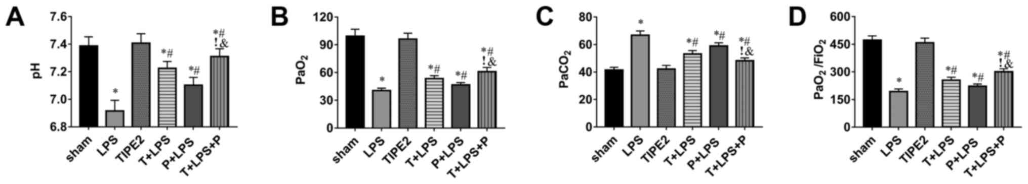
PHC pretreatment and TIPE2
overexpression improves gas exchange in the lungs of LPS-induced
mice
Gas analysis of arterial blood showed a significant
decrease in PaO2 and an increase in PaCO2 in
the LPS group (P<0.05; Fig. 2B and
C). Similarly, the pH and PaO2/FiO2 ratio
in arterial blood were decreased in the LPS group compared with the
sham group (P<0.05; Fig. 2A and
D). In the T+LPS, P+LPS and T+LPS+P groups, the dysfunction of
gas exchange induced by LPS stimulation was partially reversed
(P<0.05; Fig. 2A-D).
PHC pretreatment and TIPE2
overexpression attenuates the expression of pro-inflammatory
cytokines
As one of the most important cytokines in the
induction of ALI, TNF-α has a synergistic effect with IL-6 and
IL-1β in ALI development. According to the present study, it was
found that the expression levels of these cytokines were
significantly increased in the LPS group. Furthermore, compared
with the mice in the LPS group, the expression levels of these
cytokines were significantly downregulated in the T+LPS, P+LPS and
T+LPS+P groups (P<0.05; Fig.
3A-C).
Effects of PHC pretreatment and TIPE2
overexpression on the expression of different proteins in the lung
tissues of mice
Mice were humanely sacrificed and the expression
levels of related proteins were determined via western blotting
(Fig. 4A). As shown in Fig. 4, the expression levels of
pro-apoptotic proteins, such as Bax, cleaved caspase 3 and cleaved
caspase 9, in mice lung tissue increased in the LPS group compared
with the sham group (P<0.05; Fig.
4B, E and F), whereas the expression levels of anti-apoptotic
Bcl-2 decreased compared with the sham group (P<0.05; Fig. 4C). In addition, the ratio of
Bax/Bcl-2 increased in the LPS group compared with the sham group
(P<0.05; Fig. 4I). These
expression changes were partially reversed in the T+LPS and P+LPS
groups, and most significantly reversed in the T+LPS+P group
(P<0.05; Fig. 4B, C, E, F and
I). In addition, the expression levels of NF-κB p65 and
phosphorylated JNK increased (P<0.05; Fig. 4G and H), and the expression of TIPE2
decreased, in mice in the LPS group compared with the sham group
(P<0.05; Fig. 4D). The
expression levels of NF-κB p65 and p-JNK decreased (P<0.05;
Fig. 4G and H), and the expression
of TIPE2 increased (P<0.05; Fig.
4D), in mice in the T+LPS, P+LPS and T+LPS+P groups compared
with the LPS group.
 | Figure 4.Effects of PHC pretreatment and TIPE2
overexpression on the expression of different proteins in the lung
tissues of mice. (A) Western blotting analysis of protein
expression levels. Semi-quantification of (B) Bax, (C) Bcl-2, (D)
TIPE2, (E) cleaved caspase 3, (F) cleaved caspase 9, (G) NF-κB p65,
(H) p-JNK/total JNK and (I) Bax/Bcl-2 ratio. n=8/group. *P<0.05
vs. sham group; #P<0.05 vs. LPS group;
&P<0.05 vs. T+LPS group; !P<0.05
vs. P+LPS group. PHC/P, penehyclidine hydrochloride; TIPE2/T, tumor
necrosis factor α-induced protein 8-like protein 2; LPS,
lipopolysaccharide; p-, phosphorylated. |
Discussion
The pathological changes of ALI are mainly
associated with alveolar damage and the release of neutrophil and
inflammatory cytokines (29). At
present, no specific drugs are used for the treatment of ALI
(1). In addition to controlling the
primary disease, inhibiting systemic inflammation is an important
measure to prevent and treat ALI (30). In the present study, it was found
that LPS stimulation could worsen histological changes of lung
tissues and dysfunctions of gas exchange.
PHC is a novel anticholinergic drug that can inhibit
biomembrane lipid peroxidation and decrease the levels of cytokines
and oxyradicals in patients with sepsis (31). Previous research has demonstrated
that PHC plays a protective role against sepsis through inhibiting
the expression of inflammatory factors and inducible nitric oxide
synthase mRNA (32). Some previous
studies (33,34) have shown that it plays a protective
role in ischemia-reperfusion injury, but the role in transplant
surgery has not yet been reported in the literature, thus the
related dosage and clinical effects need to be further
investigated. A study reported that the protective effect of PHC
may involve the inhibition of NF-κB activation via inhibition of
the p38MAPK and ERK signaling pathways (35). In the present study, it was found
that PHC pretreatment ameliorated pulmonary edema in mice with
LPS-induced ALI, as evaluated by a significant decrease in the lung
wet/dry ratio. At the same time, PMNs/total cells and total protein
concentration in the BALF, which are the common indicators to
detect pulmonary vascular permeability (36), were significantly increased in LPS
group and decreased in the P+LPS group. PHC pretreatment also
alleviated lung histological damage and improved gas exchange
dysfunction induced by LPS. TNF-α is the earliest proinflammatory
factor that plays a central role in stress response and is the
initiator of the occurrence and development of ALI (37). The present results showed that PHC
could significantly inhibit the expression of TNF-α, IL-6 and
IL-1β, which indicated that PHC has anti-inflammatory properties.
At the same time, PHC pretreatment could decrease the Bax/Bcl-2
ratio, as well as the expression of cleaved caspase 3 and cleaved
caspase 9 protein, indicating a potential antiapoptotic effect.
Recent studies have also demonstrated that PHC can decrease the
expression of phosphorylated JNK in mice and rats with cerebral
ischemia-reperfusion and myocardial ischemia-reperfusion injury,
and provided protective effects in the brain (38) and heart (39), respectively, which is consistent
with the present experimental results in the lung tissues of
mice.
TIPE2 is predominantly expressed in immune cells of
the myeloid and lymphoid lineages (40), which provides TIPE2 with an
important role in the maintenance of immune homeostasis (41). Therefore, the abnormal expression of
TIPE2 can lead to diseases such as systemic autoimmunity, diabetic
nephropathy and hepatitis B in humans (42). In the present study, it was
demonstrated that TIPE2 overexpression could inhibit the expression
of TNF-α, IL-6 and IL-1β compared with the LPS group, which was
concomitant with a decrease in NF-κB activation. Some studies have
found that TIPE2 is a negative regulator of MAPK and NF-κB
signaling pathways (14,15), However, the underlying mechanisms
are currently unknown. Thus, the present study explored the
expression of related proteins and the underlying relationship
between them. It was found that TIPE2 overexpression significantly
reduced the expression of pro-apoptotic proteins such as Bax,
cleaved caspase-9, cleaved caspase-3 and p-JNK, at the same time,
the expression of anti-apoptotic protein Bcl-2 was increased. These
data suggested that the effect of TIPE2 overexpression on cell
apoptosis may be associated with suppression of JNK activation.
Furthermore, treatment with PHC and TIPE2 overexpression at the
same time led to a more obvious protective effect compared with PHC
or TIPE2 alone. In our previous experiment, we found that TIPE2
overexpression markedly inhibited inflammation and cell apoptosis
induced by LPS (20), and the
present results suggested that the expression of TIPE2 was
increased after the mice received intraperitoneal injection with
PHC. These findings indicate that TIPE2 may be linked to the
protective mechanism of PHC in mice with LPS-induced ALI.
In the present study, LPS-induced ALI in mice led to
the increased expression of NF-κB and promoted the activation of
JNK protein, subsequently causing the excessive release of
pro-inflammatory cytokines and the activation of pro-apoptotic
proteins, which was inhibited following PHC pretreatment-induced
upregulation of TIPE2 expression. TIPE2 can negatively regulate TLR
signaling, thereby inhibiting activation of NF-κB and JNK and
reducing apoptosis (20). Thus,
based on these findings, it was speculated that PHC inhibits these
effects by upregulating the expression of TIPE2, thereby
alleviating lung inflammation and apoptosis. The current study
preliminarily investigated the effects of TIPE2 and PHC drug
treatment on mice with LPS-induced ALI. However, the specific
underlying mechanism of PHC treatment on TIPE2 expression also
needs to be verified by knockout mice. At present, we have carried
out the cultivation of gene knockout mice, but the number of mice
was not enough to be included in the present study, so research on
specific pathways will be investigated in future research.
In conclusion, the present study provided evidence
that TIPE2 contributes to the protective effects of PHC
pretreatment against LPS-induced ALI through decreasing the
expression of NF-κB and inhibiting the activation of JNK in the
lungs of mice. However, the potential underlying mechanisms will be
further elucidated in depth in the future through the use of gene
knockout mice.
Acknowledgements
Not applicable.
Funding
This study was supported by the National Natural
Science Foundation of China (grant nos. 81571941 and 81901952).
Availability of data and materials
The datasets used and/or analyzed during the current
study are available from the corresponding author on reasonable
request.
Authors' contributions
XMS and XJW participated in the design of the study
and reviewed the manuscript. MY and QK carried out the experiments.
GQJ and TQM performed the data analyses. MY wrote and revised the
manuscript. XMS and XJW confirm the authenticity of all the raw
data. All authors read and reviewed the final manuscript.
Ethics approval and consent to
participate
Ethics approval was provided by the Medical Ethics
Committee of Renmin Hospital of Wuhan University (Wuhan, China).
All surgical procedures were performed in accordance with Wuhan
University Animal Care and Use committee.
Patient consent for publication
Not applicable.
Competing interests
The authors declare that they have no competing
interests.
References
|
1
|
Hodder R: Critical care in the ED:
Potentially fatal asthma and acute lung injury syndrome. Open
Access Emerg Med. 4:53–68. 2012.PubMed/NCBI
|
|
2
|
Galani V, Tatsaki E, Bai M, Kitsoulis P,
Lekka M, Nakos G and Kanavaros P: The role of apoptosis in the
pathophysiology of acute respiratory distress syndrome (ARDS): An
up-to-date cell-specific review. Pathol Res Pract. 206:145–150.
2010. View Article : Google Scholar : PubMed/NCBI
|
|
3
|
Lin S, Wu H, Wang C, Xiao Z and Xu F:
Regulatory T cells and acute lung injury: Cytokines, uncontrolled
inflammation, and therapeutic implications. Front Immunol.
9:15452018. View Article : Google Scholar : PubMed/NCBI
|
|
4
|
Bhatia M, Zemans RL and Jeyaseelan S: Role
of chemokines in the pathogenesis of acute lung injury. Am J Respir
Cell Mol Biol. 46:566–572. 2012. View Article : Google Scholar : PubMed/NCBI
|
|
5
|
Xie K, Yu Y, Huang Y, Zheng L, Li J, Chen
H, Han H, Hou L, Gong G and Wang G: Molecular hydrogen ameliorates
lipopolysaccharide-induced acute lung injury in mice through
reducing inflammation and apoptosis. Shock. 37:548–555. 2012.
View Article : Google Scholar : PubMed/NCBI
|
|
6
|
Erickson SE, Martin GS, Davis JL, Matthay
MA and Eisner MD; NIH NHLBI ARDS Network, : Recent trends in acute
lung injury mortality: 1996–2005. Crit Care Med. 37:1574–1579.
2009. View Article : Google Scholar : PubMed/NCBI
|
|
7
|
Kangelaris KN, Prakash A, Liu KD,
Aouizerat B, Woodruff PG, Erle DJ, Rogers A, Seeley EJ, Chu J, Liu
T, et al: Increased expression of neutrophil-related genes in
patients with early sepsis-induced ARDS. Am J Physiol Lung Cell Mol
Physiol. 308:L1102–L1113. 2015. View Article : Google Scholar : PubMed/NCBI
|
|
8
|
Chuang CY, Chen TL, Cherng YG, Tai YT,
Chen TG and Chen RM: Lipopolysaccharide induces apoptotic insults
to human alveolar epithelial A549 cells through reactive oxygen
species-mediated activation of an intrinsic mitochondrion-dependent
pathway. Arch Toxicol. 85:209–218. 2011. View Article : Google Scholar : PubMed/NCBI
|
|
9
|
Fonceca AM, Zosky GR, Bozanich EM, Sutanto
EN, Kicic A, McNamara PS, Knight DA, Sly PD, Turner DJ and Stick
SM: Accumulation mode particles and LPS exposure induce TLR-4
dependent and independent inflammatory responses in the lung.
Respir Res. 19:152018. View Article : Google Scholar : PubMed/NCBI
|
|
10
|
Sun K, Huang R, Yan L, Li DT, Liu YY, Wei
XH, Cui YC, Pan CS, Fan JY, Wang X and Han JY: Schisandrin
attenuates lipopolysaccharide-induced lung injury by regulating
TLR-4 and Akt/FoxO1 signaling pathways. Front Physiol. 9:11042018.
View Article : Google Scholar : PubMed/NCBI
|
|
11
|
Ding X, Tong Y, Jin S, Chen Z, Li T,
Billiar TR, Pitt BR, Li Q and Zhang LM: Mechanical ventilation
enhances extrapulmonary sepsis-induced lung injury: Role of
WISP1-αvβ5 integrin pathway in TLR4-mediated inflammation and
injury. Crit Care. 22:3022018. View Article : Google Scholar : PubMed/NCBI
|
|
12
|
Bordoloi D, Banik K, Shabnam B, Padmavathi
G, Monisha J, Arfuso F, Dharmarajan A, Mao X, Lim LHK, Wang L, et
al: TIPE family of proteins and its implications in different
chronic diseases. Int J Mol Sci. 19:29742018. View Article : Google Scholar : PubMed/NCBI
|
|
13
|
Lou Y and Liu S: The TIPE (TNFAIP8) family
in inflammation, immunity, and cancer. Mol Immunol. 49:4–7. 2011.
View Article : Google Scholar : PubMed/NCBI
|
|
14
|
Oho M, Nakano R, Nakayama R, Sakurai W,
Miyamoto A, Masuhiro Y and Hanazawa S: TIPE2 (Tumor Necrosis Factor
α-induced Protein 8-like 2) is a novel negative regulator of TAK1
signal. J Biol Chem. 291:22650–22660. 2016. View Article : Google Scholar : PubMed/NCBI
|
|
15
|
Zhang Y, Mei S, Zhou Y, Yang D, Pan T,
Chen Z and Wang Q: TIPE2 negatively regulates mycoplasma
pneumonia-triggered immune response via MAPK signaling pathway. Sci
Rep. 7:133192017. View Article : Google Scholar : PubMed/NCBI
|
|
16
|
Xiao HT, Liao Z and Tong RS: Penehyclidine
hydrochloride: A potential drug for treating COPD by attenuating
Toll-like receptors. Drug Des Devel Ther. 6:317–322. 2012.
View Article : Google Scholar : PubMed/NCBI
|
|
17
|
Sun H, Gong S, Carmody RJ, Hilliard A, Li
L, Sun J, Kong L, Xu L, Hilliard B, Hu S, et al: TIPE2, a negative
regulator of innate and adaptive immunity that maintains immune
homeostasis. Cell. 133:415–426. 2008. View Article : Google Scholar : PubMed/NCBI
|
|
18
|
Chen C, Wang Y, Zhang Z, Wang C and Peng
M: Toll-like receptor 4 regulates heme oxygenase-1 expression after
hemorrhagic shock induced acute lung injury in mice: Requirement of
p38 mitogen-activated protein kinase activation. Shock. 31:486–492.
2009. View Article : Google Scholar : PubMed/NCBI
|
|
19
|
Chambers E, Rounds S and Lu Q: Pulmonary
endothelial cell apoptosis in emphysema and acute lung injury. Adv
Anat Embryol Cell Biol. 228:63–86. 2018. View Article : Google Scholar : PubMed/NCBI
|
|
20
|
Wu X, Kong Q, Zhan L, Qiu Z, Huang Q and
Song X: TIPE2 ameliorates lipopolysaccharide-induced apoptosis and
inflammation in acute lung injury. Inflamm Res. 68:981–992. 2019.
View Article : Google Scholar : PubMed/NCBI
|
|
21
|
Han XY, Liu H, Liu CH, Wu B, Chen LF,
Zhong BH and Liu KL: Synthesis of the optical isomers of a new
anticholinergic drug, penehyclidine hydrochloride (8018). Bioorg
Med Chem Lett. 15:1979–1982. 2005. View Article : Google Scholar : PubMed/NCBI
|
|
22
|
Weng J, Chen M, Lin Q, Chen J, Wang S and
Fang D: Penehyclidine hydrochloride defends against LPS-induced ALI
in rats by mitigating endoplasmic reticulum stress and promoting
the Hes1/Notch1 pathway. Gene. 721:1440952019. View Article : Google Scholar : PubMed/NCBI
|
|
23
|
Li H, Qian Z, Li J, Han X and Liu M:
Effects of early administration of a novel anticholinergic drug on
acute respiratory distress syndrome induced by sepsis. Med Sci
Monit. 17:BR319–BR325. 2011. View Article : Google Scholar : PubMed/NCBI
|
|
24
|
Wu XJ, Liu HM, Song XM, Zhao B, Leng Y,
Wang EY, Zhan LY, Meng QT and Xia ZY: Penehyclidine hydrochloride
inhibits TLR4 signaling and inflammation, and attenuates blunt
chest trauma and hemorrhagic shock-induced acute lung injury in
rats. Mol Med Rep. 17:6327–6336. 2018.PubMed/NCBI
|
|
25
|
Li Y, Cao Y, Zeng Z, Liang M, Xue Y, Xi C,
Zhou M and Jiang W: Angiotensin-converting enzyme
2/angiotensin-(1–7)/Mas axis prevents lipopolysaccharide-induced
apoptosis of pulmonary microvascular endothelial cells by
inhibiting JNK/NF-kB pathways. Sci Rep. 5:82092015. View Article : Google Scholar : PubMed/NCBI
|
|
26
|
Zeke A, Misheva M, Remenyi A and
Bogoyevitch MA: JNK signaling: Regulation and functions based on
complex protein-protein partnerships. Microbiol Mol Biol Rev.
80:793–835. 2016. View Article : Google Scholar : PubMed/NCBI
|
|
27
|
Huang C, He J, Chen Y, Zhang Y and Chen C:
Penehyclidine hydrochloride inhibits the LPS-induced inflammatory
response in microglia. J Surg Res. 188:260–267. 2014. View Article : Google Scholar : PubMed/NCBI
|
|
28
|
National Research Council (US) Committee
for the Update of the Guide for the Care and Use of Laboratory
Animals, . Guide for the Care and Use of Laboratory Animals, 8th
edition. National Academies Press (US); Washington (DC): 2011
|
|
29
|
Z'Graggen BR, Tornic J, Muller-Edenborn B,
Reyes L, Booy C and Beck-Schimmer B: Acute lung injury: Apoptosis
in effector and target cells of the upper and lower airway
compartment. Clin Exp Immunol. 161:324–331. 2010.PubMed/NCBI
|
|
30
|
Standiford TJ and Ward PA: Therapeutic
targeting of acute lung injury and acute respiratory distress
syndrome. Transl Res. 167:183–191. 2016. View Article : Google Scholar : PubMed/NCBI
|
|
31
|
Wang Y, Gao Y and Ma J: Pleiotropic
effects and pharmacological properties of penehyclidine
hydrochloride. Drug Des Devel Ther. 12:3289–3299. 2018. View Article : Google Scholar : PubMed/NCBI
|
|
32
|
Zhan J, Wang Y, Wang C, Li J, Zhang Z and
Jia B: Protective effects of penehyclidine hydrochloride on septic
mice and its mechanism. Shock. 28:727–732. 2007. View Article : Google Scholar : PubMed/NCBI
|
|
33
|
Liu Z, Li Y, Yu L, Chang Y and Yu J:
Penehyclidine hydrochloride inhibits renal
ischemia/reperfusion-induced acute lung injury by activating the
Nrf2 pathway. Aging (Albany NY). 12:13400–13421. 2020. View Article : Google Scholar : PubMed/NCBI
|
|
34
|
Wang Y, Lin D, Tan H, Gao Y and Ma J:
Penehyclidine hydrochloride preconditioning provides pulmonary and
systemic protection in a rat model of lung ischaemia reperfusion
injury. Eur J Pharmacol. 839:1–11. 2018. View Article : Google Scholar : PubMed/NCBI
|
|
35
|
Shen W, Gan J, Xu S, Jiang G and Wu H:
Penehyclidine hydrochloride attenuates LPS-induced acute lung
injury involvement of NF-kappaB pathway. Pharmacol Res. 60:296–302.
2009. View Article : Google Scholar : PubMed/NCBI
|
|
36
|
Xing J, Yu Z, Zhang X, Li W, Gao D, Wang
J, Ma X, Nie X and Wang W: Epicatechin alleviates inflammation in
lipopolysaccharide-induced acute lung injury in mice by inhibiting
the p38 MAPK signaling pathway. Int Immunopharmacol. 66:146–153.
2019. View Article : Google Scholar : PubMed/NCBI
|
|
37
|
Liu S, Feng G, Wang GL and Liu GJ: p38MAPK
inhibition attenuates LPS-induced acute lung injury involvement of
NF-kappaB pathway. Eur J Pharmacol. 584:159–165. 2008. View Article : Google Scholar : PubMed/NCBI
|
|
38
|
Shu Y, Yang Y and Zhang P: Neuroprotective
effects of penehyclidine hydrochloride against cerebral
ischemia/reperfusion injury in mice. Brain Res Bull. 121:115–123.
2016. View Article : Google Scholar : PubMed/NCBI
|
|
39
|
Feng M, Wang L, Chang S and Yuan P:
Penehyclidine hydrochloride regulates mitochondrial dynamics and
apoptosis through p38MAPK and JNK signal pathways and provides
cardioprotection in rats with myocardial ischemia-reperfusion
injury. Eur J Pharm Sci. 121:243–250. 2018. View Article : Google Scholar : PubMed/NCBI
|
|
40
|
Padmavathi G, Banik K, Monisha J, Bordoloi
D, Shabnam B, Arfuso F, Sethi G, Fan L and Kunnumakkara AB: Novel
tumor necrosis factor-α induced protein eight (TNFAIP8/TIPE)
family: Functions and downstream targets involved in cancer
progression. Cancer Lett. 432:260–271. 2018. View Article : Google Scholar : PubMed/NCBI
|
|
41
|
Freundt EC, Bidere N and Lenardo MJ: A
different TIPE of immune homeostasis. Cell. 133:401–402. 2008.
View Article : Google Scholar : PubMed/NCBI
|
|
42
|
Zhang S, Zhang Y, Wei X, Zhen J, Wang Z,
Li M, Miao W, Ding H, Du P, Zhang W, et al: Expression and
regulation of a novel identified TNFAIP8 family is associated with
diabetic nephropathy. Biochim Biophys Acta. 1802:1078–1086. 2010.
View Article : Google Scholar : PubMed/NCBI
|