Introduction
Doxorubicin (DOX), one of the most widely used
anthracycline antibiotics, is typically used to treat various types
of cancer such as breast cancer, ovarian cancer and leukemia
(1–4). However, its side effects,
particularly cardiotoxicity, severely limit its clinical
application. The cardiotoxicity induced by DOX involves multiple
molecular mechanisms, including redox homeostasis imbalance, DNA
and RNA damage, autophagy inhibition and programmed cell death
(such as pyroptosis and apoptosis), ultimately leading to
myocardial dysfunction (5–7). The dose-dependent cardiotoxicity
associated with DOX treatment can eventually develop into
congestive heart failure or even death (8,9).
Although these mechanisms have been partially elucidated, the
corresponding effective clinical intervention measures remain
limited, and DOX-induced cardiotoxicity (DIC) has not fundamentally
been improved in clinical practice (10). Therefore, further understanding the
novel mechanisms underlying DIC is vital for the development of
novel cardioprotective strategies.
Proprotein convertase subtilisin/kexin type 9
(PCSK9), a gene typically associated with familial
hypercholesterolemia, is expressed predominantly in the liver,
kidneys and intestines (11).
PCSK9 affects the level of low-density lipoprotein cholesterol
(LDL-C) in the blood by regulating the degradation of LDL receptors
(LDLRs) (12). PCSK9 is not only
closely associated with cholesterol metabolism but also serves
important roles in cardiovascular diseases such as in
atherosclerosis and cardiac ischemia-reperfusion injury (13–15).
In addition, PCSK9 is involved in various biological processes,
such as apoptosis, autophagy, inflammation and tumor immunity, and
is closely related to diabetes and neurodegenerative diseases
(6,16–19).
Recent research has further indicated that PCSK9 also serves a
regulatory role in other physiological processes of the heart,
including influencing the stress responses and death processes of
cardiomyocytes (7,20). The expression levels of LDLR and
CD36 are increased and lipids accumulate in the cardiac tissue of
PCSK9 knockout mice (21).
Clinical studies have demonstrated that elevated expression levels
of PCSK9 in peripheral blood are independently associated with
increased disease severity and poor prognosis in patients with
heart failure (22). These
metabolic and functional disturbances suggest PCSK9 dysregulation
is closely associated with heart failure. Although the role of
PCSK9 in heart diseases has been previously investigated, the
specific mechanism through which PCSK9 affects DIC remains unclear.
DOX can cause myocardial injury through multiple pathways,
including pyroptosis (23).
Pyroptosis is a form of lytic cell death mediated by members of the
gasdermin protein family that is characterized by cell membrane
perforation, cell swelling and rupture, and the release of large
amounts of proinflammatory factors (such as IL-1β and IL-18)
(24). A study has shown that DOX
can trigger pyroptosis (25);
however, it is currently unclear whether PCSK9 is involved in this
process. Therefore, the present study aimed to explore the
regulatory role of PCSK9 in DOX-induced pyroptosis of
cardiomyocytes, thereby providing novel insights into reducing
cardiotoxicity caused by anthracyclines. These findings offer a new
theoretical basis and target for improving the quality of life and
therapeutic effect of chemotherapy.
Materials and methods
Cell culture and experimental
design
AC-16 cells were provided by the Henan Key
Laboratory of Medical Tissue Regeneration of Henan Medical
University (Xinxiang, China). The AC-16 cells from the 5th to 8th
passages were used in the present study, ensuring stable cell
conditions and a consistent phenotype. AC-16 cells were cultured in
Dulbecco's Modified Eagle's Medium/F12 (cat. no. 12634010; Gibco;
Thermo Fisher Scientific, Inc.) supplemented with 10% fetal bovine
serum and 1% penicillin-streptomycin (cat. no. 15070063; Gibco;
Thermo Fisher Scientific, Inc.). All cells were cultured at 37°C
with 5% CO2.
DOX was dissolved in sterile DMSO at a stock
concentration of 100 mM and stored at −20°C. The DOX solution was
then diluted directly with culture medium to the working
concentration, resulting in a final DMSO concentration <0.001%
in all groups. The final working concentration of DOX in the cell
culture medium was 0.2 µM, which was consistent with the present
IC50 results. Control cells received an equal volume of
culture medium containing the same concentration of DMSO without
DOX.
Animal and experimental design
The present study used two mouse models as follows:
Male C57BL/6J mice (age, 6–8 weeks; weight, 18–22 g) and male
PCSK9−/− mice (age, 6–8 weeks; weight, 18–22 g)
purchased from Shanghai Model Organisms Center. A total of 32 mice
(16 male C57BL/6J mice and 16 PCSK9−/− mice) were
included in the experiment, and each mouse model was randomly
divided into two groups with n=8 mice per group. Grouping was
performed using a random number table method to avoid grouping bias
by ensuring that the body weight, age and other baseline indicators
of the mice in each group were comparable. All the animals were
housed under standard laboratory conditions (22±2°C, 50±5% relative
humidity and 12-h light/dark cycle). Food and water were available
ad libitum throughout the experiment.
The mice were randomly divided into DOX and control
groups and subjected to intraperitoneal injections of either DOX (4
mg/kg; cat. no. D107159; Aladdin Scientific Corp.), with an
accumulated dosage of 20 mg/kg, or an equivalent volume of saline
once per week for five weeks, respectively. This is a common
modelling approach for chronic cardiotoxicity; a systematic review
has confirmed that a cumulative dose of 15–24 mg/kg reliably
induces cardiac injury, and the survival rate at a dose of ≥20
mg/kg is approximately 30% (26).
All animals were monitored twice daily (every 12 h)
for signs of pain, distress or impending death. Predefined humane
endpoints including weight loss >20%, severe matting of the fur
with reduced activity or food intake, dyspnea, inability to reach
food or water, and spontaneous recumbency were strictly applied.
Animals meeting any endpoint were immediately sacrificed under deep
isoflurane anesthesia (inhalation of 5% isoflurane until loss of
consciousness and lack of response to toe pinch, 5–10 min) by
cervical dislocation. Those exhibiting moderate distress (15–20%
weight loss, mild symptoms) received supportive care (subcutaneous
fluids, warmth and wet diet) and were re-evaluated after 12–24 h;
sacrifice followed if no improvement was observed.
Following the echocardiographic examination, the
mice were sacrificed by cervical dislocation performed under deep
anesthesia (5% isoflurane for 2–3 min), and the cardiac tissue was
rapidly collected for subsequent analysis. All animal procedures
were conducted in accordance with animal welfare guidelines and
were approved by the Institutional Animal Care and Use Committee of
Xinxiang Medical University (currently Henan Medical University;
approval no. XYLL-20220192; Xinxiang, China).
Echocardiography
Cardiac function was assessed by echocardiography
five weeks after the initiation of the injections. The cardiac
function of the mice was evaluated using a high-resolution small
animal ultrasound imaging system (Visual Sonics Vevo®
2100; FUJIFILM Corporation). Anesthesia was induced using
isoflurane (RWD Life Science Co., Ltd.) at a concentration of 3–4%,
with a maintenance concentration of 1.5–2%. Once the required level
of anesthesia was confirmed using the toe pinch reflex test, the
mice were immobilized in the supine position, with ECG electrodes
were attached to their limbs, on a constant-temperature platform
(37°C) to maintain a stable physiological state (37±0.5°C). Through
transthoracic echocardiography, the short-axis section of the left
ventricle was located using two-dimensional ultrasound, and the
inner diameters of the left ventricle during systole and diastole
were recorded using M-mode ultrasound. The ejection fraction (EF)
and left ventricular fractional shortening (FS) were calculated as
the core indicators of cardiac function.
Western blotting
AC-16 cells treated with DOX or DMSO for 48 h were
lysed on ice in RIPA buffer (cat. no. P0013B; Beyotime
Biotechnology) containing PMSF. Protein concentrations were
measured by BCA assay. Equal amounts of protein (20 µg per lane)
were separated by SDS-PAGE with 10–12% gels and transferred onto
PVDF membranes. The membranes were blocked with 5% skimmed milk
powder (cat. no. GC310001; Wuhan Servicebio Technology Co., Ltd.)
in PBS containing 0.1% Tween-20 (cat. no. P9416; MilliporeSigma)
for 1 h and then probed with specific primary antibodies against
PCSK9 (1:1,000; cat. no. CY6876; Shanghai Abways Biotechnology Co.,
Ltd.), IL-1β (1:1,000; cat. no HL1421; Thermo Fisher Scientific,
Inc.), caspase-1 (1:1,000; cat. no. 22915-1-AP; Proteintech Group,
Inc.), sterol regulatory element binding protein-1
(SREBP-1;1:1,000; cat. no. HA722160; HUABIO), SREBP-2 (1:1,000;
cat. no. 28212-1-AP, Proteintech Group, Inc.) and β-actin (1:1,000;
cat. no. AB0035; Shanghai Abways Biotechnology Co., Ltd.) overnight
at 4°C. Following primary antibody incubation, the membranes were
incubated overnight with the corresponding secondary antibodies
Goat anti-rabbit IgG HRP conjugate (1:8,000; cat. no. AB0101
Shanghai Abways Biotechnology Co., Ltd.). Secondary antibody
incubation was performed for 1 h at room temperature. Protein
signals were visualized using an enhanced chemiluminescence (ECL)
kit (cat. no. P0018FS; Beyotime Biotechnology) and imaged with the
Amersham™ Imager 600 system (Cytiva). Band intensity was analyzed
using ImageJ (version 1.54f, National Institutes of Health, USA)
and normalized to β-actin as an internal control.
Reverse transcription-qPCR
Total RNA was extracted from DOX-treated and control
AC-16 cells using RNAiso Plus (cat. no. 9108Q; Takara Bio, Inc.)
according to the manufacturer's instructions. Total RNA was reverse
transcribed to cDNA using PrimerScript RT Master Mix (cat. no.
RR036A; Takara Bio, Inc.) according to the manufacturer's
instructions, and real-time qPCR was performed using a SYBR Green
Kit (cat. no. Q221-01; Vazyme Biotech Co., Ltd.). The thermal
cycling conditions were as follows: Initial denaturation at 95°C
for 5 min, followed by 40 cycles of denaturation at 95°C for 10–15
sec and combined annealing/extension at 60°C for 30–40 sec.
Fluorescence data were collected during the annealing/extension
step. Following amplification, a melting curve analysis was
performed to verify the specificity of the PCR products. β-actin
was used as the control, and the relative expression of the genes
was quantified by the 2−ΔΔCq method (27). The primer sequences are listed in
Table SI.
Lactate dehydrogenase (LDH) assay
AC-16 cells were seeded in 96-well plates (1×10⁴
cells/well) and treated with DOX for 48 h. Blank (no cells),
negative (untreated) and positive (treated with LDH release
reagent) controls groups were also included. After centrifugation
at 400 × g for 5 min at room temperature, the supernatant was
collected and added to a new 96-well plate, after which the working
solution from the LDH Cytotoxicity Assay kit (cat. no. C0017;
Beyotime Biotechnology) was added (according to the manufacturer's
instructions). The mixture was incubated at room temperature in the
dark for 30 min. Absorbance was detected at two wavelengths (main
wavelength, 490 nm; reference wavelength, 600 nm) using a
microplate reader.
CCK-8 assay
AC-16 cells were seeded into 96-well plates
(5×10³ cells/well) and cultured overnight at 37°C and 5%
CO2. The culture medium was then replaced with fresh
medium containing a gradient concentration of DOX (0, 0.1, 0.2,
0.3, 0.4, 0.5 and 0.6 µM), and the cells were further cultured at
37°C for 24 or 48 h. Subsequently, 10 µl of CCK-8 reagent (cat. no.
C0038; Beyotime Biotechnology) was added to each well, and the
plates were incubated at 37°C in the dark for 4 h. The absorbance
was detected at 450 nm using a microplate reader.
Immunofluorescence staining
AC-16 cells were seeded onto glass slides in 12-well
plates (5×10⁵ cells/ml) and cultured at 37°C and 5% CO2
overnight. Cells were treated with DOX or DMSO for 24 h, washed
with PBS, fixed with 4% paraformaldehyde at room temperature for 15
min and permeabilized with 0.3% Triton X-100 for 10 min. The cells
were blocked with 5% BSA (cat. no. 9048-46-8; Beijing Solarbio
Science & Technology Co., Ltd.) at room temperature for 1 h and
incubated overnight at 4°C with specific primary antibodies against
PCSK9 (1:250; cat. no. HA610371; HUABIO), caspase-1 (1:200; cat.
no. MA5-16215; Thermo Fisher Scientific, Inc.) and gasdermin D
(GSDMD; 1:200; cat. no. PA5-116815; Thermo Fisher Scientific,
Inc.). Then, the cells were treated with fluorescent Alexa Fluor
488-conjugated goat anti-rabbit IgG (H+L) (1:500; cat. no. A-11008;
Thermo Fisher Scientific, Inc.) and Alexa Fluor 594-conjugated goat
anti-mouse IgG (H+L) (1:500; cat. no. A-11005; Thermo Fisher
Scientific, Inc.) for 1 h at room temperature in the dark. Finally,
nuclear staining was performed with 5 µg/ml DAPI (cat. no. D9542;
MilliporeSigma) in PBS at room temperature for 5 min. The cells
were observed and images captured using a fluorescence microscope.
All images were acquired using the same optimized imaging
parameters and were applied uniformly to all samples. Image
analysis was performed using ImageJ software (version 1.54f;
National Institutes of Health).
Lentiviral packaging and transduction
of short hairpin RNA (shRNA) in AC-16 cells
293 cells (provided by the Henan Key Laboratory of
Medical Tissue Regeneration of Henan Medical University, Xinxiang,
China) were used for lentiviral packaging. Cells were cultured in
DMEM supplemented with 10% fetal bovine serum and 1%
penicillin/streptomycin at 37°C in a 5% CO2 incubator.
293 cells were co-transfected with either sh-PCSK9 or sh-negative
control (NC) plasmids (pLKO.1 vector; cat. no. 12260; Addgene,
Inc.), a packaging plasmid (psPAX2; cat. no. 12260; Addgene, Inc.)
and an envelope plasmid (pMD2.G; cat. no. 12259; Addgene, Inc.),
which together constituted a second-generation lentiviral packaging
system.
Transfection was performed using
Lipofectamine® 3000 (cat. no. L3000015; Thermo Fisher
Scientific, Inc.) according to the manufacturer's protocol. The
following amounts of plasmids were used per 10-cm dish: 5 µg of
shRNA plasmid (pLKO.1-sh-PCSK9 or sh-NC), 3.75 µg of psPAX2 and
1.25 µg of pMD2.G (ratio of shRNA: psPAX2: pMD2.G=4:3:1), all
diluted in Opti-MEM medium. The DNA-Lipofectamine® 3000
complexes were incubated for 15 min at room temperature before
being added to the cells. Viral supernatants were collected at 48 h
and 72 h post-transfection, filtered (0.45 µm) and concentrated
using a Lenti-X Concentrator (cat. no. 631231; Takara Bio, Inc.)
according to the manufacturer's instructions. The concentrated
lentiviral pellets were resuspended in 1/100 of the original volume
of sterile PBS or culture medium and stored at −80°C until use. The
target sequences were as follows: sh-PCSK9,
5′-GCATGTCTTCCATGGCCTTCT-3′; sh-NC (negative control, scrambled)
5′-TTCTCCGAACGTGTCACGTAA-3′.
AC-16 cells were seeded in 6-well plates (5×10⁴
cells/well) and incubated overnight. The cells were then transduced
with the concentrated lentivirus (sh-PCSK9 or sh-NC) at a
multiplicity of infection (MOI) of 5 in the presence of 5 µg/ml
polybrene (cat. no. C0351; Beyotime Biotechnology) at 37°C for 24
h. After a 48 h recovery period, the cells were selected using 2
µg/ml puromycin (cat. no. ST551; Beyotime Biotechnology) for seven
days. Knockdown efficiency was validated by qPCR and western
blotting.
Masson's trichrome staining
Heart tissues were fixed in 4% paraformaldehyde
(PFA; cat. no. P0099; Beyotime Biotechnology) at 4°C for 24 h,
embedded in paraffin, and cut into 4-µm sections. Sections were
stained using a Masson's trichrome kit (cat. no. G1340; Beijing
Solarbio Science & Technology Co., Ltd.) according to the
manufacturer's protocol. Images were captured using a light
microscope (Olympus BX53; Olympus Corporation).
Proteomics analysis
AC-16 cells were divided into two groups: A control
group (treated with vehicle alone) and a DOX-treated group (treated
with DOX at 1 µM for 48 h). Cells were lysed in RIPA buffer (cat.
no. P0013B; Beyotime Biotechnology) containing protease inhibitor
cocktail (cat. no. 78430; Thermo Fisher Scientific). Proteins were
reduced with 10 mM DTT (cat. no. D0632; MilliporeSigma) at 56°C for
30 min, alkylated with 20 mM iodoacetamide (cat. no. I1149;
MilliporeSigma) in the dark at room temperature for 30 min and then
separated by SDS-PAGE (12% gel). After Coomassie Blue staining
(cat. no. P0017A; Beyotime Biotechnology) at room temperature for 1
h, the gel lanes were excised and digested overnight with trypsin
(cat. no. V5111; Promega Corporation) at 37°C. The resulting
peptides were analyzed by nano-LC-MS/MS using an EASY-nLC 1200
chromatograph coupled to a Q-Exactive HF-X mass spectrometer
(Thermo Fisher Scientific). The mass spectrometer was operated in
positive ion mode with a spray voltage of 2.1 kV, ion transfer tube
temperature of 275°C, and ion source heater temperature of 300°C.
Sheath and auxiliary gas flows were set to 35 and 10 arbitrary
units, respectively. Peptides were separated on a C18 column with a
90-min linear gradient from 3 to 35% acetonitrile in 0.1% formic
acid. MS data were acquired in data-dependent mode (scan range m/z
375–1500).
Raw data were processed using the MaxQuant software
(version 2.3.1.0; Max Planck Institute of Biochemistry) against the
UniProt human database (2023 release; http://www.uniprot.org/). Trypsin was specified as the
cleavage enzyme with a maximum of two missed cleavages allowed.
Carbamidomethylation of cysteine was set as a fixed modification,
and oxidation of methionine and acetylation of the protein
N-terminus were set as variable modifications. Protein
identification was conducted with a false discovery rate (FDR)
<1% at both the peptide and protein levels. Label-free
quantification was performed using the MaxQuant software, and
proteins with |log2(FC)| >1 and P<0.05 were defined as
differentially abundant proteins (DAPs).
DAPs were subjected to gene ontology (GO) and Kyoto
Encyclopedia of Genes and Genomes (KEGG) enrichment analyses using
DAVID bioinformatics resources (version 6.8; http://david.ncifcrf.gov/). To ensure statistical
reliability, significantly enriched GO terms and KEGG pathways were
identified using a threshold of P<0.05, FDR<0.05, and a
minimum gene count of ≥2 per term/pathway. The enrichment results
were subsequently used for bioinformatic interpretation and
subsequent experimental validation. The sequencing was performed by
Novogene (Novogene Co., Ltd.).
Data analysis
The experimental data were analyzed using the
GraphPad Prism (version 9.0; Dotmatics) software. An unpaired
Student's t-test was used for comparisons between two groups (the
data conformed to a normal distribution and were homogeneous), and
one-way ANOVA followed by Tukey's post hoc test was used for
comparisons among multiple groups. The data are expressed as the
mean ± SEM. P<0.05 was considered to indicate a statistically
significant difference.
Results
DOX treatment downregulates PCSK9
expression and induces cardiomyocyte injury
Compared with those in the control group, cells in
the DOX-treated experimental groups showed notable morphological
changes including balloon-like transformation and vacuolation,
particularly with the increase of DOX concentration at 0.4 µM
treatment for 48 h (Fig. 1A).
These morphological alterations are consistent with previous
reports showing that doxorubicin induces concentration-dependent
cytoplasmic vacuolization, nuclear swelling, and mitochondrial
disruption in cardiomyocytes (28). CCK-8 results showed that DOX
decreased the viability of AC-16 cells in a dose-dependent manner
after treatment with DOX at different concentrations for 24 and 48
h, respectively. The semi-inhibitory concentration
(IC50) value of DOX for AC-16 cells was 0.2 µM in the 48
h treatment group (Fig. 1B). LDH
is a soluble cytoplasmic enzyme; upon cell membrane damage, LDH is
released from the cytosol into the culture medium, serving as a
well-established biochemical marker of cell death and membrane
integrity impairment (29). LDH
release was significantly elevated with increasing DOX
concentration (Fig. 1C). These
results suggested that DOX has a significant damaging effect on
cardiomyocytes.
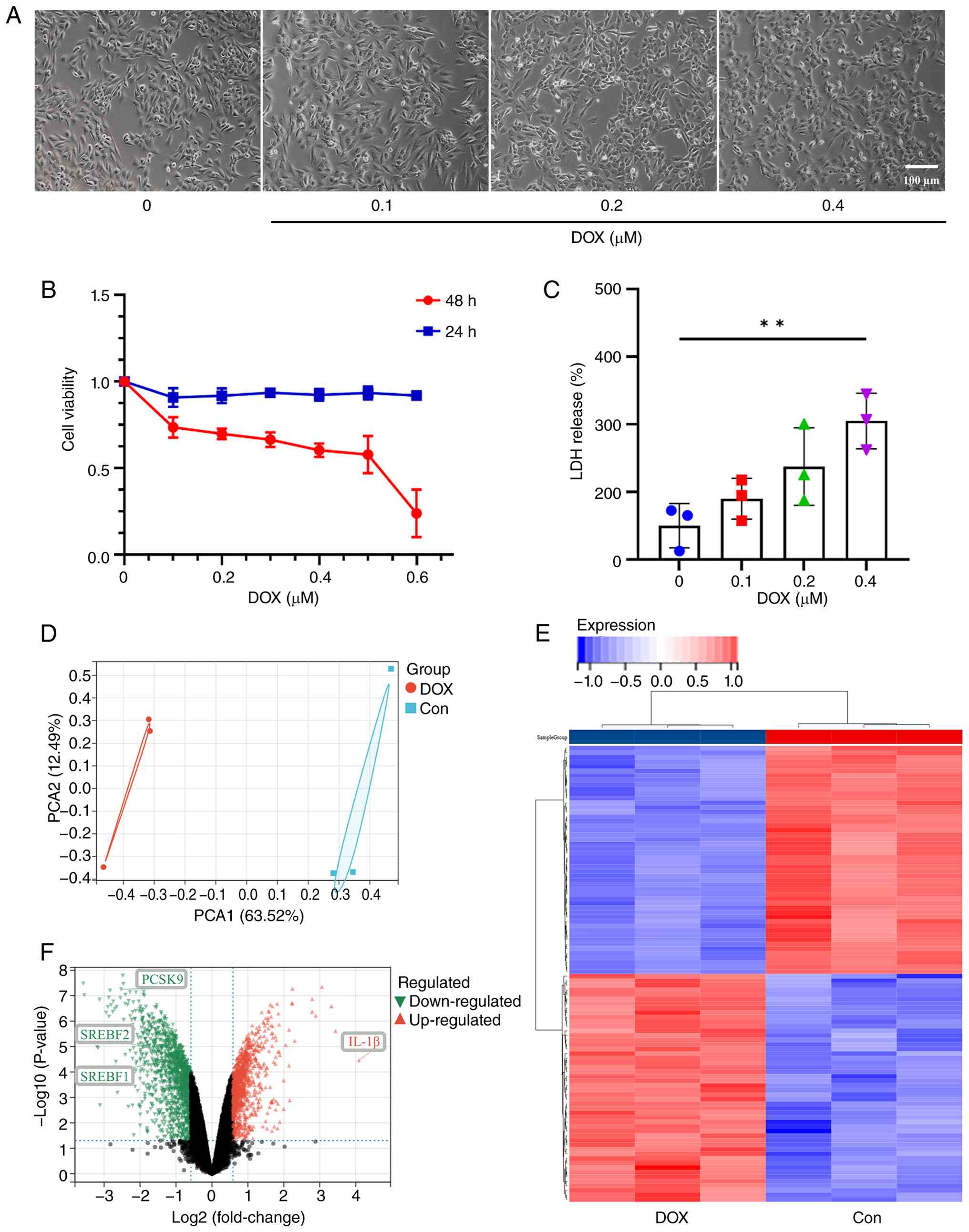 | Figure 1.Toxicity and biogenic analysis of DOX
in cardiomyocytes. (A) Cell morphology of AC-16 cells under a light
microscope after treatment with DOX for 48 h. (B) The cell
viability of AC-16 cells after treatment with increasing
concentrations of DOX for 24 and 28 h was determined using the Cell
Counting Kit-8 assay. (C) LDH release in AC-16 cells treated with
different concentrations of DOX for 48 h. (D) PCA revealed a
significant difference between the DOX and control group. (E) The
heatmap shows the cluster analysis of differentially expressed
genes, with different colors representing the level of gene
expression. (F) Key genes regulating cholesterol metabolism and
lipid synthesis, such as PCSK9, SREBF2 and SREBF1, as well as the
important proinflammatory cytokine IL-1β, showed significant
changes in abundance (outlined). Red represents upregulated
proteins and green represents downregulated proteins and black
represents the proteins with no significant changes in expression.
The differentially abundant protein volcano plot uses the fold
change (log2) value as the X-axis and the P-value
(−log10) as the Y-axis. **P<0.01; n=3. DOX,
doxorubicin; PCA, principal component analysis; LDH, lactate
dehydrogenase; Con, control; PCSK9, Proprotein convertase
subtilisin/kexin type 9; SREBF, sterol regulatory element binding
transcription factor 2. |
In accordance with the aforementioned experimental
results, AC-16 cells were treated with 0.2 µM DOX for 48 h, after
which proteomic sequencing was performed, and the results were
analyzed. First, principal component analysis was used to reduce
the dimensionality of the total protein expression profiles of the
experimental and control groups. PCA analysis demonstrated a
significant difference between the DOX and control groups (Fig. 1D). Heatmap analysis also revealed
significant differences in protein levels between the DOX-treated
and control group (Fig. 1E). To
further elucidate the potential mechanisms underlying DOX-induced
myocardial injury, an in-depth analysis based on the results of
proteomic KEGG enrichment analysis was conducted (Fig. S1). The results showed that
differentially expressed proteins were significantly enriched in
several core lipid metabolic pathways, including ‘steroid
metabolism’, ‘sterol metabolism’, ‘cholesterol metabolism’ and
‘lipid storage regulation’. This finding suggested that DOX-induced
myocardial injury is closely associated with disrupted lipid
homeostasis, and that PCSK9 may participate in this pathological
process by regulating lipid metabolism via the classical LDLR
pathway. Furthermore, proteomic results revealed a significant
enrichment of differentially expressed proteins in inflammation-
and stress-related pathways (such as ‘starvation response’ and
‘Notch signaling pathway’). This suggested that PCSK9 may also
possess non-classical functions independent of lipid metabolism,
such as the direct regulation of inflammatory or stress signaling
pathways.
Furthermore, statistical analysis of the proteomics
data was conducted using a two-tailed independent sample t-test,
and fold-change>1.2 and FDR<0.05 were set as screening
thresholds to define differentially expressed proteins. Compared
with the control group, the expression levels SREBF/SREBP-1,
SREBF-2/SREBP-2 and PCSK9, key genes associated with lipid
metabolism, were significantly downregulated in the DOX-treated
group (Fig. 1F). IL-1β, which is
closely related to inflammation and pyrosis expression, was
upregulated in the DOX-treated group compared with the control
group. Among these differentially expressed genes, PCSK9 was
selected for further investigation due to its central role in lipid
metabolism and its emerging regulatory functions in inflammatory
responses and cardiomyocyte injury. Moreover, recent studies have
suggested a potential crosstalk between PCSK9 and IL-1β-mediated
signaling pathways, although direct evidence in the context of
doxorubicin-induced cardiotoxicity remains limited. Based on these
correlative observations, it is plausible that PCSK9 may be
involved in IL-1β-mediated myocardial cell injury, but further
mechanistic studies are required to establish a causal
relationship.
PCSK9 expression in DOX-induced
cardiomyocyte pyroptosis
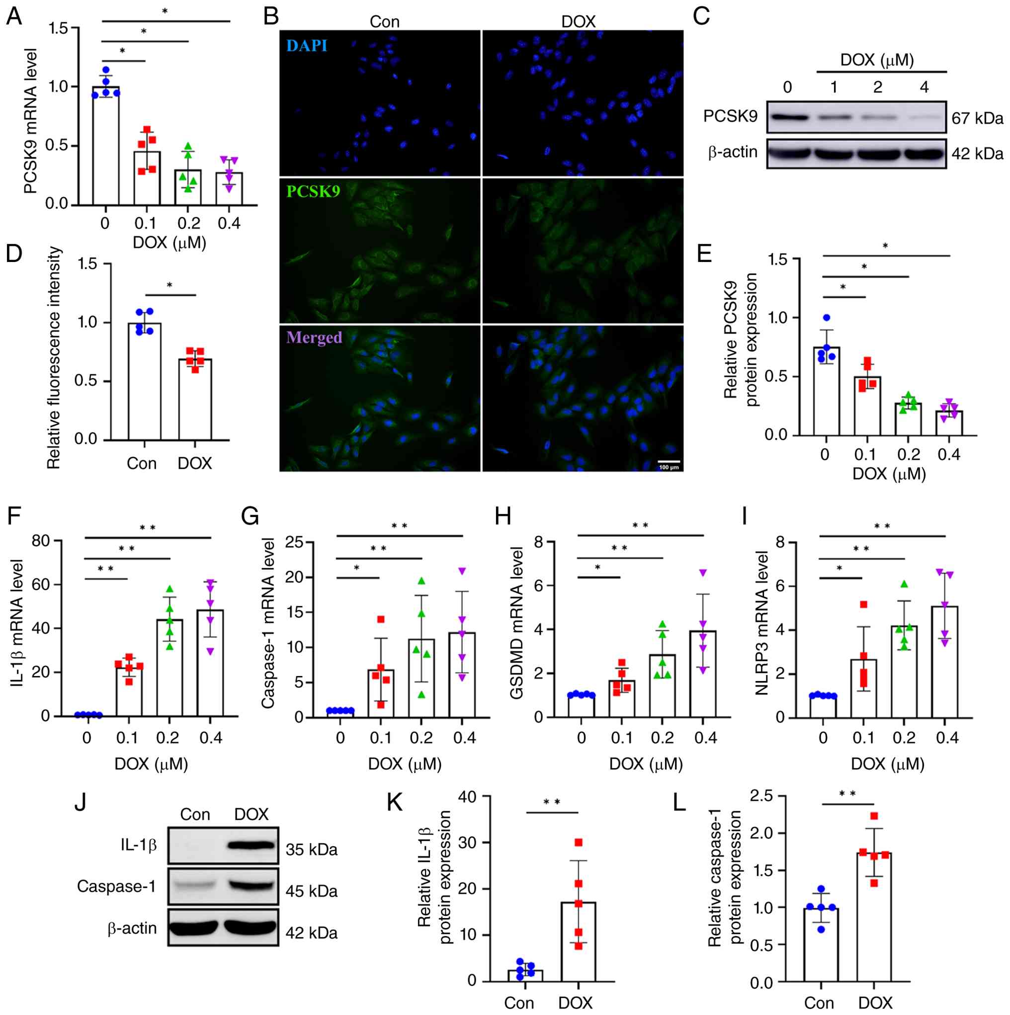
Further verification of the proteomics results by
immunofluorescence, qPCR and western blotting demonstrated that DOX
significantly reduced PCSK9 expression at both the mRNA and protein
levels in a dose-dependent manner (Fig. 2A-E). The proteomics analysis
initially identified IL-1β as a differentially expressed protein
associated with inflammation. The present study further evaluated
additional key pyroptosis-related markers including caspase-1,
GSDMD and NLRP3, based on their established roles as core
components of the canonical pyroptosis pathway downstream of IL-1β
signaling. Specifically, NLRP3 functions as an upstream
inflammasome sensor, caspase-1 serves as the executioner protease
that cleaves pro-IL-1β and pro-IL-18, and GSDMD is the pore-forming
effector that mediates membrane rupture and pyroptotic cell death
(30,31). Therefore, these molecules were
selected to comprehensively assess whether DOX induces pyroptosis
in AC-16 cells. In accordance with the proteomic sequencing
results, AC-16 cells were treated with different concentrations of
DOX, and the mRNA expression levels of IL-1β, caspase-1, GSDMD and
NLRP3 were determined. The mRNA expression levels of these genes
increased in a dose-dependent manner (Fig. 2F-I). Furthermore, the protein
expression levels of IL-1β and caspase-1 were determined by using
western blotting. The protein expression levels of IL-1β and
caspase-1 were significantly increased in AC-16 cells after
treatment with 0.2 µM DOX for 48 h, compared with that of control
cells (Fig. 2J-L).
Intervention with PCSK9 expression
alleviates DOX-induced pyroptosis
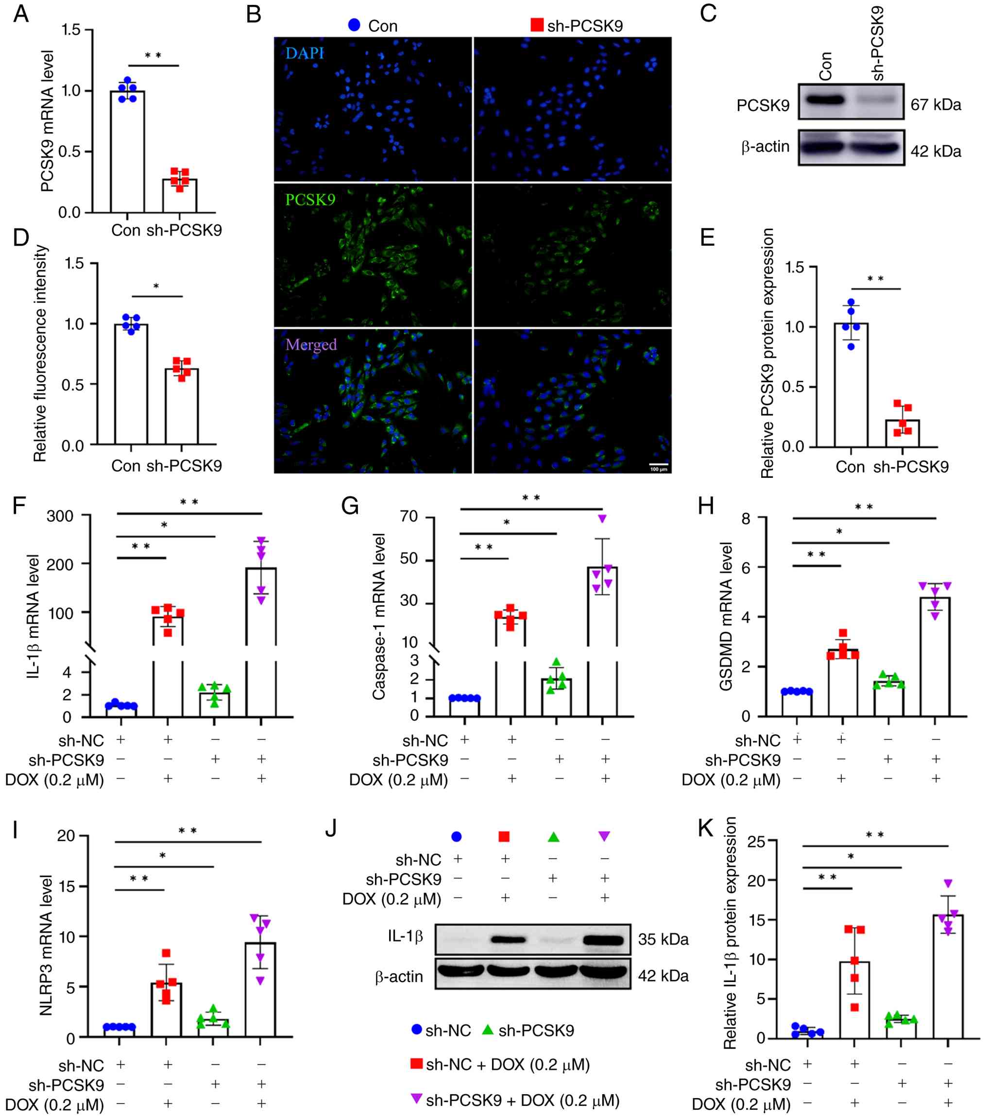
To explore the relationship between PCSK9- and
DOX-induced pyroptosis in cardiomyocytes, PCSK9-knockdown AC-16
cells were constructed by lentivirus transfection with shRNA
targeting the PCSK9 gene. The knockdown effect was verified by
determining PCSK9 mRNA and protein expression in cells. The
transcription and protein levels of PCSK9 were significantly
decreased in sh-PCK9 cells compared with control cells (Fig. 3A-E). The mRNA expression levels of
IL-1β, caspase-1, GSDMD and NLRP3 were markedly increased in the
sh-PCSK9 group compared with those in the sh-NC group, and the
increase was even more pronounced in the sh-PCSK9+DOX group
compared with the sh-NC+DOX group (Fig. 3F-I). Furthermore, the protein
expression levels of IL-1β were consistent with the changes in mRNA
expression in sh-PCSK9+DOX cells compared with sh-NC+DOX cells
(Fig. 3J-K). These results
suggested that knockdown of PCSK9 can exacerbate the increase in
myocardial pyroptosis induced by DOX.
PCSK9 knockout reduced the tolerance of mice to
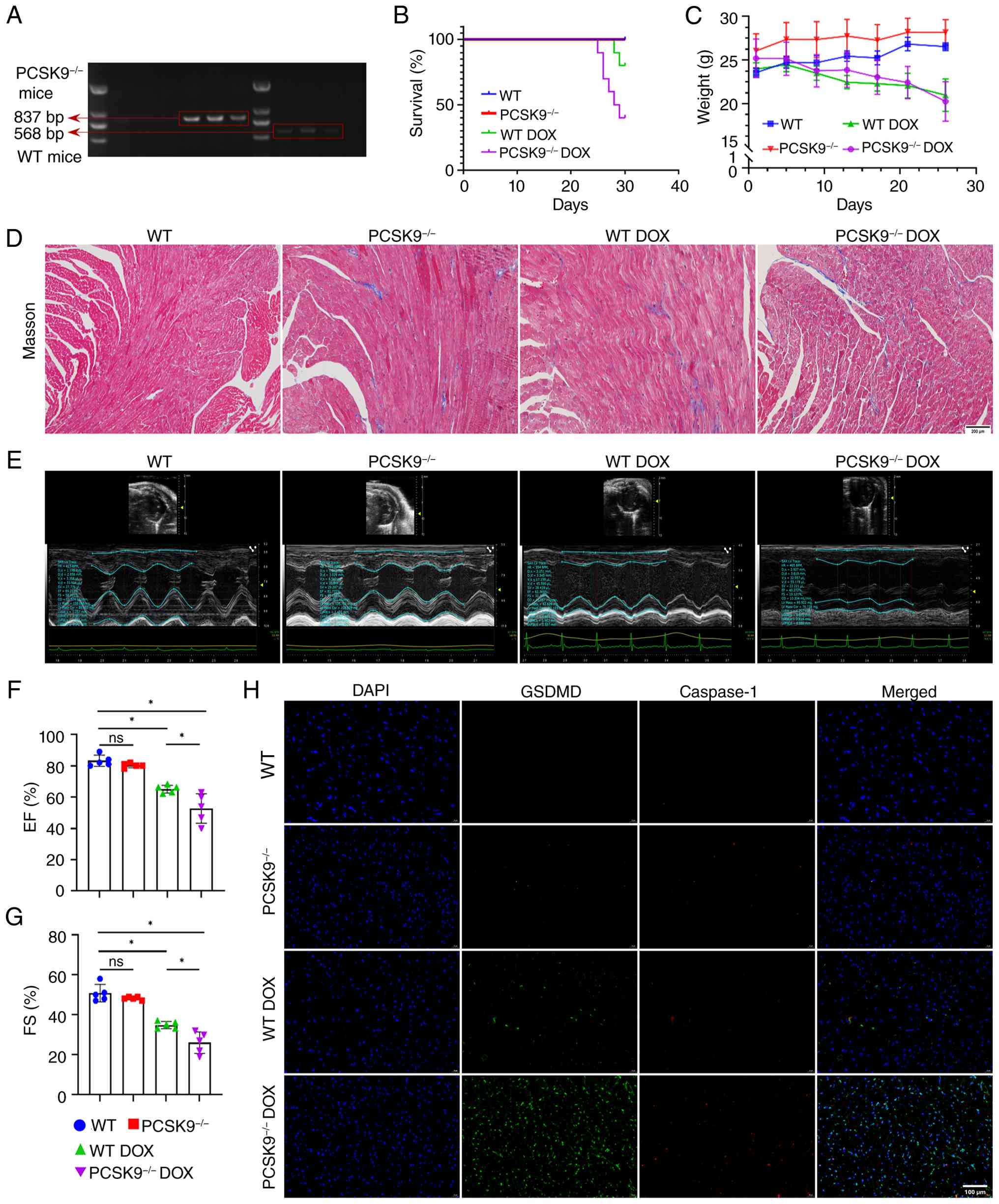
DOX. The in vivo effects of the present cell experiments
were further verified using PCSK9 knockout transgenic mice. Mouse
tail genotype identification demonstrated that WT mice presented a
single target band (568 bp), whereas PCSK9−/− mice
presented a mutant band (837 bp) (Fig.
4A). By the 30th day of modeling, the survival rate of the WT
mice decreased, and the survival rate of the PCSK9−/−
mice decreased markedly by >50% (Fig. 4B). Compared with the control group,
the mice in DOX modeling group showed dull hair, slow movement and
weight loss (Fig. 4C). Masson's
trichrome staining revealed that compared with those in the WT
group, the mice in the WT DOX group showed obvious fibrosis within
the myocardium (Fig. 4D).
Moreover, PCSK9-deficient mice exhibited more severe cardiac
fibrosis upon DOX challenge, as evidenced by the increased collagen
deposition in the PCSK9−/− DOX group compared with the
WT DOX group (Fig. 4D).
EF and FS are two important parameters commonly used
in echocardiography to evaluate the systolic function of the heart
(9). Compared with those in the WT
group, the EF and FS were decreased in the WT DOX group and were
further reduced in the PCSK9−/− DOX group (Fig. 4E-G). Consistent with the functional
deterioration observed in echocardiography, PCSK9 deficiency
exacerbated DOX-induced cardiac systolic dysfunction, leading to a
more pronounced decline in both ejection fraction and fractional
shortening. Compared with the WT group, the protein expression
levels of GSDMD and caspase-1 in the heart tissues of DOX-treated
WT mice (WT DOX group) were markedly elevated, but a notably
greater increase was observed in the PCSK9−/− DOX mice
compared with the WT DOX group (Fig.
4H). These results suggested that PCSK9 knockout renders mice
less tolerant to DOX-induced myocardial damage, possibly by
exacerbating pyroptosis-related inflammatory responses and cardiac
fibrosis.
Discussion
In current clinical practice, the cardiotoxicity
induced by chemotherapeutic agents remains an unavoidable and
challenging issue to address (32). DOX is a widely used anthracycline
chemotherapy drug (1). Although
the use of DOX has shown significant efficacy in antitumor
treatment, its clinical application is considerably restricted by
notable cardiotoxic reactions (33). In the present study, DOX was used
as a representative chemotherapeutic drug to establish a cell and
animal model of DOX-induced cardiomyocyte injury, and to further
investigate the roles of PCSK9 in DIC. The present results
demonstrated that targeting PCSK9 could effectively alleviate
DOX-induced cardiomyocyte pyroptosis and cardiac dysfunction.
It was found that DOX treatment significantly
reduced the viability of cardiomyocytes, and the cells underwent
obvious morphological changes, including balloon-like swelling and
vacuolar damage. A previous study has shown that DOX induces
cardiotoxicity through a variety of complex and interrelated
mechanisms (34). The classic
pathway involves the production of a large amount of reactive
oxygen species (ROS), leading to oxidative stress damage within
cardiomyocytes, which triggers DNA damage, lipid peroxidation and
mitochondrial dysfunction (35).
In addition to oxidative stress, DOX can directly induce apoptosis,
necrosis and inflammatory responses in cardiomyocytes and can
interfere with the transcription and repair processes of
cardiomyocytes by inhibiting topoisomerase IIβ (36–38).
In recent years, a study on the DIC have gradually shifted from
traditional mechanisms of oxidative stress and apoptosis to more
complex forms of cell death, such as pyroptosis (39). As a form of inflammatory cell
death, pyroptosis is characterized by the loss of membrane
integrity, cell swelling and the release of inflammatory factors
such as IL-1β and IL-18 (40). The
present study found that DOX could induce pyroptosis in AC-16
cardiomyocytes in a dose-dependent manner, which is consistent with
the findings of previous studies (25,41,42).
These multilevel toxicity mechanisms ultimately cause cardiomyocyte
death, cardiac function decline and potentially irreversible heart
failure.
Proteomic sequencing revealed that the expression of
SREBF and PCSK9, which are related to lipid metabolism, was
downregulated in cardiomyocytes treated with DOX. In recent years,
the role of PCSK9 in cardiovascular diseases has attracted
increasing attention (43).
Accumulating evidence has demonstrated that PCSK9 exerts
context-dependent dual effects on the cardiovascular system, which
are closely associated with experimental models, stimulating
factors, protein concentration, and the pathophysiological status
of the body. Several clinical and basic studies have demonstrated
that PCSK9 promotes the development of atherosclerosis via classic
lipid metabolic pathways (20,21,44,45).
Inhibiting PCSK9 expression or activity effectively reduces
circulating lipid levels and decreases the risk of coronary heart
disease and major adverse cardiovascular events, providing a
rationale for the clinical application of PCSK9 inhibitors in lipid
management (46).
A previous study reported that the levels of PCSK9
in the serum and hearts of mice with ischemia-reperfusion injury
and in the serum of patients with myocardial infarction are
elevated (47). Notably, excessive
PCSK9 expression significantly aggravates myocardial remodeling and
accelerates the progression of heart failure, indicating that PCSK9
serves a detrimental role in patients with myocardial injury under
pathological conditions such as chronic stress (48). Mechanistically, PCSK9 promotes
myocardial fibrosis and cardiac remodeling by activating the
TLR4/NF-κB/NLRP3 inflammatory signaling pathway and facilitating
the proliferation and excessive collagen deposition of cardiac
fibroblasts. Furthermore, PCSK9 also facilitates the fibrotic
process via the TGF-β1/Smad3 pathway, thereby further impairing
cardiac function (48,49).
PCSK9 can also exert noncanonical cardioprotective
effects within the myocardial microenvironment, in contrast to its
primarily atherogenic role in peripheral blood vessels. A recent
study confirmed that cardiomyocyte-specific PCSK9 deficiency leads
directly to abnormalities in the assembly and function of
mitochondrial respiratory chain complexes. This results in reduced
ATP production and increased ROS generation. It also triggers a
shift in myocardial metabolism toward glycolysis, as well as
cardiac hypertrophy, fibrosis and impaired cardiac function
(20). Whole-body PCSK9 knockout
leads to reprogrammed myocardial lipid metabolism, mitochondrial
dysfunction and abnormal accumulation of lipid droplets. This
ultimately results in heart failure with a preserved EF (21).
In the present study, the expression of PCSK9
decreased after cardiomyocytes were treated with DOX. These
findings reflect the discourse previously noted clinical research;
although PCSK9 inhibitors are widely used to treat
hypercholesterolemia, their role in cardiomyo-protection remains
controversial. Studies have shown that PCSK9 inhibitors can reduce
myocardial damage by lowering cholesterol levels and inhibiting
inflammatory responses (47),
whereas PCSK9 inhibitors may increase the risk of cardiovascular
events while lowering cholesterol levels (50). Therefore, the role of PCSK9 in
myocardial protection may be pleiotropic. PCSK9 protects
cardiomyocytes by regulating cholesterol metabolism and inhibiting
the inflammatory response; conversely, excessive inhibition of
PCSK9 may aggravate myocardial injury. However, how PCSK9 regulates
this process in the case of anthracycline-induced cardiomyocyte
death remains unclear.
Proteomic sequencing of cardiomyocytes treated with
DOX also revealed that the expression of IL-1β, which is related to
pyroptosis, was upregulated. Therefore, it was speculated that
PCSK9 may cause myocardial injury through IL-1β mediated pyroptosis
of myocardial cells. To further verify the role of PCSK9 in DIC,
models of AC-16 cells with reduced expression of PCSK9 and mice
with PCSK9 gene knockout were established. The results showed that
PCSK9 deficiency or deletion significantly aggravated DOX-induced
myocardial injury, as indicated by increased cardiomyocyte
pyroptosis and further deterioration of cardiac function.
A limitation of the present study is the lack of
rescue experiments (such as PCSK9 replenishment in
PCSK9−/− mice). However, the present loss-of-function
data from both in vitro knockdown and in vivo
knockout consistently demonstrated the protective role of
endogenous PCSK9 against DOX-induced pyroptosis and cardiac
dysfunction. DOX significantly downregulated the expression level
of PCSK9 in AC-16 cells in a dose-dependent manner. By using an
in vitro cardiomyocyte model and an in vivo PCSK9
gene knockout mouse model, it was verified that PCSK9 deletion
aggravated myocardial pyroptosis induced by DOX, emphasizing the
important role of PCSK9 in myocardial protection, and the specific
regulatory molecular mechanisms need to be further studied. The
results of the present study provided a novel perspective on the
mechanism of the DIC.
Based on these findings, the present study revealed
the protective role of PCSK9 in DIC. These results may have
considerable translational value and clinical implications; these
findings suggest that maintaining or moderately increasing the
expression or function of PCSK9 could be a new cardioprotective
strategy. In patients with cancer receiving anthracycline-based
chemotherapy, maintaining steady-state levels of PCSK9 in
cardiomyocytes during treatment may effectively reduce the
incidence of myocardial pyroptosis, delaying or even preventing the
onset of cardiac dysfunction and heart failure. Furthermore,
significant changes in PCSK9 expression levels during myocardial
injury suggested that PCSK9 may serve as a potential biomarker for
the early identification of individuals at high risk of
anthracycline-induced cardiotoxicity. Patients with low baseline
PCSK9 levels or a significant decrease in PCSK9 levels during
chemotherapy should undergo more frequent cardiac function
monitoring and receive cardioprotective interventions early if
necessary.
Supplementary Material
Supporting Data
Supporting Data
Acknowledgements
Not applicable.
Funding
The present study was sponsored by the Natural Science
Foundation of Henan (grant no. 202300410309), the National Natural
Science Foundation of China (grant no. 81902442), the Henan Medical
University Grant (grant no. XYBSKYZZ201828) and the Graduate
Education Reform Project of Henan Province (grant no.
2025SJGLX441Y).
Availability of data and materials
The mass spectrometry proteomics data generated in
the present study may be found in the ProteomeXchange Consortium
via the iProX partner repository (https://www.iprox.cn) under accession number PXD077062
or at the following URL: https://www.iprox.cn//page/project.html?id=IPX0016598000.
All other data generated in the present study may be requested from
the corresponding author.
Authors' contributions
CC and JZ conceived and designed the study. SH, XL,
GL, XX, CG and SL performed experiments and data analysis. XL
conducted a comprehensive secondary review of the manuscript and
participated in the supplementary experiments during the revision
process. JD provided critical reagents and participated in the
interpretation of western blotting and qPCR data. TZ contributed to
the interpretation of immunofluorescence staining and western
blotting results. XW contributed to the analysis of
echocardiography parameters (EF/FS) and data interpretation. JY
contributed to the interpretation of pyroptosis-related protein
expression data and made critical revisions to the manuscript for
important intellectual content. TZ, XW and JY supervised the
project and revised the manuscript. CC and JY confirm the
authenticity of all the raw data. All authors read and approved of
the final manuscript.
Ethics approval and consent to
participate
The present study was conducted with ethics approval
from the Ethics Committee of Xinxiang Medical University (currently
Henan Medical University; approval no. XYLL-20220192; Xinxiang,
China).
Patient consent for publication
Not applicable.
Competing interests
The authors declare that they have no competing
interests.
References
|
1
|
Gergely S, Hegedűs C, Lakatos P, Kovács K,
Gáspár R, Csont T and Virág L: High throughput screening identifies
a novel compound protecting cardiomyocytes from doxorubicin-induced
damage. Oxid Med Cell Longev. 2015:1785132015. View Article : Google Scholar : PubMed/NCBI
|
|
2
|
Bisht A, Avinash D, Sahu KK, Patel P, Das
Gupta G and Kurmi BD: A comprehensive review on doxorubicin:
Mechanisms, toxicity, clinical trials, combination therapies and
nanoformulations in breast cancer. Drug Deliv Transl Res.
15:102–133. 2025. View Article : Google Scholar : PubMed/NCBI
|
|
3
|
Newhouse R, Nelissen E, El-Shakankery KH,
Rogozińska E, Bain E, Veiga S and Morrison J: Pegylated liposomal
doxorubicin for relapsed epithelial ovarian cancer. Cochrane
Database Syst Rev. 7:CD0069102023.PubMed/NCBI
|
|
4
|
Zeng Y, Zhang X, Lin D, Feng X, Liu Y,
Fang Z, Zhang W, Chen Y, Zhao M, Wu J and Jiang L: A
lysosome-targeted dextran-doxorubicin nanodrug overcomes
doxorubicin-induced chemoresistance of myeloid leukemia. J Hematol
Oncol. 14:1892021. View Article : Google Scholar : PubMed/NCBI
|
|
5
|
Abdel-Daim MM, Kilany OE, Khalifa HA and
Ahmed AAM: Allicin ameliorates doxorubicin-induced cardiotoxicity
in rats via suppression of oxidative stress, inflammation and
apoptosis. Cancer Chemother Pharmacol. 80:745–753. 2017. View Article : Google Scholar : PubMed/NCBI
|
|
6
|
Xu R, Li T, Luo J, Zhang X, Wang T, Wang
Y, Ma Y, Yang B, Jia J, Dmytriw AA, et al: PCSK9 increases
vulnerability of carotid plaque by promoting mitochondrial
dysfunction and apoptosis of vascular smooth muscle cells. CNS
Neurosci Ther. 30:e146402024. View Article : Google Scholar : PubMed/NCBI
|
|
7
|
D'Onofrio N, Prattichizzo F, Marfella R,
Sardu C, Martino E, Scisciola L, Marfella L, Grotta R, Frige C,
Paolisso G, et al: SIRT3 mediates the effects of PCSK9 inhibitors
on inflammation, autophagy, and oxidative stress in endothelial
cells. Theranostics. 13:531–542. 2023. View Article : Google Scholar : PubMed/NCBI
|
|
8
|
Hamo CE, Bloom MW, Cardinale D, Ky B,
Nohria A, Baer L, Skopicki H, Lenihan DJ, Gheorghiade M, Lyon AR
and Butler J: Cancer Therapy-related cardiac dysfunction and heart
failure: Part 2: Prevention, treatment, guidelines, and future
directions. Circ Heart Fail. 9:e0028432016. View Article : Google Scholar : PubMed/NCBI
|
|
9
|
Antonini-Canterin F, Pavan D and Nicolosi
GL: Echocardiographic evaluation of the volumes and global systolic
function of the left ventricle. Ital Heart J Suppl. 1:1261–1272.
2000.(In Italian). PubMed/NCBI
|
|
10
|
Sperling W and Rafferty CN: Relationship
between absorption spectrum and molecular conformations of
11-cis-retinal. Nature. 224:590–594. 1969. View Article : Google Scholar : PubMed/NCBI
|
|
11
|
Seidah NG, Benjannet S, Wickham L,
Marcinkiewicz J, Jasmin SB, Stifani S, Basak A, Prat A and Chretien
M: The secretory proprotein convertase neural apoptosis-regulated
convertase 1 (NARC-1): Liver regeneration and neuronal
differentiation. Proc Natl Acad Sci USA. 100:928–933. 2003.
View Article : Google Scholar : PubMed/NCBI
|
|
12
|
Tremblay F, Xiong Q, Shah SS, Ko CW, Kelly
K, Morrison MS, Giancarlo C, Ramirez RN, Hildebrand EM, Voytek SB,
et al: A potent epigenetic editor targeting human PCSK9 for durable
reduction of low-density lipoprotein cholesterol levels. Nat Med.
31:1329–1338. 2025. View Article : Google Scholar : PubMed/NCBI
|
|
13
|
Dogan Z, Senyigit A, Durmus S, Duvarcı C,
Gelişgen R, Uzun H and Tabak O: The relationship between oxLDL,
sLOX-1, PCSK9 and carotid intima-media thickness in patients with
prediabetes and type 2 diabetes. Sci Rep. 15:45542025. View Article : Google Scholar : PubMed/NCBI
|
|
14
|
Li Z, Zhu L, Xu Y, Zhang Y, Liu Y, Sun H,
Li S, Wang M, Jiang T, Zhou J and Deng Q: Pleiotropic effects of
PCSK9 inhibitors on Cardio-cerebrovascular diseases. Biomedicines.
12:27292024. View Article : Google Scholar : PubMed/NCBI
|
|
15
|
Liu G, Yu X, Cui C, Li X, Wang T, Palade
PT, Mehta JL and Wang X: The pleiotropic effects of PCSK9 in
cardiovascular diseases beyond cholesterol metabolism. Acta Physiol
(Oxf). 241:e142722025. View Article : Google Scholar : PubMed/NCBI
|
|
16
|
Cui C, Yan A, Huang S, Chen Y, Zhao J, Li
C, Wang X and Yang J: PCSK9 manipulates lipid metabolism and the
immune microenvironment in cancer. Onco Targets Ther. 18:411–427.
2025. View Article : Google Scholar : PubMed/NCBI
|
|
17
|
Gu Y, Lin X, Dong Y, Wood G, Seidah NG,
Werstuck G, Major P, Bonert M, Kapoor A and Tang D: PCSK9
facilitates melanoma pathogenesis via a network regulating tumor
immunity. J Exp Clin Cancer Res. 42:22023. View Article : Google Scholar : PubMed/NCBI
|
|
18
|
Gao JJ, Wu FY, Liu YJ, Li L, Lin YJ, Kang
YT, Peng YM, Liu YF, Wang C, Ma ZS, et al: Increase of PCSK9
expression in diabetes promotes VEGFR2 ubiquitination to inhibit
endothelial function and skin wound healing. Sci China Life Sci.
67:2635–2649. 2024. View Article : Google Scholar : PubMed/NCBI
|
|
19
|
Vilella A, Bodria M, Papotti B, Zanotti I,
Zimetti F, Remaggi G, Elviri L, Poti F, Ferri N, Lupo MG, et al:
PCSK9 ablation attenuates Aβ pathology, neuroinflammation and
cognitive dysfunctions in 5XFAD mice. Brain Behav Immun.
115:517–534. 2024. View Article : Google Scholar : PubMed/NCBI
|
|
20
|
Laudette M, Lindbom M, Arif M, Cinato M,
Ruiz M, Doran S, Miljanovic A, Rutberg M, Andersson L, Klevstig M,
et al: Cardiomyocyte-specific PCSK9 deficiency compromises
mitochondrial bioenergetics and heart function. Cardiovasc Res.
119:1537–1552. 2023. View Article : Google Scholar : PubMed/NCBI
|
|
21
|
Da Dalt L, Castiglioni L, Baragetti A,
Audano M, Svecla M, Bonacina F, Pedretti S, Uboldi P, Benzoni P,
Giannetti F, et al: PCSK9 deficiency rewires heart metabolism and
drives heart failure with preserved ejection fraction. Eur Heart J.
42:3078–3090. 2021. View Article : Google Scholar : PubMed/NCBI
|
|
22
|
Xu Q, Zhao YM, He NQ, Gao R, Xu WX, Zhuo
XJ, Ren Z, Wu CY and Liu LS: PCSK9: A emerging participant in heart
failure. Biomed Pharmacother. 158:1141062023. View Article : Google Scholar : PubMed/NCBI
|
|
23
|
Fang G, Li X, Yang F, Huang T, Qiu C, Peng
K, Wang Z, Yang Y and Lan C: Amentoflavone mitigates
doxorubicin-induced cardiotoxicity by suppressing cardiomyocyte
pyroptosis and inflammation through inhibition of the STING/NLRP3
signalling pathway. Phytomedicine. 117:1549222023. View Article : Google Scholar : PubMed/NCBI
|
|
24
|
Zhu C, Xu S, Jiang R, Yu Y, Bian J and Zou
Z: The gasdermin family: Emerging therapeutic targets in diseases.
Signal Transduct Target Ther. 9:872024. View Article : Google Scholar : PubMed/NCBI
|
|
25
|
Zhang L, Jiang YH, Fan C, Zhang Q, Jiang
YH, Li Y and Xue YT: MCC950 attenuates doxorubicin-induced
myocardial injury in vivo and in vitro by inhibiting NLRP3-mediated
pyroptosis. Biomed Pharmacother. 143:1121332021. View Article : Google Scholar : PubMed/NCBI
|
|
26
|
Li M, Zhang Y, Wu B, Qiu R, Zhao C, Chen B
and Shang H: Optimizing dose selection for doxorubicin-induced
cardiotoxicity in mice: A comprehensive analysis of single and
multiple-dose regimens. Eur J Pharmacol. 1003:1778832025.
View Article : Google Scholar : PubMed/NCBI
|
|
27
|
Livak KJ and Schmittgen TD: Analysis of
relative gene expression data using real-time quantitative PCR and
the 2(−Delta Delta C(T)) method. Methods. 25:402–408. 2001.
View Article : Google Scholar : PubMed/NCBI
|
|
28
|
Sardão VA, Oliveira PJ, Holy J, Oliveira
CR and Wallace KB: Morphological alterations induced by doxorubicin
on H9c2 myoblasts: Nuclear, mitochondrial, and cytoskeletal
targets. Cell Biol Toxicol. 25:227–243. 2009. View Article : Google Scholar : PubMed/NCBI
|
|
29
|
Méry B, Guy JB, Vallard A, Espenel S,
Ardail D, Rodriguez-Lafrasse C, Rancoule C and Magné N: In vitro
cell death determination for drug discovery: A landscape review of
real issues. J Cell Death. 10:11796707176912512017. View Article : Google Scholar : PubMed/NCBI
|
|
30
|
Vande Walle L and Lamkanfi M: Drugging the
NLRP3 inflammasome: From signalling mechanisms to therapeutic
targets. Nat Rev Drug Discov. 23:43–66. 2024. View Article : Google Scholar : PubMed/NCBI
|
|
31
|
Man SM and Kanneganti TD: Converging roles
of caspases in inflammasome activation, cell death and innate
immunity. Nat Rev Immunol. 16:7–21. 2016. View Article : Google Scholar : PubMed/NCBI
|
|
32
|
Madrigal-Burgaleta R, Banerji A,
Vazquez-Revuelta P and Guyer AC: New approaches to chemotherapeutic
drug allergy: From drug challenge to desensitization. J Allergy
Clin Immunol Pract. 13:955–966. 2025. View Article : Google Scholar : PubMed/NCBI
|
|
33
|
Dempke WCM, Zielinski R, Winkler C,
Silberman S, Reuther S and Priebe W: Anthracycline-induced
cardiotoxicity-are we about to clear this hurdle? Eur J Cancer.
185:94–104. 2023. View Article : Google Scholar : PubMed/NCBI
|
|
34
|
Kciuk M, Gielecinska A, Mujwar S, Kolat D,
Kaluzinska-Kolat Z, Celik I and Kontek R: Doxorubicin-An agent with
multiple mechanisms of anticancer activity. Cells. 12:6592023.
View Article : Google Scholar : PubMed/NCBI
|
|
35
|
Hu Y, Yu J, He W, Qiao Y, Cheng X, Huang
H, Lai S, Yin D and He H: Astragaloside IV intervenes
multi-regulatory cell death forms against doxorubicin-induced
cardiotoxicity by regulating AMPKα2 pathway. Int Immunopharmacol.
142:1130782024. View Article : Google Scholar : PubMed/NCBI
|
|
36
|
Tian W, Zhang P, Yang L, Song P, Zhao J,
Wang H, Zhao Y and Cao L: Astragaloside IV alleviates
Doxorubicin-Induced cardiotoxicity by inhibiting cardiomyocyte
pyroptosis through the SIRT1/NLRP3 Pathway. Am J Chin Med.
52:453–469. 2024. View Article : Google Scholar : PubMed/NCBI
|
|
37
|
Ipek E and Tunca R: Silymarin protects
against doxorubicin induced cardiotoxicity by down-regulating
topoisomerase IIbeta expression in mice. Biotech Histochem.
98:412–423. 2023. View Article : Google Scholar : PubMed/NCBI
|
|
38
|
Yu J, Qi HL, Zhang H, Zhao ZY, Jing Z and
Nie ZY: Morin inhibits Dox-induced vascular inflammation by
regulating PTEN/AKT/NF-κB pathway. Inflammation. 45:2406–2418.
2022. View Article : Google Scholar : PubMed/NCBI
|
|
39
|
Zhang W, Wang X, Tang Y and Huang C:
Melatonin alleviates doxorubicin-induced cardiotoxicity via
inhibiting oxidative stress, pyroptosis and apoptosis by activating
Sirt1/Nrf2 pathway. Biomed Pharmacother. 162:1145912023. View Article : Google Scholar : PubMed/NCBI
|
|
40
|
Dai S, Chen Y, Fan X, Han J, Zhong L,
Zhang Y, Liu Q, Lin J, Huang W, Su L, et al: Emodin attenuates
cardiomyocyte pyroptosis in doxorubicin-induced cardiotoxicity by
directly binding to GSDMD. Phytomedicine. 121:1551052023.
View Article : Google Scholar : PubMed/NCBI
|
|
41
|
Chen XT, Yu YH, Du YH, Xu L and Meng XP:
MiR-145-5p attenuates Doxorubicin-induced heart injury through
targeting cardiomyocyte pyroptosis. Kaohsiung J Med Sci.
42:e701262026. View Article : Google Scholar : PubMed/NCBI
|
|
42
|
Huang X, Chen Y, Zhang Y, Hu X, Yang W,
Pan T, Gao W, Eom K, Chen P, Dong J and Li L: Pterostilbene
alleviates doxorubicin-induced cardiotoxicity by inhibiting
cardiomyocytes pyroptosis mediated by the
IL-6/STAT3-caspase-3/GSDME axis and M1 polarization of macrophages.
Pharmacol Res. 225:1081292026. View Article : Google Scholar : PubMed/NCBI
|
|
43
|
Ajoolabady A, Pratico D, Mazidi M, Davies
IG, Lip GYH, Seidah N, Libby P, Kroemer G and Ren J: PCSK9 in
metabolism and diseases. Metabolism. 163:1560642025. View Article : Google Scholar : PubMed/NCBI
|
|
44
|
Shin D, Kim S, Lee H, Lee HC, Lee J, Park
HW, Fukai M, Choi E, Choi S, Koo BJ, et al: PCSK9 stimulates Syk,
PKCδ, and NF-κB, leading to atherosclerosis progression
independently of LDL receptor. Nat Commun. 15:27892024. View Article : Google Scholar : PubMed/NCBI
|
|
45
|
Bao H, Wang X, Zhou H, Zhou W, Liao F, Wei
F, Yang S, Luo Z and Li W: PCSK9 regulates myofibroblast
transformation through the JAK2/STAT3 pathway to regulate fibrosis
after myocardial infarction. Biochem Pharmacol. 220:1159962024.
View Article : Google Scholar : PubMed/NCBI
|
|
46
|
Liu Y, Zhao Y, Feng P and Jiang H: PCSK9
inhibitor attenuates atherosclerosis by regulating
SNHG16/EZH2/TRAF5-mediated VSMC proliferation, migration, and foam
cell formation. Cell Biol Int. 47:1267–1280. 2023. View Article : Google Scholar : PubMed/NCBI
|
|
47
|
Li ZZ, Guo L, An YL, Yu WJ, Shi DY, Lin QY
and Zhang B: Evolocumab attenuates myocardial ischemia/reperfusion
injury by blocking PCSK9/LIAS-mediated cuproptosis of
cardiomyocytes. Basic Res Cardiol. 120:301–320. 2025. View Article : Google Scholar : PubMed/NCBI
|
|
48
|
Chung CC, Kao YH, Chen YC, Lin YK, Higa S,
Hsu KC and Chen YJ: PCSK9 enhances cardiac fibrogenesis via the
activation of Toll-like receptor and NLRP3 inflammasome signaling.
Int J Mol Sci. 26:19212025. View Article : Google Scholar : PubMed/NCBI
|
|
49
|
Huang Q, Zhou Z, Xu L, Zhan P and Huang G:
PCSK9 inhibitor attenuates cardiac fibrosis in reperfusion injury
rat by suppressing inflammatory response and TGF-β1/Smad3 pathway.
Biochem Pharmacol. 230:1165632024. View Article : Google Scholar : PubMed/NCBI
|
|
50
|
Steffens D, Bramlage P, Scheeff C, Kasner
M, Hassanein A, Friebel J and Rauch-Kröhnert U: PCSK9 inhibitors
and cardiovascular outcomes. Expert Opin Biol Ther. 20:35–47. 2020.
View Article : Google Scholar : PubMed/NCBI
|