Introduction
Hepatic fibrosis (HF) represents a maladaptive
wound-healing response triggered by recurrent hepatocyte injury,
leading to excessive extracellular matrix (ECM) deposition and its
aberrant distribution (1). This
process is a universal feature of chronic liver diseases and
constitutes the pivotal bridge linking persistent inflammation to
cirrhosis and, ultimately, hepatocellular carcinoma (2). Given the decisive influence of HF on
long-term prognosis, halting or reversing fibrogenesis is a
therapeutic priority. Viral, parasitic, chemical, pharmaceutical,
alcoholic and autoimmune insults can all incite necroinflammatory
cascades that converge on hepatic stellate cell (HSC) activation,
the central effector of ECM overproduction and progressive scarring
(3). Consequently, beyond
etiology-specific therapy, pharmacological reversal of fibrosis is
now regarded as the cornerstone of chronic liver disease
management. Substantial experimental evidence has confirmed that
suppression of HSC activation markedly attenuates fibrotic
progression (4).
The circadian clock is an evolutionarily conserved
endogenous timing system that coordinates physiology, metabolism
and behavior with the 24-h light/dark cycle (5). In mammals, the master pacemaker
resides in the suprachiasmatic nuclei (SCN) of the hypothalamus.
Light input via retinal photoreceptors entrains SCN neuronal
oscillators and, in turn, aligns subsidiary clocks in virtually
every peripheral tissue to the 24-h solar cycle (6). These peripheral oscillators operate
autonomously in organs such as the liver, kidney, pancreas,
skeletal and cardiac muscle; these peripheral clocks sustain
rhythmic gene expression through transcriptional-translational
feedback loops (7). Core
components include the heterodimeric transcription factors clock
circadian regulator (CLOCK) and basic helix-loop-helix
ARNT-like 1 (BMAL1), the period circadian protein homolog
(PER)1-3 and cryptochrome (CRY)1/2 proteins, and nuclear receptors
such as Rev-Erbα. The hepatic clock integrates systemic cues with
local metabolic demands (8). Its
architecture comprises input pathways (9), a central oscillator (comprising a
core loop and a stabilizing loop), and output arms that gate
liver-specific rhythms (10).
Disruption of this network has increasingly been recognized as a
pathogenic driver of hepatitis (11), steatosis (12), fibrosis (13), cirrhosis (14) and hepatocellular carcinoma
(15). As a basic
helix-loop-helix-PER-ARNT-SIM transcription factor, CLOCK
dimerizes with BMAL1 to drive rhythmic expression of
CLOCK-controlled genes governing metabolism, redox balance
and cell proliferation (16).
Rodent models bearing germline CLOCK mutations exhibit
dampened circadian gene expression, metabolic syndrome, cognitive
deficits and multi-organ pathology, including accelerated HF,
highlighting the non-redundant role of CLOCK in hepatic
homeostasis (17). Similarly,
genetic ablation of PER1/2 or BMAL1 perturbs hepatic
circadian function, resulting in systemic metabolic disturbances,
including fasting hyperglycemia, hyperlipidemia, hepatomegaly, and
progressive liver pathology characterized by cholangiocyte
hyperplasia, chronic inflammation, hepatocyte apoptosis and
collagen deposition (18).
Ginsenosides (GSS) are a standardized saponin-rich
extract predominantly derived from the roots of Panax
ginseng (19), and represent
one of the most pharmacologically important bioactive fractions of
this medicinal herb. The synergistic actions of these compounds
underlie a number of documented physiological effects, including
immunomodulation, anti-fatigue activity, neuroprotection, glucose
and blood pressure homeostasis, and potent antioxidant capacity.
Among these properties, the anti-fibrotic potential of individual
GSS has received notable attention; for example, Rg3 (20) suppresses HSC activation and
inflammatory cascades (21),
whereas Rg1 activates the Nrf2-ARE pathway to counteract oxidative
stress-driven ECM accumulation (22). Building on this foundation, the
present study aimed to systematically evaluate the therapeutic
impact of GSS in carbon tetrachloride (CCl4)-provoked
murine fibrosis and in TGF-β1-challenged LX-2 cells, while
assessing the functional involvement of the core circadian
transcription factor CLOCK in the fibrotic program, thereby
defining a previously uncharacterized chronotherapeutic axis in
fibrosis suppression.
Materials and methods
Chemicals and reagents
GSS was purchased from Shanghai Yuanye
Bio-Technology Co., Ltd. (UV ≥80%) (23). CCl4 solution was
obtained from Sinopharm Chemical Reagent Co., Ltd. Olive oil was
sourced from MilliporeSigma. Human TGF-β1 Recombinant Protein was
purchased from Wuhan Sanying Biotechnology. The selective CLOCK
inhibitor (CLK8) was purchased from MedChemExpress.
Animals
A total of 60 male C57BL/6 mice (age, 6–8 weeks;
weight, 18–22 g) were purchased from Shanghai Model Organisms
Center, Inc. The mice were acclimated under specific pathogen-free
conditions at Shanghai Hospital of Traditional Chinese Medicine
(Shanghai, China) under the following conditions: Adaptation period
of 1 week, ambient temperature maintained at 20–25°C, humidity at
40–70%, 12-h light/dark cycle, quiet environment, and free access
to food and water. All procedures conformed to National Standards
for Laboratory Animals of China and were approved by the Animal
Ethics and Welfare Committee of Shanghai Municipal Hospital of
Traditional Chinese Medicine (ethics approval no. 2025051;
Shanghai, China).
The mice were randomly assigned to six groups
(n=10/group): i) Control group (vehicle only), ii) CCl4
model group, iii) GSS low-dose group (50 mg/kg), iv) GSS
medium-dose group (100 mg/kg), v) GSS high-dose group (200 mg/kg)
and vi) resveratrol(Macklin Inc.) positive control group (30
mg/kg). Based on the preliminary experimental foundation of our
research group, the present study established three gradient doses
(low, medium and high) encompassing the effective concentrations of
GSS, aiming to comprehensively evaluate its dose-dependent effects.
HF was induced by intraperitoneal injection of 20% CCl4
in olive oil (v/v) twice weekly for 8 weeks in all groups except
for the control group. The control group received equivalent
volumes of olive oil on the same schedule. The GSS low, medium and
high dose groups were administered GSS via intraperitoneal
injection once daily for 4 weeks. The positive control group
received resveratrol diluted in normal saline to a final
concentration of 30 mg/kg via intraperitoneal injection once daily
for 4 weeks.
Cell culture and treatment
LX-2 human HSCs were purchased from Shanghai Zhong
Qiao Xin Zhou Biotechnology. Cells were maintained at 37°C in a 5%
CO2 incubator using high-glucose DMEM (Gibco; Thermo
Fisher Scientific, Inc.) supplemented with 10% fetal bovine serum
(Shanghai Yeasen Biotechnology Co., Ltd.). For experiments, LX-2
cells were seeded into 6-well plates at a density of
3×10⁵ well and allowed to adhere overnight; they were
then divided into the following sixdi) Control (untreated; 37°C),
ii) TGF-β1 (10 µg/ml; 6 h; 37°C), iii) TGF-β1 + GSS (10 µg/ml
TGF-β1 for 6 h followed by 1 mg/ml GSS for 24 h; 37°C), iv) CLK8 (5
µM; 12 h; 37°C), v) CLK8 + TGF-β1 (10 µg/ml TGF-β1 for 6 h followed
by 5 µM for 12 h; 37°C), vi) TGF-β1 + GSS + CLK8 (10 µg/ml TGF-β1
for 6 h; then 1 mg/ml GSS for 24 h; followed by 5 µM CLK8 for 12 h;
37°C). At the same time, to investigate cell activation conditions,
activation was modeled by treating HSCs with TGF-β1, TGF-β1 was
administered at concentrations of 0, 5, 10, 20 and 40 µg/ml and
treated for 0, 6, 12 and 24 h.
Measurement of serum levels of alanine
aminotransferase (ALT), aspartate aminotransferase (AST), alkaline
phosphatase (ALP), lactate dehydrogenase (LDH), laminin (LN),
hyaluronan (HA), Procollagen type III (PC III) and Procollagen type
IV (PC IV)
At the end of the 8-week experimental period, mouse
blood sampling was performed via retro-orbital bleeding. Each mouse
was subjected to blood collection (~0.2 ml), with the collected
volume not exceeding 20% of the total blood volume. The maximum
single blood collection volume was ~0.25 ml, as the total blood
volume of a 30 g mouse is ~1.5 ml. Anesthesia was induced by
intraperitoneal injection of 1% pentobarbital sodium (45 mg/kg),
corresponding to an injection volume of ~0.135 ml/30 g mouse.
Immediately after blood collection, sterile cotton balls were used
to achieve hemostasis by compression and antibiotic ophthalmic
ointment was applied. Subsequently, the mice were administered a
pentobarbital sodium (90 mg/kg) and euthanized by cervical
dislocation. The humane endpoints included body weight loss >20%
of the initial body weight, inability to consume food and water by
themselves, no mice required early euthanasia in the study. The
blood samples were kept undisturbed and allowed to clot at room
temperature for 30 min and centrifuged at 3,500 × g for 10 min at
4°C. Serum was then aliquoted and stored at −20°C for subsequent
analysis. Commercially available assay kits were used to measure
the serum levels of ALT (cat. no. BY-BC-K021), AST (cat. no.
BY-BC-K022), LDH (cat. no. BY-BC-K045) and ALP (cat. no.
BY-BC-K091) (Shanghai Baipu Biotechnology Co., Ltd.), and the serum
levels of LN (cat. no. BY-EM220216), HA (cat. no. BY-WJZF0179), PC
III (cat. no. BY-EM220067) and PC IV(cat. no. BY-EM228277) were
quantified using ELISA kits (Shanghai Baipu Biotechnology Co.,
Ltd.). All kits were used strictly according to the manufacturers'
protocols.
Histopathological examination
At the end of the 8-week experimental period, the
mice euthanized by cervical dislocation and tissues were harvested
from all liver lobes. The tissues were fixed in 4% paraformaldehyde
at room temperature for 24 h and paraffin-embedded sections were
prepared using conventional methods with a thickness of 5 µm. The
sections were then stained with hematoxylin and eosin (H&E),
Masson's trichrome and Sirius red dyes following standard
protocols: for H&E staining, the sections were stained with
0.5% hematoxylin at room temperature (25±2°C) for 5 min, followed
by differentiation and counterstaining with 0.5% eosin at room
temperature for 3 min. For Masson's trichrome staining, the
sections were stained with 0.1% hematoxylin at room temperature for
8 min, followed by staining with 0.5% Masson's trichrome dye at
room temperature for 15 min. for Sirius red staining, the sections
were stained with 0.1% Sirius red dye at room temperature for 30
min. After staining, all sections were observed for pathological
alterations and the degree of HF under a light microscope. The
stained areas were semi-quantitatively analyzed using ImagePro Plus
software (version 5.1; Media Cybernetics, Inc.).
Immunohistochemical staining
Liver tissue samples from mice were fixed in 4%
paraformaldehyde at 4°C for 24 h and routinely processed into
paraffin-embedded sections with a thickness of 5 µm. The sections
were first dewaxed with xylene (washing reagent) for 5 min twice,
followed by rehydration in a descending alcohol series (100%
ethanol for 3 min; 95% ethanol for 3 min; 80% ethanol for 3 min;
70% ethanol for 3 min) and rinsed with distilled water. For antigen
retrieval, the sections were immersed in citrate buffer (pH 6.0)
and heated in a water bath at 95–8°C for 20 min, then cooled to
room temperature naturally. Since the target antigens were
intracellular antigens, permeabilization was performed with 0.1%
Triton X-100 (permeabilization reagent) at room temperature for 10
min. Endogenous peroxidase activity was quenched with 3% hydrogen
peroxide in methanol for 10 min at room temperature. Subsequently,
the sections were blocked with 10% normal goat serum (Beijing
Solarbio Science & Technology Co., Ltd.) at room temperature
(25±2°C) for 30 min, then incubated overnight at 4°C with primary
antibodies diluted in antibody diluent containing 1% BSA
(MilliSigma). Subsequently, HRP-conjugated goat anti-rabbit IgG
secondary antibodies were applied at a dilution of 1:500 and
incubated at room temperature for 1 h. After chromogenic
development and counterstaining, the sections were mounted with
neutral balsam, avoiding bubble formation and observed under a
microscope for imaging.
Triple immunofluorescence
staining
LX-2 cells were seeded on glass coverslips at a cell
density of 5×10³ cells and fixed with 4%
paraformaldehyde in PBS for 10 min at room temperature,
permeabilized with 0.1% Triton X-100 (or 0.2% Tween-20) for 10 min
at room temperature, and blocked with 5% BSA + 0.1% Tween-20 at
room temperature (25±2°C) for 15 min. The cells were sequentially
incubated with primary antibodies against CLOCK (1:500; cat. no.
18094-1; Proteintech Group, Inc.), BMAL1 (1:500; cat. no. ab230822;
Abcam) and CRY2 (1:500; cat. no. 13997-1; Proteintech Group, Inc.)
at 4°C overnight. After washing with PBS, the cells were incubated
with fluorescent-conjugated goat anti-rabbit IgG secondary
antibodies (1:500; cat. no. ab150077; Abcam) for 1 h at room
temperature. HRP-conjugated secondary antibodies are not suitable
for immunofluorescence analysis (HRP is mainly used for chromogenic
reactions in immunohistochemistry), so fluorescent-conjugated
secondary antibodies were specifically used in this experiment to
ensure the accuracy and reliability of fluorescence imaging
results. Nuclei were stained with 1 µg/ml DAPI for 5 min at room
temperature and washed with PBS. Finally, the samples were mounted
with anti-fade mounting medium and imaged under a confocal
microscope.
Western blotting
Briefly ~100 mg snap-frozen liver tissue/sample was
homogenized in lysis buffer (cat. no. P0013B; Beyotime
Biotechnology) for thorough grinding and complete lysis. For
protein extraction from cells, the cells were washed twice with
pre-cooled PBS, then lysed with the same lysis buffer (cat. no.
P0013B; Beyotime Biotechnology) on ice for 30 min, followed by
centrifugation at 12,000 × g for 15 min at 4°C to collect the
supernatant (total cellular protein). Protein quantification was
performed using the BCA assay kit (cat. no. P0012; Beyotime
Biotechnology) and the protein concentration of each sample was
calculated for subsequent sample preparation. A total of 20 µg of
protein per lane was loaded for electrophoresis. The PAGE gel was
prepared using the Epizyme Fast PAGE Gel Preparation Kit (Ipsen
Pharma) according to the manufacturer's instructions. Subsequent
steps included electrophoresis, transfer onto a PVDF membrane
(polyvinylidene fluoride membrane), blocking with 5% BSA at room
temperature (25±2°C) for 1 h, followed by overnight incubation at
4°C with the following primary antibodies: Anti-α-smooth muscle
actin (α-SMA; cat. no. 19245; Cell Signaling Technology, Inc.),
anti-collagen type 1 (COL-1; cat. no. 67288; Proteintech Group,
Inc.), anti-CLOCK (cat. no. 18094-1-AP; Proteintech Group, Inc.),
anti-BMAL1 (cat. no. ab230822; Abcam), anti-CRY1 (cat. no. ab54649;
Abcam), anti-CRY2 (cat. no. 13997-1-AP; Proteintech Group, Inc.),
anti-PER1 (cat. no. 13463-1-AP; Proteintech Group, Inc.; all
1:1,000). In addition, the membranes were incubated for 1 h at room
temperature with anti-GAPDH (1:10,000; cat. no. 10494-1-AP;
Proteintech Group, Inc.). Finally, the membranes were developed
using the ECL chemiluminescence visualization reagent (cat. no.
P0018M; Beyotime Biotechnology), and grayscale analysis of the
bands was performed using ImageJ software (version 1.8.0_172;
National Institutes of Health).
Reverse transcription-quantitative PCR
(RT-qPCR)
Total RNA was extracted from mouse liver tissues and
LX-2 cells using the EZ-press RNA Purification Kit (cat. no.
B004DP; EZBioscience) according to the manufacturer's instructions.
No additional reagents or additives were used during RNA extraction
with the EZ-press RNA Purification Kit from EZBioscience. RNA
concentration and purity were measured with a NanoDrop 2000
instrument (NanoDrop; Thermo Fisher Scientific, Inc.). cDNA was
synthesized in a 20-µl RT reaction system using the ChamQ RT Master
Mix (cat. no. Q311-02; Vazyme Biotech Co., Ltd.). The temperature
protocol for RT was as follows: 42°C for 15 min (cDNA synthesis)
and 85°C for 5 sec (reverse transcriptase inactivation). qPCR
amplification was performed using ChamQ Universal SYBR qPCR Master
Mix (Vazyme Biotech Co., Ltd.) under the following cycling
conditions: Amplification: 95°C for 5 min, followed by 40 cycles at
95°C for 10 sec and 60°C for 30 sec; dissociation curve: 95°C for
15 sec, 60°C for 1 min and 95°C for 30 sec. The 2−ΔΔCq
method was used for the quantification of target gene expression
(24). We confirm that the primer
sequences for all target genes and internal reference gene are
accurately listed in Table I.
 | Table I.Primer sequences used for reverse
transcription-quantitative PCR. |
Table I.
Primer sequences used for reverse
transcription-quantitative PCR.
| Gene | Forward
(5′-3′) | Reverse
(5′-3′) |
|---|
| Human |
|
|
|
ACTIN (ACTB) |
CCACCATGTACCCTGGCATT |
GTCCTCGGCCACATTGTGAA |
|
CLOCK |
GATCACAGCCCAACCCCTTC |
CTGGGTGGAGTGCTCGTATC |
|
BMAL1 |
ACTGTGCTAAGGATGGCTGT |
GCTGCCCTGAGAATGAGGTG |
|
CRY1 |
CCTGGAATGCACCAGAAGGT |
TCCACTGCTGCTACAACCTG |
|
CRY2 |
CCAGCCAGGGAAACCATTCT |
CTCTGGGTCAAACCTCCCAC |
|
PER1 |
GACCCCTCATGCTGGCTATC |
ACAGAAGCGGATAGGGGAGT |
|
ACTA2d |
AAAAGACAGCTACGTGGGTGA |
GCCATGTTCTATCGGGTACTTC |
|
COL1A1 |
TCTGGAGCAAGTGGTGAACG |
CCCCTCACGTCCAGATTCAC |
| Mouse |
|
|
|
ACTIN |
TGAGCTGCGTTTTACACCCT |
TTTGGGGGATGTTTGCTCCA |
|
CLOCK |
CATCAACTCAGCAGAGTCAACA |
AGGCTGGGAAATCACCATCG |
|
BMAL1 |
AAAGATGGGGCTGGACGAAG |
ACCCGTATTTCCCCGTTCAC |
|
CRY2 |
TGGACAAGCACTTGGAACGGAAG |
GTAGAAGAGGCGGCAGGAGAGG |
|
α-SMA |
GTCCCAGACATCAGGGAGTAA |
CGGATACTTCAGCGTCAGGA |
|
COL1A1 |
GAGACAGGCGAACAAGGTGA |
GGGAGACCGTTGAGTCCATC |
Transcriptome sequencing
Total RNA was isolated from LX-2 cells using the
RNeasy Mini Kit (Cat. no. 74104; Qiagen), followed by on-column
DNase digestion and eukaryotic mRNA was enriched using magnetic
beads with Oligo (dT) included in the NEBNext Ultra RNA Library
Prep Kit for Illumina (cat no. E7530L; New England BioLabs, Inc.).
For prokaryotic samples, prokaryotic ribosomal RNA (rRNA) was
removed using the Ribo-Zero rRNA Removal Kit (Bacteria) (cat. no.
MRZB12424, Illumina, Inc.) to enrich mRNA). Fragmentation reagents
were added to decompose the mRNA into small fragments, and these
cleaved mRNAs were used as a template for first-strand cDNA
synthesis with random hexamer primers. Subsequently, a
second-strand synthesis reaction system was used to produce
double-stranded cDNA, which was purified using the purification
module included in the NEBNext library preparation kit. The
purified double-stranded cDNA was subjected to end repair,
A-tailing and adapter ligation. Subsequently, size selection was
performed, followed by PCR amplification. The quality and integrity
of the final sequencing library were verified using the Agilent
2100 Bioanalyzer (Agilent Technologies, Inc.) to confirm the
fragment size distribution and library quality. The quantified
library was normalized to a loading concentration of 2 nM, and
sequenced on the Illumina HiSeq X-ten platform (Illumina, Inc.).
The sequencing was performed in paired-end mode, with a read length
of 150 bp for each end. To ensure the quality of the data, before
performing bioinformatics analyses, FASTQC (http://www.bioinformatics.babraham.ac.uk/projects/fastqc/)
was used to evaluate the raw data, and poor quality data at the
first reading were eliminated by pre-processing. Analyses were then
performed, including gene expression determination, gene structure
refinement, variable splice detection, identification of new
transcripts, coding potential prediction and single nucleotide
polymorphism detection. Specifically, clean reads were aligned to
the reference genome using HISAT2 (v2.2.1; http://ccb.jhu.edu/software/hisat2/index.shtml),
and transcript assembly and expression quantification were
performed using StringTie (v2.2.1; http://ccb.jhu.edu/software/stringtie/). Differently
expressed genes (DEGs) between the treatment and control groups of
LX-2 cell samples were identified from the gene expression results.
Genes with |log2 fold change|≥1 and
Benjamini-Hochberg-adjusted P-value (false discovery rate) <0.05
were considered statistically significant. Based on these DEGs,
cluster analysis, Gene Ontology (GO) functional enrichment analysis
and pathway enrichment analysis were performed. A negative binomial
distribution test was used to assess the significance of
differences between readings and counts, and the baseline mean was
used to estimate the level of gene expression.
Molecular docking analysis
To investigate the binding potential between the
active ingredients of GSS and the core target CLOCK protein,
molecular docking analysis was performed following a standard
protocol. Briefly, 8 major active ingredients of GSS, including MC,
Rb1, Rb2, Rg1, Rg3, Rg5, Rh1 and Rh2, were selected as the docking
ligands based on our component characterization results. The 2D
structural files of these small-molecule ligands were downloaded
from the PubChem database (https://pubchem.ncbi.nlm.nih.gov/). Subsequently, the
2D structures were converted to optimized 3D structures, and the
protonation state of each ligand was adjusted and optimized using
OpenBabel (version 3.1.1; http://openbabel.org/). For the target protein
preparation, the crystal structure of the human CLOCK protein was
downloaded from the RCSB Protein Data Bank (PDB ID: 4h10;
http://www.rcsb.org/), which was preprocessed to
remove redundant water molecules and heteroatoms before docking.
The protein-ligand blind docking was performed using the CB-Dock2
web server (version 1.0; http://cadd.labshare.cn/cb-dock2/), a comprehensive
online tool that integrates structure-based docking and
template-based docking to improve the accuracy of binding site
prediction and binding conformation prediction. Default parameters
of the web server were adopted for the whole docking analysis.
After the docking calculation, the binding affinities of each
ligand with the CLOCK protein were calculated via the built-in
AutoDock Vina scoring function, with the unit of kcal/mol. Finally,
the docking results were visualized using PyMOL software (version
2.5.0; Schrödinger, LLC; http://pymol.org/2/), to generate the protein-ligand
interaction diagrams and binding heatmap.
Statistical analysis
All experiments were independently repeated at least
three times with consistent results, and all statistical analyses
were performed based on the biological replicates. Experimental
data were analyzed using ImageJ and GraphPad Prism 9.0 (Dotmatics),
and are presented as the mean ± SD. For normally distributed data,
significant differences were determined by t-test (for comparisons
between two groups) or one-way analysis of variance with Tukey's
test (for comparisons among multiple groups). P<0.05 was
considered to indicate a statistically significant difference.
Results
GSS improves liver function and HF in
a CCl4-induced model
A murine model of HF was established by induction
with CCl4 to evaluate the extent of liver injury and
fibrosis, as well as the reversive effect of GSS treatment. GSS was
administered at gradient concentrations to establish the
dose-response relationship and to determine the optimal therapeutic
dosage. In addition, resveratrol was selected as the positive
control drug based on our preliminary findings, aiming to validate
the successful establishment of the experimental model and enhance
the credibility of the results. After 8 weeks, liver tissue samples
were collected for histopathological examination using H&E,
Masson's trichrome and Sirius red staining. After GSS
administration, these pathological and biochemical abnormalities
were markedly ameliorated in a concentration-dependent manner.
Low-dose GSS partially alleviated inflammatory infiltration and
reduced collagen deposition by ~25%. Medium-dose GSS achieved more
prominent therapeutic effects, with obviously recovered
hepatocellular structure, 50% reduction in fibrotic area. Notably,
high-dose GSS exhibited a robust hepatoprotective and anti-fibrotic
effect, which was comparable to that of the positive control
resveratrol, the liver lobular structure was nearly restored to
normal, the collagen deposition was largely resolved with only mild
residual fibrosis (Fig. 1A). Serum
levels of hepatic function and fibrosis markers were also measured,
including AST, ALT, ALP, LDH, HA, LA, PC III and PC IV. Low-dose
GSS accompanied by a mild reduction in serum injury and fibrosis
markers. Medium-dose GSS significantly downregulated serum
biomarker levels. Notably, the serum levels of all detected hepatic
function and fibrosis markers were significantly reversed in
high-dose GSS, close to the levels of the normal control group
(Fig. 1B). The results
demonstrated that GSS effectively attenuated hepatic inflammation,
improved liver function and ameliorated HF in model mice in a
concentration-dependent manner, exhibiting notable hepatoprotective
effects.
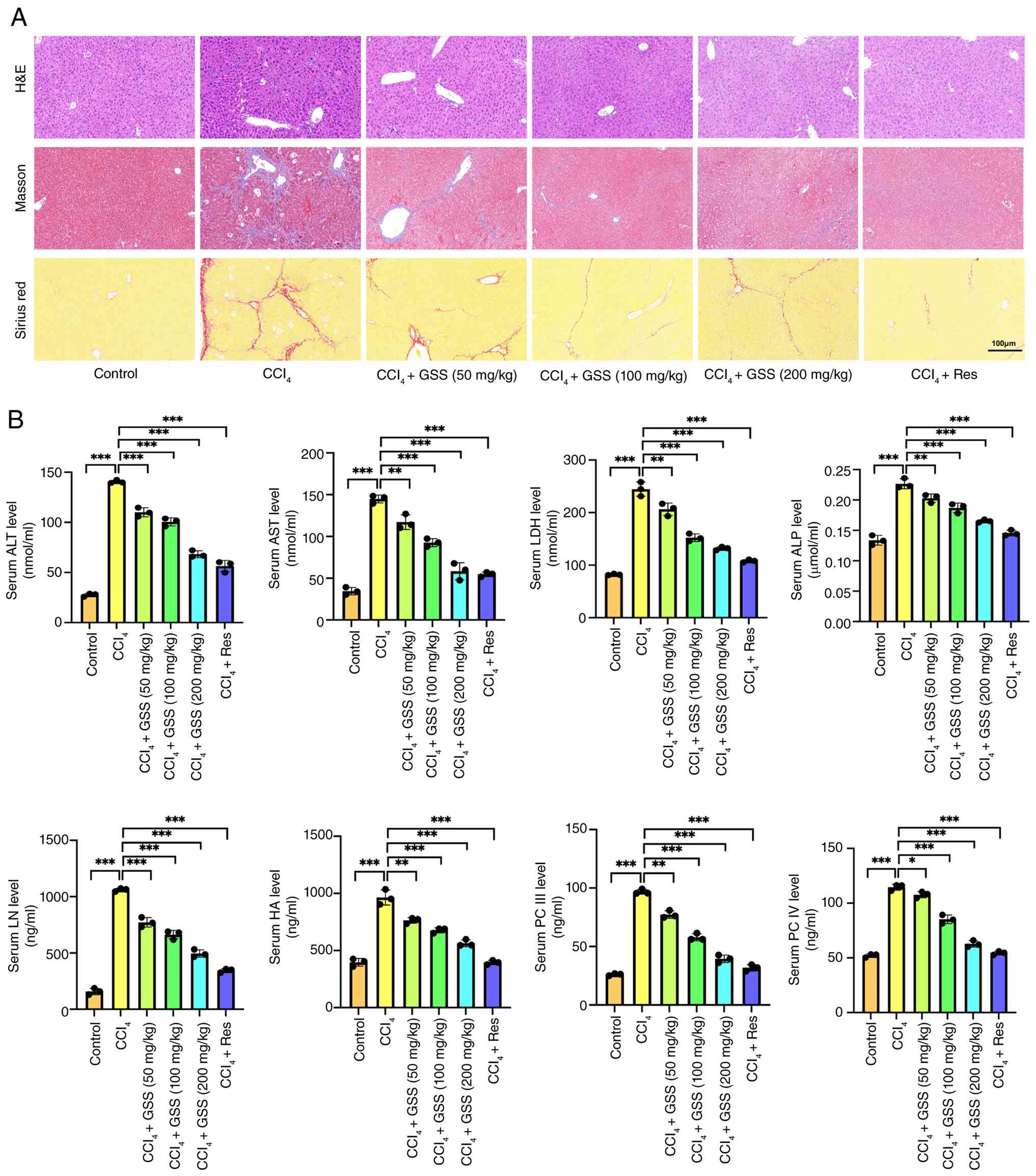 | Figure 1.GSS improves liver function and
hepatic fibrosis levels in CCl4-induced modeling. GSS
alleviated the progression of liver fibrosis in mice with
CCl4-induced modeling. (A) Histological examination of
mouse liver tissues. (B) Serum parameters of liver function and
fibrosis in mice. Compared with the control group (within the
normal range of serum levels), the serum levels of liver function
and hepatic fibrosis indices were significantly elevated in the
model group mice, while the ginsenoside group dose-dependently
reversed the serum levels of liver function and hepatic fibrosis in
mice. Data are presented as the mean ± SD from three independent
replicates. *P<0.05, **P<0.01, ***P<0.001. ALP, alkaline
phosphatase; PC, procollagen; ALT, alanine aminotransferase; AST,
aspartate aminotransferase; CCl4, carbon tetrachloride;
GSS, ginsenosides; H&E, hematoxylin and eosin; HA, hyaluronan;
LDH, lactate dehydrogenase; LN, laminin; Res, resveratrol. |
GSS reverses CCl4-induced
expression of α-SMA and COL-1 in model mice
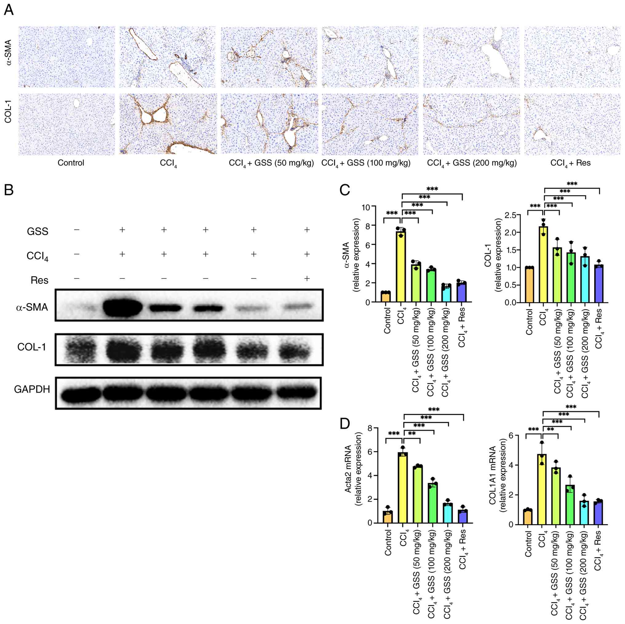
α-SMA and COL-1, key indicators of HF, were examined
in the present study, since α-SMA serves as a marker of activated
HSCs (25) and COL-1 is a major
component of the ECM; elevated expression of both is closely
associated with fibrosis progression. Therefore,
immunohistochemical analysis (Fig.
2A), western blotting (Fig. 2B and
C) and RT-qPCR (Fig. 2D) were
performed on liver tissues to assess the protein and mRNA
expression levels of α-SMA and COL-1. The CCl4-treated
group exhibited increased protein and mRNA expression levels of
α-SMA and COL-1, whereas intervention with different concentrations
of GSS exhibited a dose-dependent reduction on the expression
levels of α-SMA and COL-1. Notably, high concentrations of GSS and
treatment with resveratrol demonstrated the strongest antifibrotic
effect. These results indicated that CCl4 resulted in
notable progression of HF, whereas GSS effectively inhibited the
development of fibrosis.
GSS suppresses TGF-β1-induced
activation of HSCs
HSC activation is a critical driver of HF; the
process begins with HSC transformation into myofibroblasts, a key
event leading to ECM deposition and liver damage (4). To investigate this, activation was
modeled by treating HSCs with TGF-β1, and monitoring α-SMA and
COL-1 expression. TGF-β1 was administered at concentrations of 0,
5, 10, 20 and 40 µg/ml (Fig. 3A and
B), and the optimal concentration of 10 µg/ml was used to
stimulate HSCs for 0, 6, 12 and 24 h (Fig. 3C and D). The results showed that
TGF-β1 at 10 µg/ml induced maximum activation of HSCs after 6 h of
stimulation, with significant pro-fibrotic effects observed. Under
these conditions, HSCs were markedly activated, and the protein and
mRNA expression levels of α-SMA and COL-1 were considerably
increased. This effect was effectively reversed by GSS treatment
(Fig. 3E-G).
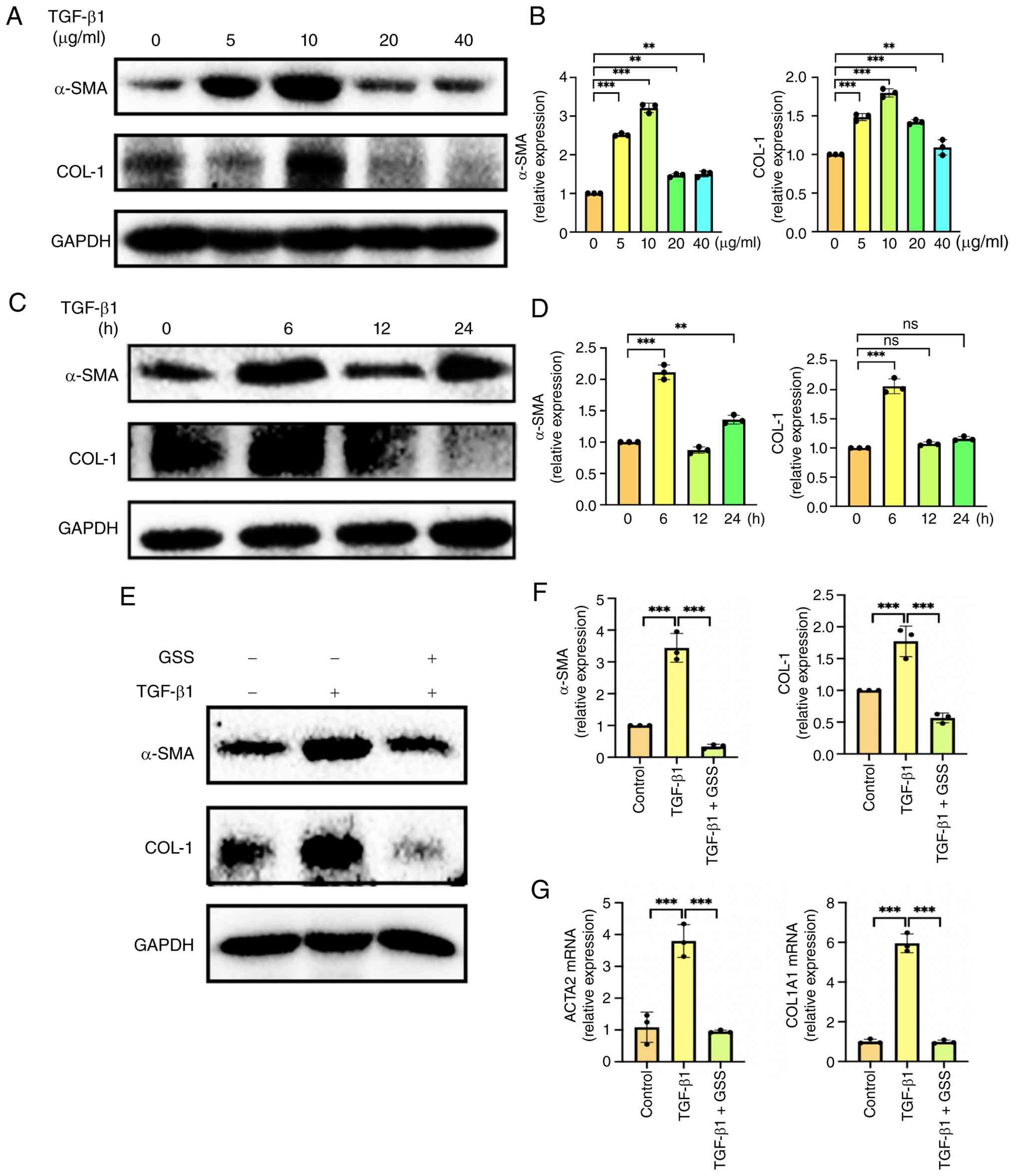 | Figure 3.GSS suppresses TGF-β1-induced
activation of LX-2 cells. (A) The concentration conditions of
TGF-b1-induced cell activation. LX-2 cells were treated with
increasing concentrations (0, 5, 10, 20 and 40 µg/ml) of TGF-β1.
(B) Dose-dependent expression of α-SMA and COL-1 proteins in
TGF-β1-induced cell activation. At a TGF-β1 stimulation
concentration of 10 µg/ml, the protein expression of α-SMA and
COL-1 was most significant compared with the unstimulated group.
(C) Time-course of TGF-b1-induced cell activation. LX-2 cells were
treated with 10 µg/ml TGF-β1 for 0, 6, 12 and 24 h. (D) Time-course
analysis of α-SMA and COL-1 protein expression in TGF-b1-induced
cell activation. Hepatic stellate cell activation was induced by
TGF-b1 stimulation at 10 mg/ml for 6 h. (E) Western blot analysis
of α-SMA and COL-1 in activated cells. (F) The protein and mRNA
expression levels of α-SMA and COL-1 in response to TGF-β1 and GSS
treatment. (G) mRNA expression levels of α-SMA and COL-1 in
response to TGF-β1 and GSS treatment. Data are presented as the
mean ± SD from three independent replicates. **P<0.01,
***P<0.001. α-SMA, α-smooth muscle actin; COL-1, collagen type
1; GSS, ginsenosides; ns, not significant. |
Investigation of the targets
underlying the anti-fibrotic effects of GSS
To delineate the role of circadian clock genes in
the anti-fibrotic mechanism of GSS, RNA sequencing (RNA-seq) was
conducted on LX-2 cells. The cells were split into the following
treatment groups: Control, TGF-β1 (model) and TGF-β1 + GSS
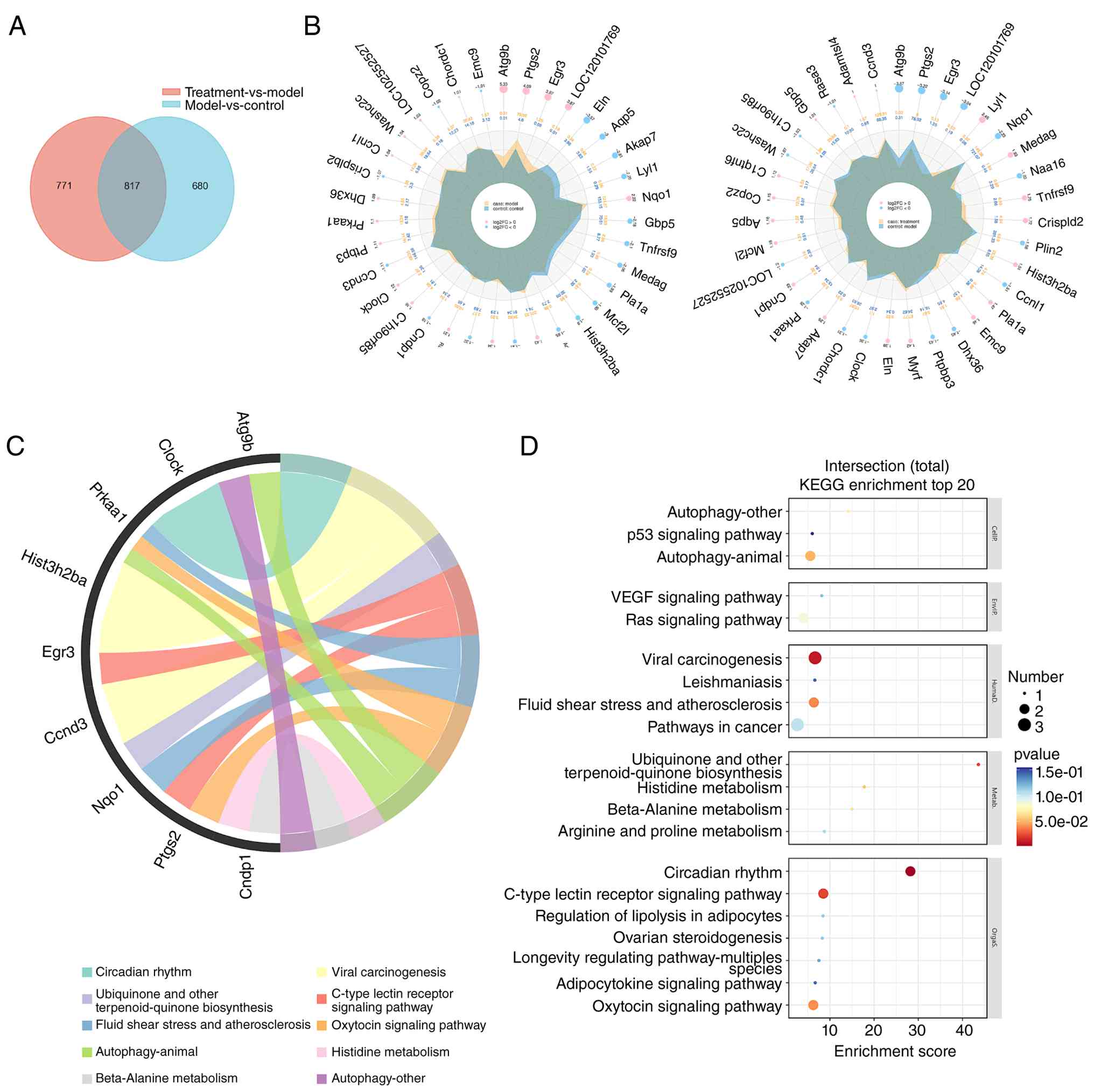
(treatment). Analysis of 2,268 genes revealed 817 genes were
differentially expressed between the groups (Fig. 4A). Subsequent analysis of the top
significantly upregulated and downregulated DEGs across the two
comparisons, visualized via radar maps, confirmed that the CLOCK
gene was significantly upregulated in the model group compared with
the control group. Notably, CLOCK was significantly downregulated
in the GSS treatment group compared with the model group; this
expression trend was fully consistent with our prior phenotypic
observations of CLOCK protein expression in preliminary
experiments, which validated the regulatory effect of GSS on CLOCK
at the protein level (Fig. 4B).
Analysis of the transcriptome by intensive GO analysis showed a
significant enrichment of CLOCK in ‘Circadian rhythm’, and
the top 10 pathways are shown in Fig.
4C. Kyoto Encyclopedia of Genes and Genomes (KEGG) pathway
analysis further supported this, showing the ‘Circadian rhythm’
pathway with an enrichment score close to 30, with the size of the
bubble indicating a substantial number of associated genes,
underscoring its importance in the mechanism of GSS; the top 20
terms are shown in Fig. 4D. The
GSEA results for the CLOCK-BMAL1 transcription complex are also
presented in the supplementary Fig.
S1.
Interaction potential between active
constituents of GSS and the core circadian regulator CLOCK
Based on RNA-seq results, CLOCK was shown to be a
putative central target for GSS. Molecular docking was subsequently
performed to validate interactions between CLOCK and key active
constituents of GSS. The CB-Dock2 web server (version 1.0;
http://cadd.labshare.cn/cb-dock2/)
(26) was employed to as this tool
leverages integrated structural models to accurately predict the
binding conformation and interaction state of the target CLOCK
protein with the input small-molecule ligands. From the mass
spectrometry data of GSS performed in the referenced study
(27), several major active
ingredients of GSS were screened and molecular docking of these
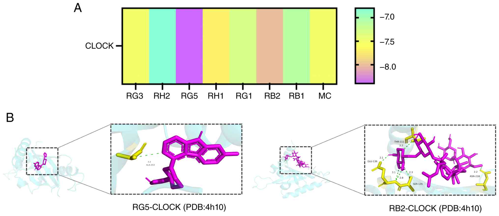
active components was performed with the CLOCK protein. Among them,
two GSS active ingredients, RB2 and RG5, exhibited high binding
affinities, with binding scores of −8.0 kcal/mol and −8.4 kcal/mol
(Fig. 5A and B). The docking
results for the remaining six GSS active ingredients with CLOCK are
shown in Fig. S2.
GSS modulates circadian clock gene
expression in vivo and in vitro
To further investigate alterations in hepatic
circadian clock genes during fibrogenesis, the expression levels of
these genes in mouse liver samples and cultured cells were
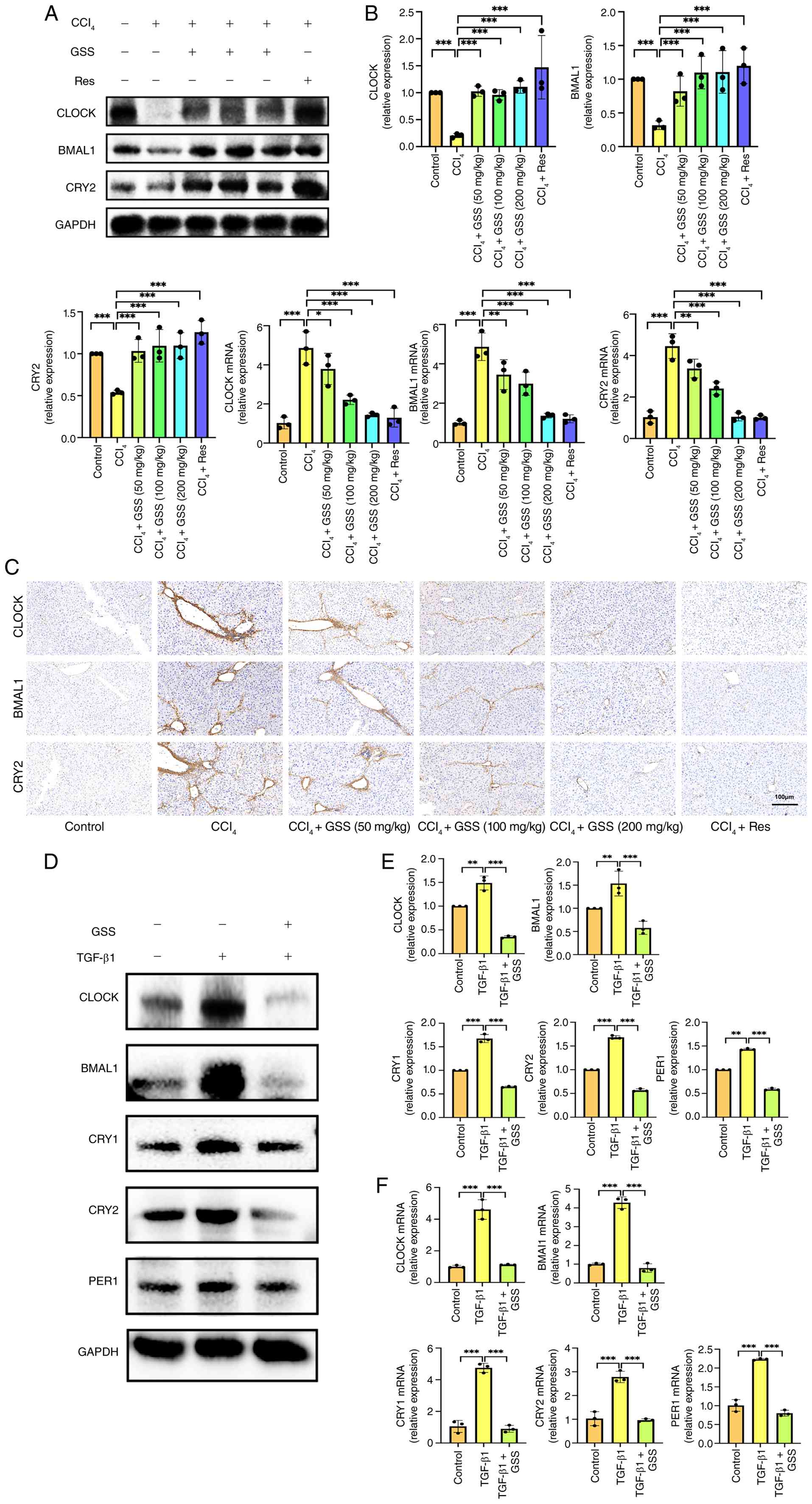
evaluated. In CCl4-induced fibrotic mice, the protein
expression of CLOCK, BMAL1 and CRY2 was significantly
downregulated, whereas their mRNA expression showed upward trend,
both protein and mRNA expression were reversed upon GSS
intervention (Fig. 6A and B). The
differential expression of circadian clock genes at the protein and
mRNA levels reflects the dynamic nature of circadian rhythmic
expression. As the peak times of mRNA and protein differ, their
expression inevitably exhibits an inherent phase separation. The
results of immunohistochemical analysis showed a consistent trend
with the pathological staining results of mouse liver (Fig. 6C). Furthermore, it was observed
that circadian gene expression was altered in activated HSCs.
Previous studies have shown that circadian disorders due to
prolonged jet lag or shift work are a direct risk factor for
diseases such as obesity and metabolic syndrome, and are associated
with increased incidence of non-alcoholic fatty liver disease
(NAFLD) (28) and related fibrosis
(29). In accordance with this
view, the present in vitro experiments demonstrated that the
protein and mRNA expression levels of CLOCK, BMAL1, CRY1, CRY2 and
PER1 was significantly upregulated by TGF-β1, thus indicating HSC
activation, whereas they were downregulated by GSS treatment
(Fig. 6D-F). These results
suggested that the anti-fibrotic effect of GSS may be associated to
the correction of circadian disorders.
Role of the CLOCK inhibitor in the
anti-fibrotic mechanism of GSS
Building on the findings of the current study (that
CLOCK is a target gene in the GSS treatment of HF), this
role was validated using the CLOCK inhibitor CLK8. CLK8 was
administered simultaneously in the control group, the model group
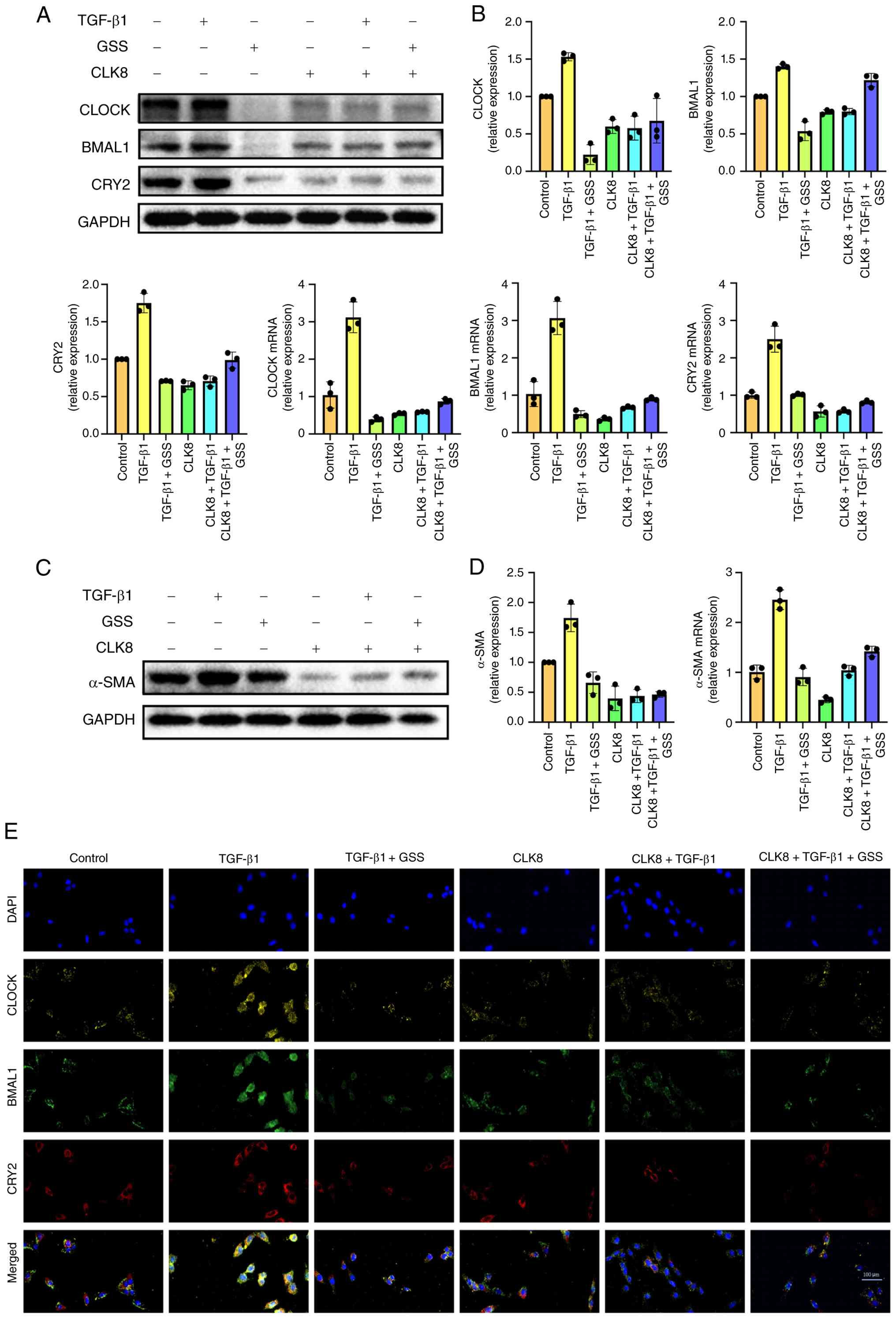
(TGF-β1) and the GSS treatment group. The results demonstrated that
CLK8 treatment (vs. the TGF-β1 model group) reduced the expression
of core circadian genes (including CLOCK, BMAL1 and CRY2) at both
the protein and mRNA levels (Fig. 7A
and B). However, Compared with the TGF-β1 + GSS group, the
TGF-β1 + GSS + CLK8 group showed an upward trend, indicating that
the combined use of CLK8 led to a relative increase in the
expression of these circadian rhythm genes. Based on the above
results, inhibition of Clock expression effectively restores the
oscillation of circadian rhythms, maintains circadian clock
stability, and significantly suppresses the progression of hepatic
fibrosis, this also effectively validated that targeting the clock
gene effectively inhibits the development of hepatic fibrosis.
The present study further examined the effect of
clock inhibitors on α-SMA protein expression. The experimental
results showed that, compared with the TGF-β1 + GSS group, α-SMA
protein expression in the TGF-β1 + GSS + CLK8 group exhibited a
downward trend, indicating that CLK8 inhibited α-SMA protein
expression (Fig. 7C and D).
Immunofluorescence analysis confirmed that in HSCs activated by
TGF-β1, the phenomenon of enhanced fluorescence intensity of
circadian rhythm proteins was weakened after GSS intervention or
CLK8 treatment (Fig. 7E).
Discussion
The circadian clock system is an intrinsic
timekeeping mechanism capable of harmonizing various physiological
processes (30). The positive
regulators BMAL1 and CLOCK are at the core of this
system and form a heterodimer that activates the transcription of
rhythm genes, such as PER, CRY and Rev-erbα (Rev-erbα
is encoded by the NR1D1 gene). Subsequently, the negative
regulators PER and CRY accumulate and inhibit
BMAL1/CLOCK activity, creating a feedback loop that
drives the oscillating expression of the circadian rhythm-related
gene. In the liver, this molecular oscillator serves a central role
in maintaining homeostasis of glucose, lipid and bile acid
metabolism. The current study demonstrated that the clock gene
CLOCK serves a critical role in the anti-fibrotic action of
GSS. GSS may regulate the circadian rhythm in the mouse liver and
could inhibit HF by inhibiting HSCs activation through CLOCK gene
targeting, thus supporting the hypothesis that circadian disorders
promote HF. These results highlight the importance of circadian
regulation in the prevention and treatment of liver disease,
including fibrosis.
The precise regulation of circadian rhythms
synchronizes hepatic metabolic activities with feeding cycles. For
example, the liver prioritizes glycogen synthesis and fat storage
during the day, while switching to glyconeogenesis and β-oxidation
of fatty acids at night. This disruption of metabolic rate is a key
factor in the pathogenesis of chronic liver diseases, including
fatty liver disease and fibrosis. Studies have shown that night
shifts, sleep deprivation and jet lag may exacerbate the
progression of NAFLD to non-alcoholic steatohepatitis (NASH), and
even hepatocellular carcinoma, by dysregulating the hepatic
circadian transcriptome-metabolome network (31). Numerous investigations have linked
circadian disruption to hepatitis, hepatic steatosis, fibrosis and
hepatocellular carcinoma. For example, the core clock protein BMAL1
has been shown to regulate mitochondrial dynamics, leading to
enhanced oxidative stress and sustained activation of profibrotic
TGF-β signaling (32).
Furthermore, the Rev-Erbα agonist SR9009 restores liver-gut
circadian rhythms, reduces lipopolysaccharide translocation, and
ameliorates NASH and early fibrosis (33). BMAL1 deficiency can disrupt
the rhythmic coordination of corticosteroids with insulin, cause
systemic insulin resistance and exacerbate diet-induced
non-alcoholic steatohepatitis (NASH) (34). A previous 4-year prospective cohort
study of 7,236 railway workers found a dose-response relationship
between exposure to night work and the incidence of NAFLD (35). Overall, these results highlight the
strong link between circadian disorders and the pathogenesis of
chronic liver disease. The present study established a mouse model
of HF and administered GSS therapeutic intervention. The results
demonstrated the effective reversal of fibrosis and the
hepatoprotective effects of GSS. GSS significantly reduced serum
levels of ALT, AST, ALP, LDH and HF marker levels in model mice,
and ameliorated hepatic histopathological alterations.
Concurrently, through in vivo experimental investigations,
circadian rhythm disruption was identified during disease
progression in the model mice, and this disruption was gradually
reversed following GSS intervention. As natural rhythms are
increasingly disrupted by modern lifestyle, targeting circadian
rhythms is becoming a promising therapeutic strategy to restore
metabolic homeostasis. Therefore, we propose elevating ‘circadian
disruption’ from a associated phenomenon to a modifiable causal
factor in liver fibrogenesis, offering a novel paradigm in the
interdisciplinary field of circadian, metabolic-inflammatory
rhythms, and a circadian treatment strategy for liver disease.
The progression of HF is a coordinated imbalance
within multicellular networks, wherein the activation of HSCs is
the central driving force. Hematopoietic stem cells, the main
effector cells of fibrosis, accelerate the process of fibrosis when
their circadian rhythm is disrupted. Under physiological
conditions, clock genes in quiescent HSCs help maintain ECM
homeostasis by regulating the TGF-β signaling pathway. These
quiescent HSCs store vitamin A, and express markers such as glial
fibrillary acidic protein and desmin; they also have metabolic
regulatory functions, including the secretion of R-spondin 3 to
maintain lobular architecture. Upon injury, HSCs rapidly
proliferate, migrate and transdifferentiate into myofibroblasts
that highly express α-SMA, COL-1 and fibronectin, leading to scar
formation (36,37). In biliary injury, portal
fibroblasts are activated and act in synergy with the tubular
response to cause biliary fibrosis; this process is regulated by
multiple signaling networks, of which the TGF-β/Smad pathway forms
the core pro-fibrotic axis (37).
TGF-β1 signaling directly activates Smad proteins and upregulates
fibrogenic genes. In line with this, Chen et al (38) demonstrated that targeting the
TGF-β/Smad pathway via ECM receptors is capable of inhibiting HSC
activation and attenuates fibrosis. In the present study, impaired
expression of circadian clock genes in TGF-β1-activated HSCs was
observed in vitro and circadian disruption in liver tissues
from fibrotic mice was confirmed in vivo. In vivo,
compared with the control group, the expression of circadian clock
genes Clock, Bmal1, and Cry2 was significantly decreased in the
model group mice, which was reversed upon GSS intervention. In
vitro, the protein expression of circadian clock genes Clock,
Bmal1, Cry1, Cry2, and Per1 was significantly increased in the
model group cells, and was markedly reversed by GSS treatment.
These findings led to the hypothesis that circadian dysregulation
may promote HSC activation and HF, a premise for which the
underlying mechanisms were subsequently analyzed.
Through RNA-seq, the present study focused on the
CLOCK gene. The sequencing results revealed that
CLOCK serves as a target for the anti-fibrotic effects of
GSS, and the ‘Circadian rhythm’ signaling pathway was also
identified among the top 20 enriched pathways in KEGG analysis.
These sequencing findings were consistent with the observations
from in vivo and in vitro experiments. Furthermore, a
CLOCK inhibitor, CLK8, was used for in vitro target
validation, and the results were consistent with our mechanistic
expectation. Treatment with CLK8 alone downregulated α-SMA
expression in the TGF-β1-induced fibrotic model group. Moreover,
combined intervention with GSS and CLK8 produced a further
reduction in α-SMA level relative to GSS single treatment. These
findings indicated that pharmacological inhibition of CLOCK
augmented the anti-fibrotic capacity of GSS, accompanied by
coordinated expression changes of other core circadian clock genes.
with concomitant synergistic effects on other circadian clock
genes. To the best of our knowledge, Chen et al (39) was the first study to reveal a link
between the circadian clock and HSC activation; Zhang et al
(40) further showed that NOX4 is
modulated by knock-out of the BMAL1 gene, resulting in an
accumulation of reactive oxygen species (ROS) in the mitochondria.
Another study (41) demonstrated
substantial dysregulation of hepatic clock genes, particularly
NR1D1, in a CCl4-induced murine model;
NR1D1-deficient mice exhibited greater susceptibility to
CCl4-induced liver fibrosis, supporting its protective
effect on fibrogenesis. Notably, the expression patterns of
circadian genes in whole mouse liver tissue does not exactly
correspond to the results observed in the isolated HSCs. In the
present study, CLOCK protein expression was significantly decreased
in the model group in animal experiments; however, it was markedly
increased in the TGF-β1-stimulated LX-2 cell model group. These
findings indicate that CLOCK displays divergent expression changes
under in vivo and in vitro conditions. Despite this
discrepancy, both experimental settings confirm that progression of
fibrosis is accompanied by marked circadian disorders. The observed
differences are likely attributable to the cellular heterogeneity
of liver tissue, where hepatocytes are the majority and may
dominate the overall pattern of expression, thus masking the
contribution of smaller populations of HSC.
The current study employed RNA transcriptome
sequencing to screen and identify CLOCK as a key circadian gene
target. In 1994, Vitaterna et al (42) identified a heterozygous Clock
mutant mouse through N-ethyl-N-nitrosourea (ENU) mutagenesis
screening, which exhibited an extended circadian period of
approximately 1.1 h. The gene was subsequently cloned and
designated as CLOCK (42)
in 1997 by the laboratories of King and Antoch. The human
CLOCK gene is situated on chromosome 4q12, spans ~117 kb and
comprises 20 exons; its murine ortholog resides on chromosome 5 and
exhibits a high degree of structural conservation. The CLOCK
protein encompasses several functional domains: bHLH (for DNA
binding), PAS-A/B (for protein interaction) and Q-rich (for
transcriptional activation), and forms a heterodimer with BMAL1 to
bind E-box elements (CACGTG), thus initiating the transcription of
the target genes of the rhythm regulation. A study published in
2025 in Nature Neuroscience reported persistently elevated,
non-circadian expression of CLOCK in the human neocortex;
humanized CLOCK knock-in mice showed long-term improved hippocampal
synaptic plasticity and 30% improved spatial memory capacity. These
phenotypes, which are independent of circadian rhythmicity,
suggesting that the CLOCK acquired new functions novel
neurodevelopmental functions during human brain evolution, likely
mediated by its sustained cortical expression and enhanced synaptic
protein interactions (43). Lu
et al proposed that CLOCK knockdown can suppress the
differentiation of mouse induced pluripotent stem cells (iPSCs)
into the three germ layers while sustaining high Oct4/Sox2
expression, indicating a role for the circadian clock in regulating
the exit from pluripotency and guiding iPSC differentiation
(44). In metabolic studies,
CLOCKΔ19 mutant mice have been reported to
develop obesity and hyperlipidemia (45), and the human 3111T/C polymorphism
has been associated with night-eating behavior and increased BMI
(46). Subsequently, the
CLOCK-specific inhibitor CLK8 was utilized in the current study to
examine the suppression of this predicted target gene on markers of
liver fibrosis, as well as the expression of other circadian genes.
The results demonstrated that compared with the GSS treatment alone
group, the GSS treatment group with CLOCK inhibitor intervention
showed a decrease expression of the fibrotic marker α-SMA,
indicating a stronger therapeutic efficacy of the pharmacological
intervention. This is further evidence that CLOCK is a
promising therapeutic target.
Currently, there is a substantial body of research
on GSS for the treatment of HF. The primary active extracts from
Panax plants contain >20 monomeric saponins, among which
Rg1 (47), Rd (48), Rh1 and Rh2 (49) have been proven to exert notable
hepatoprotective effects. The effects of GSS on liver protection,
enzyme reduction, anti-inflammatory activity and antioxidant
capacity have been extensively investigated. As the principal
active component of the traditional Chinese medicine ginseng, GSS
exhibits multi-target effects in anti-fibrotic therapies. For
example, GSS (primarily containing Rb1 and Rg1) can inhibit HSC
activation, downregulate α-SMA and COL1A1 protein expression in
LX-2 cells at concentrations of 10–40 µg/ml, and reduce ROS
stimulated by platelet-derived growth factor (50). Li et al (51) utilized GSS to alleviate liver
inflammation and impede the progression from fibrosis to cirrhosis.
Yang et al (52) employed
data mining to elucidate and validate the mechanisms through which
GSS ameliorates alcoholic hepatitis. Although, to the best of our
knowledge, there are no reports on the direct interaction between
GSS and circadian clock genes, studies have indicated that GSS
reduces ROS via the Nrf2 signaling pathway and ROS has been proven
to disrupt BMAL1/CLOCK oscillations (53), suggesting that its anti-fibrotic
effects may be partially related to the restoration of redox
rhythms. Furthermore, ginsenoside Rb1 (54) has been shown to synchronize hepatic
lipid metabolic rhythms in a high-fat diet model by activating AMPK
phosphorylation and upregulating the circadian expression of PPAR-α
and SREBP-1c. Additionally, it can improve diurnal fluctuations in
lipid metabolism by modulating AMPK phosphorylation, in parallel
with the rhythm of CLOCK-regulated SREBP-1c. These findings
imply a potential direct connection between GSS and circadian clock
genes. The current study directly applied GSS to mice with
CCl4-induced fibrosis and to activated HSCs. The results
demonstrated that GSS may effectively reverse liver fibrosis and
modulate circadian rhythms, which is consistent with the initial
hypothesis.
In recent years, traditional Chinese medicine has
been reported to serve an increasingly notable role in the field of
anti-fibrosis treatment, with an expanding number of traditional
Chinese medicine monomers and compound formulas being incorporated
into clinical practice. For example, salvianolic acid B, a major
water-soluble active monomer isolated from Salvia miltiorrhiza
Bge., has been extensively confirmed to exert significant
anti-fibrotic effects in various fibrotic diseases, such as
alleviating hepatic fibrosis by targeting PDGFRβ to inhibit hepatic
stellate cell activation and proliferation (55). Another classic TCM monomer,
astragaloside IV from Astragalus mongholicus Bunge., can relieve
liver, renal and cardiac fibrosis by suppressing the TGF-β1/Smad
signaling pathway and inhibiting epithelial-mesenchymal transition
(56). The hepatoprotective and
anti-HF effects of GSS have been extensively investigated; however,
their underlying mechanisms still require substantial research
support. The present study focused on the adverse effects of the
hepatic circadian clock on HF. Following the observation of global
alterations in circadian clock gene expression, an inhibitor
targeting the CLOCK protein was employed to validate the findings
through suppression of target gene expression. The results
demonstrated that inhibition of CLOCK expression effectively
ameliorated circadian rhythm disruption and suppressed the
progression of HF. Therefore, based on these experimental findings,
it may be concluded that GSS targets the CLOCK gene to
regulate circadian rhythm, thus ameliorating the development of
HF.
Supplementary Material
Supporting Data
Acknowledgements
Not applicable.
Funding
The present study was financially supported by the National
Natural Science Foundation of China Project (grant no.
82204816).
Availability of data and materials
The RNA-seq data generated in the present study may
be found in the NCBI Sequence Read Archive under accession number
PRJNA1436159 or at the following URL: https://www.ncbi.nlm.nih.gov/bioproject/PRJNA1436159.
The other data generated in this study are available from the
corresponding author upon request.
Authors' contributions
RQ conceived and designed the experiments. YZ
designed the experimental framework for this study and proposed the
core hypotheses, revised the manuscript, provided guiding
suggestions and approved the final version of the manuscript for
publication. CQ acquired, analyzed and interpreted working data and
experimental results. JY performed the experiments, analyzed the
data and wrote the manuscript. YZ and CQ confirm the authenticity
of all the raw data. All authors read and approved the final
manuscript.
Ethics approval and consent to
participate
All procedures conformed to National Standards for
Laboratory Animals of China and were approved by the Animal Ethics
and Welfare Committee of Shanghai Municipal Hospital of Traditional
Chinese Medicine (ethics approval no. 2025051; Shanghai,
China).
Patient consent for publication
Not applicable.
Competing interests
The authors declare that they have no competing
interests.
References
|
1
|
Roehlen N, Crouchet E and Baumert TF:
Liver fibrosis: Mechanistic concepts and therapeutic perspectives.
Cells. 9:8752020. View Article : Google Scholar : PubMed/NCBI
|
|
2
|
De Siervi S, Cannito S and Turato C:
Chronic liver disease: Latest research in pathogenesis, detection
and treatment. Int J Mol Sci. 24:106332023. View Article : Google Scholar : PubMed/NCBI
|
|
3
|
Kisseleva T and Brenner D: Molecular and
cellular mechanisms of liver fibrosis and its regression. Nat Rev
Gastroenterol Hepatol. 18:151–166. 2021. View Article : Google Scholar : PubMed/NCBI
|
|
4
|
Tsuchida T and Friedman SL: Mechanisms of
hepatic stellate cell activation. Nat Rev Gastroenterol Hepatol.
14:397–411. 2017. View Article : Google Scholar : PubMed/NCBI
|
|
5
|
Yu Q: Biological clock: The oscillator of
gene expression. Sci China Life Sci. 61:128–130. 2018. View Article : Google Scholar : PubMed/NCBI
|
|
6
|
Brooks JF II and Hooper LV: Interactions
among microbes, the immune system, and the circadian clock. Semin
Immunopathol. 42:697–708. 2020. View Article : Google Scholar : PubMed/NCBI
|
|
7
|
Richards J and Gumz ML: Advances in
understanding the peripheral circadian clocks. FASEB J.
26:3602–3613. 2012. View Article : Google Scholar : PubMed/NCBI
|
|
8
|
Bolshette N, Ibrahim H, Reinke H and Asher
G: Circadian regulation of liver function: From molecular
mechanisms to disease pathophysiology. Nat Rev Gastroenterol
Hepatol. 20:695–707. 2023. View Article : Google Scholar : PubMed/NCBI
|
|
9
|
Mukherji A, Bailey SM, Staels B and
Baumert TF: The circadian clock and liver function in health and
disease. J Hepatol. 71:200–211. 2019. View Article : Google Scholar : PubMed/NCBI
|
|
10
|
Farshadi E, van der Horst GTJ and Chaves
I: Molecular links between the circadian clock and the cell cycle.
J Mol Biol. 432:3515–3524. 2020. View Article : Google Scholar : PubMed/NCBI
|
|
11
|
Skrlec I and Talapko J: Hepatitis B and
circadian rhythm of the liver. World J Gastroenterol. 28:3282–3296.
2022. View Article : Google Scholar : PubMed/NCBI
|
|
12
|
Frazier K, Manzoor S, Carroll K, DeLeon O,
Miyoshi S, Miyoshi J, St George M, Tan A, Chrisler EA, Izumo M, et
al: Gut microbes and the liver circadian clock partition glucose
and lipid metabolism. J Clin Invest. 133:e1625152023. View Article : Google Scholar : PubMed/NCBI
|
|
13
|
Crouchet E, Dachraoui M, Jühling F,
Roehlen N, Oudot MA, Durand SC, Ponsolles C, Gadenne C,
Meiss-Heydmann L, Moehlin J, et al: Targeting the liver clock
improves fibrosis by restoring TGF-β signaling. J Hepatol.
82:120–133. 2025. View Article : Google Scholar : PubMed/NCBI
|
|
14
|
Mawatari K, Koike N, Nohara K, Wirianto M,
Uebanso T, Shimohata T, Shikishima Y, Miura H, Nii Y, Burish MJ, et
al: The polymethoxyflavone sudachitin modulates the circadian clock
and improves liver physiology. Mol Nutr Food Res. 67:e22002702023.
View Article : Google Scholar : PubMed/NCBI
|
|
15
|
Radi SH, Vemuri K, Martinez-Lomeli J and
Sladek FM: HNF4α isoforms: The fraternal twin master regulators of
liver function. Front Endocrinol (Lausanne). 14:12261732023.
View Article : Google Scholar : PubMed/NCBI
|
|
16
|
Rashed N, Liu W, Zhou X, Bode AM and Luo
X: The role of circadian gene CLOCK in cancer. Biochim Biophys Acta
Mol Cell Res. 1871:1197822024. View Article : Google Scholar : PubMed/NCBI
|
|
17
|
Boiko DI, Chopra H, Bilal M, Kydon PV,
Herasymenko LO, Rud VO, Bodnar LA, Vasylyeva GY, Isakov RI,
Zhyvotovska LV, et al: Schizophrenia and disruption of circadian
rhythms: An overview of genetic, metabolic and clinical signs.
Schizophr Res. 264:58–70. 2024. View Article : Google Scholar : PubMed/NCBI
|
|
18
|
Kettner NM, Voicu H, Finegold MJ, Coarfa
C, Sreekumar A, Putluri N, Katchy CA, Lee C, Moore DD and Fu L:
Circadian homeostasis of liver metabolism suppresses
hepatocarcinogenesis. Cancer Cell. 30:909–924. 2016. View Article : Google Scholar : PubMed/NCBI
|
|
19
|
Li J, Zhao J, Wang X and Lin Z, Lin H and
Lin Z: Ginsenoside-a promising natural active ingredient with
steroidal hormone activity. Food Funct. 15:1825–1839. 2024.
View Article : Google Scholar : PubMed/NCBI
|
|
20
|
Hu Y, Lang Z, Li X, Lin L, Li Y, Zhang R,
Zheng J and Yu Z: Ginsenoside Rg3 promotes hepatic stellate cell
ferroptosis by epigenetically regulating ACSL4 to suppress liver
fibrosis progression. Phytomedicine. 124:1552892024. View Article : Google Scholar : PubMed/NCBI
|
|
21
|
Liu X, Mi X, Wang Z, Zhang M, Hou J, Jiang
S, Wang Y, Chen C and Li W: Ginsenoside Rg3 promotes regression
from hepatic fibrosis through reducing inflammation-mediated
autophagy signaling pathway. Cell Death Dis. 11:4542020. View Article : Google Scholar : PubMed/NCBI
|
|
22
|
Zhou H, Liu Y, Su Y, Ji P, Kong L, Sun R,
Zhang D, Xu H and Li W and Li W: Ginsenoside Rg1 attenuates
lipopolysaccharide-induced chronic liver damage by activating Nrf2
signaling and inhibiting inflammasomes in hepatic cells. J
Ethnopharmacol. 324:1177942024. View Article : Google Scholar : PubMed/NCBI
|
|
23
|
Zhang K, Li J, Shi Z, Zhu Y, Yang J, Liu
X, Que R, Lin L, Chen Y and Li Y: Ginsenosides regulates innate
immunity to affect immune microenvironment of AIH through
hippo-YAP/TAZ signaling pathway. Front Immunol. 13:8515602022.
View Article : Google Scholar : PubMed/NCBI
|
|
24
|
Livak KJ and Schmittgen TD: Analysis of
relative gene expression data using real-time quantitative PCR and
the 2(−Delta Delta C(T)) method. Methods. 25:402–408. 2001.
View Article : Google Scholar : PubMed/NCBI
|
|
25
|
Luo L, Wang S, Du R, Zhong M, Zhu W, Huang
F, Ouyang W, Huang J and Tong G: HBHP ameliorates CCL4-induced
liver fibrosis in rats by inhibiting HMGB1-mediated inflammatory
response through binding to the HMGB1-A box. Eur J Pharmacol.
1004:1779712025. View Article : Google Scholar : PubMed/NCBI
|
|
26
|
Liu Y, Yang X, Gan J, Chen S, Xiao ZX and
Cao Y: CB-Dock2: Improved protein-ligand blind docking by
integrating cavity detection, docking and homologous template
fitting. Nucleic Acids Res. 50((W1)): W159–W164. 2022. View Article : Google Scholar : PubMed/NCBI
|
|
27
|
Li ZY, Dai YX, Wu ZM, Li G, Pu PM, Hu CW,
Zhou LY, Zhu K, Shu B, Wang YJ, et al: Network pharmacology
analysis and animal experiment validation of neuroinflammation
inhibition by total ginsenoside in treating CSM. Phytomedicine.
126:1550732024. View Article : Google Scholar : PubMed/NCBI
|
|
28
|
Zheng R, Xiang X, Shi Y, Qiu A, Luo X, Xie
J, Russell R and Zhang D: Chronic jet lag alters gut microbiome and
mycobiome and promotes the progression of MAFLD in HFHFD-fed mice.
Front Microbiol. 14:12958692023. View Article : Google Scholar : PubMed/NCBI
|
|
29
|
Arab A, Karimi E, Garaulet M and Scheer
FAJL: Social jetlag and obesity: A systematic review and
meta-analysis. Obes Rev. 25:e136642024. View Article : Google Scholar : PubMed/NCBI
|
|
30
|
Zhang R, Lahens NF, Ballance HI, Hughes ME
and Hogenesch JB: A circadian gene expression atlas in mammals:
Implications for biology and medicine. Proc Natl Acad Sci USA.
111:16219–16224. 2014. View Article : Google Scholar : PubMed/NCBI
|
|
31
|
Singh A, Anjum B, Naz Q, Raza S, Sinha RA,
Ahmad MK, Mehdi AA and Verma N: Night shift-induced circadian
disruption: Links to initiation of non-alcoholic fatty liver
disease/non-alcoholic steatohepatitis and risk of hepatic cancer.
Hepatoma Res. 10:882024.
|
|
32
|
Peng D, Fu M, Wang M, Wei Y and Wei X:
Targeting TGF-β signal transduction for fibrosis and cancer
therapy. Mol Cancer. 21:1042022. View Article : Google Scholar : PubMed/NCBI
|
|
33
|
Ni Y, Zhao Y, Ma L, Wang Z, Ni L, Hu L and
Fu Z: Pharmacological activation of REV-ERBα improves nonalcoholic
steatohepatitis by regulating intestinal permeability. Metabolism.
114:1544092021. View Article : Google Scholar : PubMed/NCBI
|
|
34
|
Jouffe C, Weger BD, Martin E, Atger F,
Weger M, Gobet C, Ramnath D, Charpagne A, Morin-Rivron D, Powell
EE, et al: Disruption of the circadian clock component BMAL1
elicits an endocrine adaption impacting on insulin sensitivity and
liver disease. Proc Natl Acad Sci USA. 119:e22000831192022.
View Article : Google Scholar : PubMed/NCBI
|
|
35
|
Xu J, Ni S, Wang Y, Yan M, Yang X, Ge H,
Jia Z, Yang Z, Shan A, Liu H and Tang NJ: Shift work and
nonalcoholic fatty liver disease incidence among Chinese rail
workers: A 4-year longitudinal cohort study. Int Arch Occup Environ
Health. 96:179–190. 2023. View Article : Google Scholar : PubMed/NCBI
|
|
36
|
Jokl E, Llewellyn J, Simpson K, Adegboye
O, Pritchett J, Zeef L, Donaldson I, Athwal VS, Purssell H, Street
O, et al: Circadian disruption primes myofibroblasts for
accelerated activation as a mechanism underpinning fibrotic
progression in non-alcoholic fatty liver disease. Cells.
12:15822023. View Article : Google Scholar : PubMed/NCBI
|
|
37
|
Hu HH, Chen DQ, Wang YN, Feng YL, Cao G,
Vaziri ND and Zhao YY: New insights into TGF-β/Smad signaling in
tissue fibrosis. Chem Biol Interact. 292:76–83. 2018. View Article : Google Scholar : PubMed/NCBI
|
|
38
|
Chen C, Chen J, Wang Y, Fang L, Guo C,
Sang T, Peng H, Zhao Q, Chen S, Lin X and Wang X: Ganoderma lucidum
polysaccharide inhibits HSC activation and liver fibrosis via
targeting inflammation, apoptosis, cell cycle, and ECM-receptor
interaction mediated by TGF-β/Smad signaling. Phytomedicine.
110:1546262023. View Article : Google Scholar : PubMed/NCBI
|
|
39
|
Chen P, Kakan X and Zhang J: Altered
circadian rhythm of the clock genes in fibrotic livers induced by
carbon tetrachloride. FEBS Lett. 584:1597–1601. 2010. View Article : Google Scholar : PubMed/NCBI
|
|
40
|
Zhang D, Tong X, Arthurs B, Guha A, Rui L,
Kamath A, Inoki K and Yin L: Liver clock protein BMAL1 promotes de
novo lipogenesis through insulin-mTORC2-AKT signaling. J Biol Chem.
289:25925–25935. 2014. View Article : Google Scholar : PubMed/NCBI
|
|
41
|
Chen L, Xia S, Wang F, Zhou Y, Wang S,
Yang T, Li Y, Xu M, Zhou Y, Kong D, et al: m6A
methylation-induced NR1D1 ablation disrupts the HSC circadian clock
and promotes hepatic fibrosis. Pharmacol Res. 189:1067042023.
View Article : Google Scholar : PubMed/NCBI
|
|
42
|
Vitaterna MH, King DP, Chang AM,
Kornhauser JM, Lowrey PL, McDonald JD, Dove WF, Pinto LH, Turek FW
and Takahashi JS: Mutagenesis and mapping of a mouse gene, Clock,
essential for circadian behavior. Science. 264:719–725. 1994.
View Article : Google Scholar : PubMed/NCBI
|
|
43
|
Sun Y, Shui K, Li Q, Liu C, Jin W, Ni JQ,
Lu J and Zhang L: Upstream open reading frames dynamically modulate
CLOCK protein translation to regulate circadian rhythms and sleep.
PLoS Biol. 23:e30031732025. View Article : Google Scholar : PubMed/NCBI
|
|
44
|
Lu C, Yang Y, Zhao R, Hua B, Xu C, Yan Z,
Sun N and Qian R: Role of circadian gene Clock during
differentiation of mouse pluripotent stem cells. Protein Cell.
7:820–832. 2016. View Article : Google Scholar : PubMed/NCBI
|
|
45
|
Turek FW, Joshu C, Kohsaka A, Lin E,
Ivanova G, McDearmon E, Laposky A, Losee-Olson S, Easton A, Jensen
DR, et al: Obesity and metabolic syndrome in circadian Clock mutant
mice. Science. 308:1043–1045. 2005. View Article : Google Scholar : PubMed/NCBI
|
|
46
|
Monteleone P, Tortorella A, Docimo L,
Maldonato MN, Canestrelli B, De Luca L and Maj M: Investigation of
3111T/C polymorphism of the CLOCK gene in obese individuals with or
without binge eating disorder: Association with higher body mass
index. Neurosci Lett. 435:30–33. 2008. View Article : Google Scholar : PubMed/NCBI
|
|
47
|
Gao Q, Li G, Zu Y, Xu Y, Wang C, Xiang D,
He W, Shang T, Cheng X, Liu D and Zhang C: Ginsenoside Rg1
alleviates ANIT-induced cholestatic liver injury by inhibiting
hepatic inflammation and oxidative stress via SIRT1 activation. J
Ethnopharmacol. 319:1170892024. View Article : Google Scholar : PubMed/NCBI
|
|
48
|
Yang X, Gao M, Miao M, Jiang C, Zhang D,
Yin Z, Ni Y, Chen J and Zhang J: Combining combretastatin A4
phosphate with ginsenoside Rd synergistically inhibited
hepatocellular carcinoma by reducing HIF-1α via PI3K/AKT/mTOR
signalling pathway. J Pharm Pharmacol. 73:263–271. 2021. View Article : Google Scholar : PubMed/NCBI
|
|
49
|
Chen X, Yang S, Nan B, Ma J and Wang Y:
Mechanisms of the anti-fibrotic effect of ginsenoside
Rh1 on hepatic fibrosis. Sichuan Da Xue Xue Bao Yi Xue
Ban. 56:120–128. 2025.(In Chinese). PubMed/NCBI
|
|
50
|
Jiang L, Yin X, Chen YH, Chen Y, Jiang W,
Zheng H, Huang FQ, Liu B, Zhou W, Qi LW and Li J: Proteomic
analysis reveals ginsenoside Rb1 attenuates myocardial
ischemia/reperfusion injury through inhibiting ROS production from
mitochondrial complex I. Theranostics. 11:1703–1720. 2021.
View Article : Google Scholar : PubMed/NCBI
|
|
51
|
Li N, Zhu C, Fu R, Ma X, Duan Z and Fan D:
Ginsenoside Rg5 inhibits lipid accumulation and hepatocyte
apoptosis via the Notch1 signaling pathway in NASH mice.
Phytomedicine. 124:1552872024. View Article : Google Scholar : PubMed/NCBI
|
|
52
|
Yang C, He X, Zhao J and Huang W:
Hepatoprotection by ginsenoside Rg1 in alcoholic liver disease. Int
Immunopharmacol. 92:1073272021. View Article : Google Scholar : PubMed/NCBI
|
|
53
|
Jin W, Li C, Yang S, Song S, Hou W, Song Y
and Du Q: Hypolipidemic effect and molecular mechanism of
ginsenosides: A review based on oxidative stress. Front Pharmacol.
14:11668982023. View Article : Google Scholar : PubMed/NCBI
|
|
54
|
Gao Y, Chu S, Zhang Z and Chen N:
Hepataprotective effects of ginsenoside Rg1-A review. J
Ethnopharmacol. 206:178–183. 2017. View Article : Google Scholar : PubMed/NCBI
|
|
55
|
Liu F, Li S, Chen P, Gu Y, Wang S, Wang L,
Chen C, Wang R and Yuan Y: Salvianolic acid B inhibits hepatic
stellate cell activation and liver fibrosis by targeting PDGFRβ.
Int Immunopharmacol. 122:1105502023. View Article : Google Scholar : PubMed/NCBI
|
|
56
|
Wu M, Li K, Wu J, Zhang Q, Ma X, Dai W,
Gao H, Ding X, Wang W and Xiao W: Astragaloside IV: A multipotent
phytochemical for treating fibrotic diseases (review). Int J Mol
Med. 57:502026.PubMed/NCBI
|