Introduction
Acute liver injury (ALI) is a common and potentially
life-threatening clinical syndrome characterized by rapid
hepatocellular damage and abrupt deterioration of liver function.
It can have diverse etiologies, including drug-induced toxicity,
ischemia-reperfusion injury (IRI), viral infection, alcohol
exposure and immune-mediated insults (1). In severe cases, ALI can progress to
acute liver failure, which is associated with high morbidity and
mortality rates. Supportive interventions, such as withdrawal of
offending agents, hepatoprotective therapies, artificial liver
support systems and liver transplantation, have improved clinical
outcomes to some extent; however, effective mechanism-based
therapies that directly promote hepatocellular recovery remain
limited (2,3). Therefore, elucidating the cellular
and molecular mechanisms underlying liver injury and repair is
crucial.
Mitochondria serve a key role in hepatocyte
physiology by regulating energy metabolism, redox balance and
responses to cellular stress (4).
Notably, mitochondrial dysfunction is a major driver of hepatocyte
injury and death in ALI (5). Loss
of mitochondrial membrane potential, excessive production of
reactive oxygen species (ROS), impaired oxidative phosphorylation
and mitochondrial DNA damage collectively contribute to metabolic
failure and the amplification of cellular injury (6). The degree of mitochondrial
dysfunction is closely associated with disease severity and
recovery outcomes in experimental models of ALI, whereas
preservation or restoration of mitochondrial function can attenuate
liver injury and promote hepatic recovery, underscoring the pivotal
role of mitochondrial homeostasis in hepatocyte survival and repair
(7,8).
Mitochondrial quality control (MQC) is an integrated
regulatory system that preserves mitochondrial integrity and
function through three interconnected processes: Mitophagy,
mitochondrial biogenesis and mitochondrial dynamics. Mitophagy
selectively removes damaged mitochondria to limit cellular stress,
whereas mitochondrial biogenesis replenishes mitochondrial content
and restores bioenergetic capacity, and mitochondrial dynamics
remodel the mitochondrial network through coordinated fission and
fusion (9). Together, these
processes maintain mitochondrial homeostasis under physiological
and pathological conditions (10).
Accumulating evidence has indicated that MQC
dysregulation contributes to the initiation and progression of ALI,
whereas appropriately regulated MQC responses facilitate
mitochondrial turnover, network remodeling and hepatocellular
recovery (11,12). Notably, MQC activation is not
uniform across different forms of ALI. Variations in the etiology,
severity and temporal stage of the injury influence the engagement
of mitophagy, mitochondrial biogenesis and mitochondrial dynamics.
Specifically, mild or early-stage injury may induce adaptive
mitophagy, compensatory mitochondrial biogenesis and balanced
mitochondrial fission-fusion remodeling, whereas severe or
prolonged injury may lead to impaired mitophagic clearance,
insufficient mitochondrial biogenesis and excessive mitochondrial
fragmentation. These context-dependent differences may partly
explain the heterogeneous disease outcomes of ALI (13). Rather than representing isolated
protective mechanisms, MQC is increasingly recognized as a dynamic
and context-dependent regulatory network in which individual
components are temporally and functionally integrated to adapt
mitochondrial homeostasis to distinct cellular stresses (14,15).
The novelty of the present narrative review lies in
the integration of MQC as a temporally organized and
etiology-dependent repair system in ALI, rather than as a set of
isolated mitochondrial pathways. Compared with previous reviews
(7,8) that have primarily focused on
mitochondrial dysfunction or individual MQC components in liver
diseases, the current review emphasizes the coordinated
‘clearance-remodeling-regeneration’ sequence, compares MQC patterns
across different ALI etiologies and discusses the translational
barriers that limit MQC-targeted therapies. Here, the
‘clearance-remodeling-regeneration’ sequence refers to a
coordinated MQC process in which damaged mitochondria are first
segregated and removed through mitochondrial fission and mitophagy,
followed by mitochondrial network remodeling and mitochondrial
biogenesis to restore mitochondrial mass and function. The present
review summarizes the current advances in understanding the
molecular mechanisms governing MQC in ALI, with an emphasis on
mitophagy, mitochondrial biogenesis and mitochondrial dynamics. It
further discusses etiology-dependent MQC patterns and therapeutic
challenges to provide a comprehensive and balanced overview of MQC
in the context of ALI.
Core modules and regulatory mechanisms of
MQC
MQC is a fundamental regulatory system that
maintains mitochondrial homeostasis and supports cell survival. It
is composed of three core modules: Mitophagy, mitochondrial
biogenesis and mitochondrial dynamics, which operate in a
coordinated rather than independent manner. Under stress
conditions, these modules are temporally organized and functionally
integrated through shared signaling pathways, including AMPK-PGC-1α
signaling, which links mitochondrial biogenesis to energy stress
responses, and PINK1/Parkin signaling, which couples mitochondrial
damage sensing to mitophagy and network remodeling (16,17).
The core components and coordinated regulation of MQC are
illustrated in Fig. 1. The present
review outlines the basic regulatory features of each MQC module
and their coordinated interactions, providing a unified framework
for understanding etiology-specific MQC patterns in ALI.
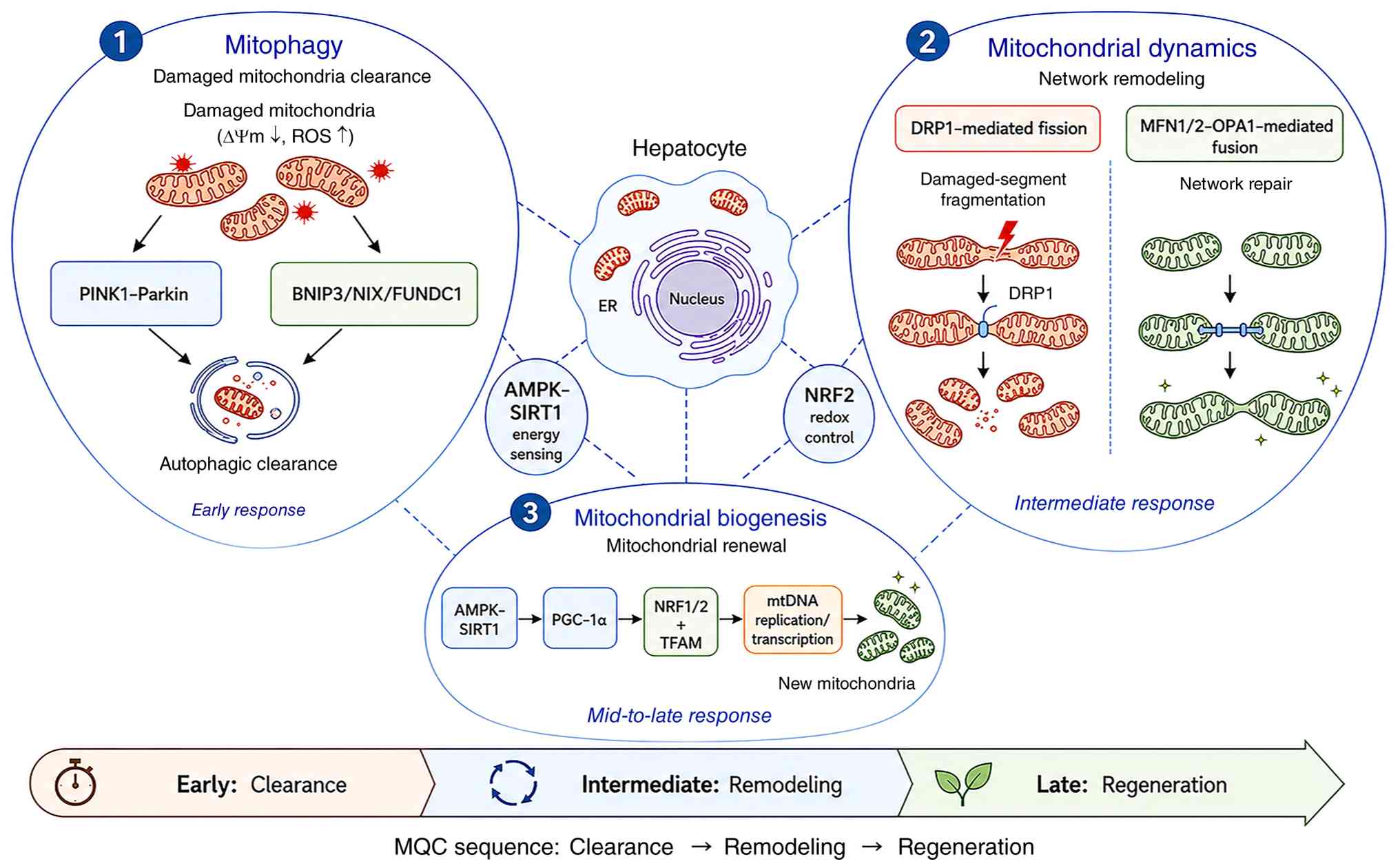 | Figure 1.Core framework of MQC in ALI. MQC is
an integrated repair network that occurs in hepatocytes during ALI.
Three major MQC modules are illustrated in the figure: Mitophagy,
mitochondrial dynamics and mitochondrial biogenesis. Mitophagy
primarily acts as an early clearance response by removing damaged
mitochondria characterized by reduced ΔΨm and increased ROS. This
process may be mediated by the ubiquitin-dependent PINK1-Parkin
pathway or by receptor-mediated mitophagy involving BNIP3, NIX and
FUNDC1. Mitochondrial dynamics represent the remodeling component
of MQC. DRP1-mediated fission facilitates the fragmentation and
segregation of damaged mitochondrial segments, whereas
MFN1/2-OPA1-mediated fusion contributes to mitochondrial network
repair and functional complementation. Mitochondrial biogenesis
functions as a mid-to-late regenerative response and replenishes
mitochondrial mass through the AMPK-SIRT1-PGC-1α-NRF1/2-TFAM axis,
which promotes mtDNA replication/transcription and the formation of
new mitochondria. AMPK-SIRT1 and NRF2 are representative energy-
and redox-sensing regulators that coordinate MQC responses.
Collectively, these processes form a temporally organized MQC
sequence in ALI: Clearance, remodeling and regeneration. ΔΨm,
mitochondrial membrane potential; ALI, acute liver injury; AMPK,
AMP-activated protein kinase; BNIP3, BCL2/adenovirus E1B 19-kDa
interacting protein 3; DRP1, dynamin-related protein 1; ER,
endoplasmic reticulum; FUNDC1, FUN14 domain containing 1; MFN,
mitofusin; MQC, mitochondrial quality control; mtDNA, mitochondrial
DNA; NIX/ BNIP3L, BCL2/adenovirus E1B 19-kDa-interacting protein
3-like; NRF, nuclear factor erythroid 2-related factor; OPA1, optic
atrophy 1; PGC-1α, peroxisome proliferator-activated receptor γ
coactivator 1α; PINK1, PTEN-induced kinase 1; ROS, reactive oxygen
species; SIRT1, sirtuin 1; TFAM, mitochondrial transcription factor
A. |
Mitophagy: Selective elimination of
damaged mitochondria
Mitophagy is a selective autophagic process that
identifies and eliminates dysfunctional mitochondria during
cellular stress. Its primary function is to limit the excessive
production of ROS and the release of pro-apoptotic signals from
damaged mitochondria. Thus, mitophagy prevents the amplification of
mitochondrial injury and reduces cellular toxicity (18). Simultaneously, the degradation
products generated through mitophagy can be reutilized to support
cellular energy metabolism and biosynthesis, rendering mitophagy
the first line of defense in the MQC system (19).
Mitophagy is primarily mediated via two
complementary pathways: Ubiquitin-dependent and receptor-mediated
ubiquitin-independent mechanisms (20). The ubiquitin-dependent pathway is
characterized by activation of the PTEN-induced kinase 1
(PINK1)-Parkin axis, and is primarily triggered by mitochondrial
depolarization and related damage signals (21,22).
By contrast, receptor-mediated mitophagy relies on outer
mitochondrial membrane receptors, including BCL2/adenovirus E1B
19-kDa interacting protein 3 (BNIP3), BCL2/adenovirus E1B
19-kDa-interacting protein 3-like (BNIP3L; also known as NIX) and
FUN14 domain containing 1 (FUNDC1), which directly interact with
LC3 and serve prominent roles under hypoxic and oxidative stress
conditions (23,24). These pathways function in a
complementary manner depending on the nature and intensity of
cellular stress (25).
From a regulatory perspective, mitophagy exhibits
pronounced temporal dependence, selectivity and a context-dependent
effect. During the early phase of mitochondrial injury, mitophagy
is rapidly activated to selectively eliminate severely damaged
mitochondria (26). As injury
resolves, mitophagy activity gradually declines and becomes
coordinated with subsequent mitochondrial biogenesis, forming a
continuous ‘clearance-regeneration’ regulatory sequence (27). Appropriately regulated mitophagy
promotes hepatocyte survival and tissue repair, whereas excessive
or prolonged activation may result in aberrant degradation of
functional mitochondria, leading to metabolic imbalance and
exacerbation of liver injury (28).
Notably, mitophagy should not be interpreted as
uniformly protective in ALI. The net effect of mitophagy depends on
injury severity, timing and the capacity of mitochondrial
biogenesis to replenish mitochondrial content. Insufficient
mitophagy permits the accumulation of damaged mitochondria and
amplifies ROS production (13),
whereas excessive or prolonged mitophagy may deplete functional
mitochondria and aggravate energy failure (29). Therefore, the therapeutic goal
should be to restore balanced mitochondrial turnover rather than
indiscriminately activate mitophagy.
Mitochondrial biogenesis:
Replenishment of mitochondrial content and functional recovery
Mitochondrial biogenesis refers to the generation of
new mitochondria through the coordinated regulation of nuclear
genes and the mitochondrial genome [mitochondrial DNA (mtDNA)],
followed by the integration of newly formed mitochondria into the
existing network. As the regenerative arm of MQC, mitochondrial
biogenesis serves a critical role in replenishing mitochondrial
mass, restoring oxidative phosphorylation capacity, and supporting
hepatocyte repair and regeneration (30).
Mitochondrial biogenesis is gradually activated
during the intermediate to late stages of ALI, and its activation
strength is closely associated with tissue repair capacity
(31). The central regulatory axis
of mitochondrial biogenesis is organized around peroxisome
proliferator-activated receptor γ (PPARγ) coactivator 1α (PGC-1α).
PGC-1α cooperates with transcriptional regulators, such as nuclear
factor erythroid 2-related factor (NRF)1/2 and mitochondrial
transcription factor A (TFAM) to promote the expression of
nuclear-encoded mitochondrial proteins, mtDNA replication and mtDNA
transcription (32,33). Energy-sensing pathways, including
the AMP-activated protein kinase (AMPK)-sirtuin 1 (SIRT1) pathway,
modulate PGC-1α activity, thereby tightly coupling cellular
metabolic status to mitochondrial regenerative responses (34).
Mitochondrial biogenesis is characterized by clear
temporal and stress dependence (35). Its activation typically lags behind
mitophagy and proceeds in parallel with hepatocellular recovery
(36). Moreover, mitochondrial
biogenesis is closely coordinated with mitophagy and mitochondrial
dynamics. Autophagic degradation products, including amino acids,
fatty acids and nucleotides, serve as substrates for macromolecular
biosynthesis, whereas dynamic remodeling of the mitochondrial
network provides a structural framework that promotes the
incorporation of newly generated mitochondria (37).
A major unresolved issue is whether mitochondrial
biogenesis drives hepatocellular recovery or is merely a secondary
marker of tissue repair. Most current evidence is derived from
changes in PGC-1α, NRF1/2 or TFAM expression, whereas direct
functional validation of newly generated mitochondria remains
limited (38,39). Future studies should combine
molecular marker detection with analyses of mitochondrial
respiratory function, including oxygen consumption and oxidative
phosphorylation capacity, mtDNA replication and longitudinal
assessment of mitochondrial recovery.
Mitochondrial dynamics: Structural
remodeling of the mitochondrial network
Mitochondrial dynamics describe the continuous
regulation of mitochondrial morphology and network architecture
through the balance between fission and fusion, and represent the
structural remodeling component of MQC (40). Their core function is to segregate
locally damaged mitochondria through fission, thereby generating
substrates for subsequent mitophagy, whereas fusion enables
complementation of mitochondrial contents to preserve network
integrity and metabolic efficiency (41).
Mitochondrial fission is primarily mediated by
dynamin-related protein 1 (DRP1), whereas fusion is coordinated by
mitofusin (MFN)1/2 and optic atrophy 1 (42). Under conditions of mitochondrial
damage and oxidative stress, the dynamic balance generally shifts
toward fission, facilitating the isolation and clearance of
dysfunctional mitochondria (17).
During the recovery phase, fusion activity progressively increases,
supporting the reconstruction of the mitochondrial network and
functional compensation. Dysregulation of mitochondrial dynamics
may lead to excessive fragmentation or abnormal elongation, thereby
amplifying oxidative stress and metabolic dysfunction, and
accelerating hepatocellular injury (43).
Although excessive mitochondrial fission is commonly
associated with the progression of injury, mitochondrial
fragmentation may also represent an adaptive step that segregates
damaged mitochondrial segments for mitophagy. Thus, the complete
inhibition of fission may be detrimental in certain contexts. The
key issue is not whether fission or fusion is beneficial but
whether mitochondrial dynamics are appropriately synchronized with
mitophagy and biogenesis.
Coordinated regulation of the three
core MQC modules
Mitophagy, mitochondrial biogenesis and
mitochondrial dynamics form a highly interconnected regulatory
network. These modules collectively maintain mitochondrial
homeostasis through temporal coordination, functional
complementarity and signal integration (44). At a systems level, these modules
follow a common regulatory sequence of ‘clearance first, remodeling
next and regeneration last’ (45,46).
In the early phase of ALI, mitochondrial dynamics shift toward
fission and cooperate with mitophagy to eliminate damaged
mitochondria (47). Subsequently,
these dynamics progressively favor fusion to remodel the
mitochondrial network. During the intermediate to late phases,
mitochondrial biogenesis is activated, enabling the generation and
integration of new mitochondria and promoting functional recovery
(48).
At the signaling level, multiple pathways
participate in coordinating these three MQC modules. Among them,
AMPK-SIRT1 functions as a central energy-sensing hub that
integrates mitophagy activation, dynamic balance adjustment and the
initiation of mitochondrial biogenesis (49). NRF2 indirectly modulates
mitochondrial dynamics and promotes regenerative responses by
alleviating oxidative stress (50). Mitochondrial-nuclear communication
may also contribute to the adjustment of MQC activation strength
and timing according to the extent of mitochondrial damage
(14,51).
Although MQC operates within a unified regulatory
framework, its activation patterns exhibit substantial
heterogeneity across different ALI etiologies and among distinct
hepatocyte subpopulations (52).
This heterogeneity directly influences mitochondrial repair
efficiency and cell fate decisions, and provides a theoretical
basis for subsequent etiology-oriented precision intervention
strategies.
Etiology-dependent regulation of MQC in
ALI
A fundamental manifestation of mitochondrial
functional heterogeneity is etiology-dependent regulation. ALI
caused by distinct pathogenic factors differs markedly in the
nature of the initial insult, mode of mitochondrial damage, injury
severity and temporal progression (52,53).
These differences directly result in pronounced heterogeneity in
activation timing, dominant regulatory pathways and coordination of
the three core modules of MQC, namely mitophagy, mitochondrial
biogenesis and mitochondrial dynamics (Fig. 2). In the present review,
drug-induced liver injury (DILI), IRI and viral ALI were used as
representative models to systematically compare etiology-specific
MQC response patterns, thereby providing a mechanistic basis for
precision-targeted therapeutic interventions.
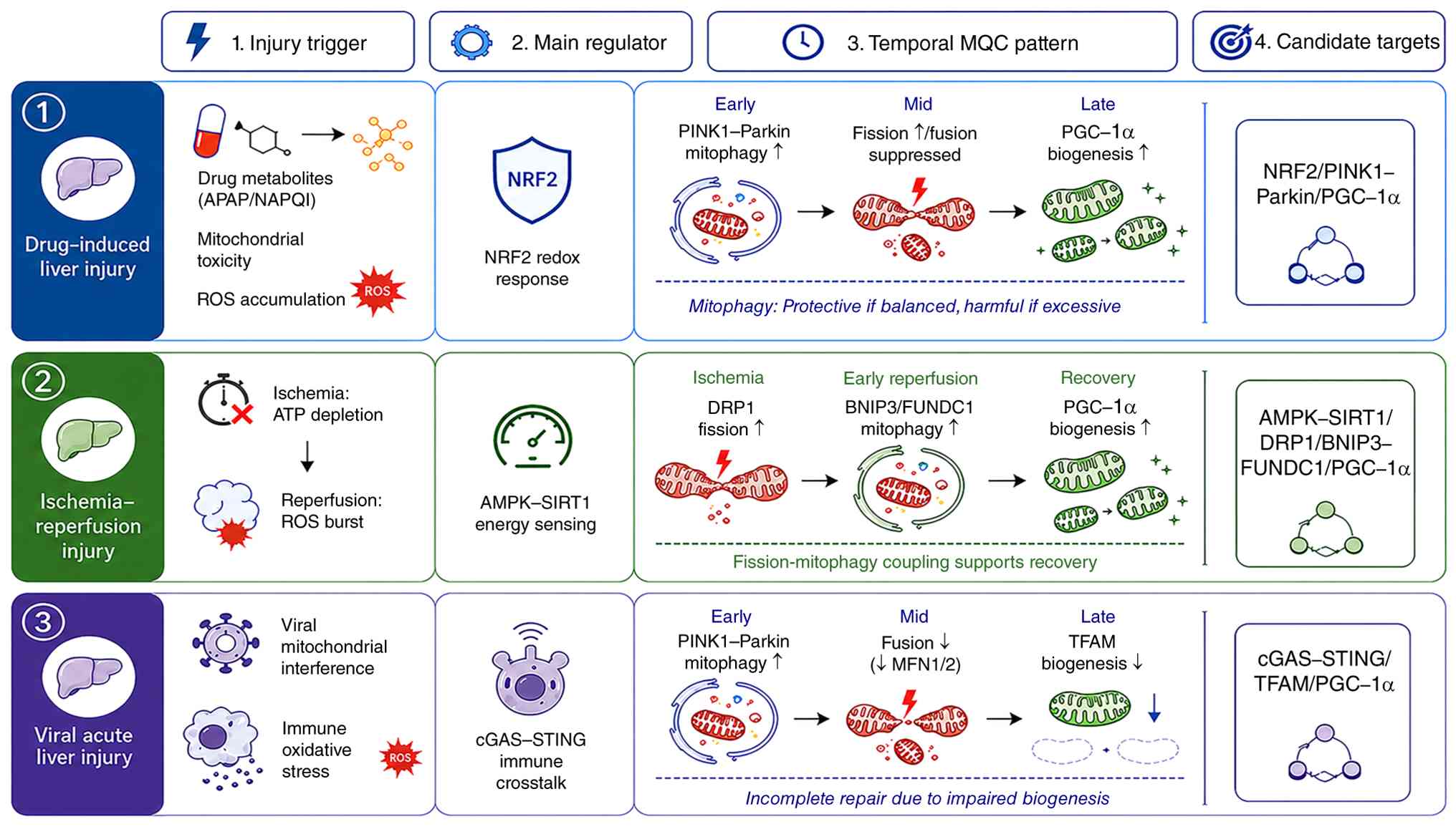 | Figure 2.Etiology-dependent patterns of MQC in
ALI. This schematic diagram summarizes the etiology-dependent MQC
response patterns in three representative forms of ALI: DILI, IRI
and viral acute liver injury. In DILI, drug metabolites, such as
APAP-derived NAPQI, induce mitochondrial toxicity and the
accumulation of ROS. NRF2-mediated redox response represents a
major regulatory context, accompanied by early
PINK1-Parkin-mediated mitophagy, mid-stage mitochondrial fission
with suppressed fusion, and late PGC-1α-associated mitochondrial
biogenesis. Mitophagy in DILI is context-dependent; balanced
activation may support mitochondrial clearance and hepatocyte
protection, whereas excessive or prolonged activation may aggravate
mitochondrial depletion and energy failure. In IRI,
ischemia-induced ATP depletion, followed by a
reperfusion-associated ROS burst, activates AMPK-SIRT1-mediated
energy sensing. DRP1-dependent fission, BNIP3/FUNDC1-associated
mitophagy and PGC-1α-driven mitochondrial biogenesis are
sequentially engaged, and efficient fission-mitophagy coupling
supports mitochondrial recovery. In acute viral liver injury, the
illustrated pattern is a representative mechanism rather than a
universal response across all viral etiologies. Viral mitochondrial
interference and immune oxidative stress promote early
PINK1-Parkin-associated mitophagy and innate immune crosstalk
involving cGAS-STING. However, fusion suppression, reduced MFN1/2
activity and TFAM-related impairment of mitochondrial biogenesis
may prevent completion of the clearance-remodeling-regeneration
repair sequence. Candidate intervention targets shown in the figure
include NRF2, PINK1-Parkin and PGC-1α in DILI; AMPK-SIRT1, DRP1,
BNIP3-FUNDC1 and PGC-1α in IRI; and cGAS-STING, TFAM and PGC-1α in
viral acute liver injury. APAP, acetaminophen; ALI, acute liver
injury; AMPK, AMP-activated protein kinase; BNIP3, BCL2/adenovirus
E1B 19-kDa interacting protein 3; cGAS, cyclic GMP-AMP synthase;
DILI, drug-induced liver injury; DRP1, dynamin-related protein 1;
FUNDC1, FUN14 domain containing 1; IRI, ischemia-reperfusion
injury; MFN, mitofusin; MQC, mitochondrial quality control; NAPQI,
N-acetyl-p-benzoquinone imine; NRF, nuclear factor erythroid
2-related factor; PGC-1α, peroxisome proliferator-activated
receptor γ coactivator 1α; PINK1, PTEN-induced kinase 1; ROS,
reactive oxygen species; SIRT1, sirtuin 1; STING, stimulator of
interferon genes; TFAM, mitochondrial transcription factor A. |
MQC patterns in DILI
DILI is one of the most common causes of ALI, with
acetaminophen (APAP) overdose and injury associated with herbal
medicines or dietary supplements being the most common (54). Mitochondrial damage in DILI is
direct and dose-dependent. It mainly results from reactive drug
metabolites, such as N-acetyl-p-benzoquinone imine, generated
during APAP metabolism, which directly interacts with mitochondrial
proteins (55,56). These interactions lead to the
collapse of mitochondrial membrane potential, excessive ROS
production and mtDNA damage. Under these conditions, MQC activation
in DILI is characterized by a pronounced context-dependent role of
mitophagy, with NRF2 signaling acting as the dominant upstream
regulator (57). Reduced NRF2
activity markedly impairs PINK1 expression and delays mitophagy
activation in APAP-induced liver injury models, thus resulting in
excessive accumulation of damaged mitochondria and the expansion of
hepatocellular necrosis (58,59).
By contrast, enhanced NRF2 activation promotes mitophagy and
mitochondrial biogenesis, thereby improving hepatocyte survival and
alleviating tissue injury (60).
MQC activation in DILI generally follows a
sequential pattern consisting of early mitophagy activation,
intermediate remodeling of mitochondrial dynamics and late
induction of mitochondrial biogenesis (46,52).
During the early phase after injury, rapid ROS accumulation
activates the PINK1-Parkin pathway and NRF2 signaling, inducing
mitophagy to eliminate severely damaged mitochondria (61). In the intermediate phase,
mitochondrial dynamics shift toward enhanced fission accompanied by
suppressed fusion, facilitating further segregation and clearance
of dysfunctional mitochondria (47). At later stages, NRF2-driven
upregulation of PGC-1α initiates mitochondrial biogenesis,
replenishing mitochondrial content and gradually restoring
oxidative phosphorylation capacity (33). Mitophagy exhibits the most
prominent dual effect in DILI; moderate activation under low-dose
exposure is protective, whereas excessive activation under
high-dose conditions may result in aberrant degradation of
functional mitochondria, leading to energy collapse and aggravated
liver injury (62).
MQC patterns in IRI
IRI commonly occurs during liver surgery,
transplantation or hemorrhagic shock. Pathophysiologically, it
involves energy deprivation during ischemia, followed by an abrupt
oxidative burst upon reperfusion (63). Unlike in DILI, mitochondrial damage
in IRI is characterized by the coexistence of energy-sensing
dysregulation and structural disruption. Accordingly, MQC
regulation in IRI is functionally dominated by the AMPK-SIRT1 axis
and features a tight coupling between mitochondrial fission and
mitophagy (64). Experimental
evidence indicates impaired mitophagy, including inhibition of
DRP1-Beclin-1-dependent mitophagy, compromises the clearance of
damaged mitochondria and exacerbates ROS-mediated hepatocellular
injury in ischemia-reperfusion models (65,66).
By contrast, activation of the AMPK-SIRT1 pathway accelerates the
initiation of mitochondrial biogenesis and facilitates the recovery
of cellular ATP levels (67).
MQC activation in IRI follows a continuous sequence
consisting of ischemia-associated remodeling of mitochondrial
dynamics, reperfusion-driven mitophagy and recovery-phase
mitochondrial biogenesis (64).
During the ischemic phase, rapid ATP depletion increases the
AMP/ATP ratio and activates AMPK, which phosphorylates DRP1 to
promote mitochondrial fission and preemptively segregate vulnerable
mitochondria (65). In the early
reperfusion phase, massive oxygen influx induces a burst of ROS,
and AMPK further activates receptor-mediated mitophagy pathways
involving BNIP3, enabling the rapid removal of severely damaged
mitochondria (64,65). During the subsequent recovery
phase, sustained activation of the AMPK-SIRT1 axis promotes
PGC-1α-dependent mitochondrial biogenesis, gradually restoring
hepatocellular energy homeostasis (49). Collectively, BNIP3-mediated
mitophagy and DRP1-dependent mitochondrial fission constitute the
key rate-limiting steps in MQC regulation during IRI, and their
coordinated efficiency largely determines injury severity and
repair capacity.
MQC patterns in viral ALI
Viral ALI results from hepatotropic viral
infections, such as hepatitis A virus and hepatitis E virus, and is
characterized by a dual origin of mitochondrial damage: Direct
viral interference with mitochondrial structure and function, and
secondary amplification driven by immune-mediated inflammation and
oxidative stress (68). In this
context, MQC regulation is characterized by extensive crosstalk
between innate immune signaling and MQC, along with persistent
suppression of mitochondrial biogenesis (69).
During viral infection, hepatotropic viruses such as
hepatitis A and E viruses consume substantial mitochondrial energy
to support viral replication, and viral proteins directly
compromise mitochondrial membrane integrity (69,70).
Simultaneously, cytokines released by natural killer cells and
cytotoxic T lymphocytes promote excessive ROS production, further
aggravating mitochondrial injury (71–73).
Consequently, MQC activation follows a characteristic pattern
consisting of early mitophagy activation, intermediate disruption
of mitochondrial dynamics and late-stage impairment of
mitochondrial biogenesis (74). In
the early phase of infection, virus-induced oxidative stress
activates the PINK1-Parkin pathway, thereby promoting mitophagy to
eliminate damaged mitochondria (75,76).
During the intermediate phase, viral proteins suppress MFN1/2
expression, resulting in impaired mitochondrial fusion and
fragmentation of the mitochondrial network (74,77).
In the later phase, viral interference with PGC-1α signaling
inhibits mitochondrial biogenesis, thereby constraining the
hepatocellular repair capacity (78).
At the molecular level, the cyclic GMP-AMP synthase
(cGAS)-stimulator of interferon genes (STING)-PINK1 axis represents
a critical intersection between MQC regulation and the innate
immune response in viral ALI. mtDNA released from damaged
mitochondria activates cGAS-STING signaling, which amplifies
inflammatory responses and enhances PINK1 expression and mitophagy
activation (79,80). In contrast to DILI and IRI, the
sustained suppression of mitochondrial biogenesis is a defining
feature of viral ALI. Viral downregulation of TFAM directly impairs
mtDNA transcription and replication, restricting the generation of
new mitochondria and preventing completion of the MQC
‘clearance-regeneration’ repair cycle (81). These findings suggest that the
disruption of mitochondrial biogenesis and TFAM-related mtDNA
maintenance impairs mitochondrial recovery during viral infection.
However, whether the direct restoration of TFAM improves hepatocyte
survival in viral ALI remains unclear (81,82).
Comparative overview and implications
of etiology-specific MQC regulation
Taken together, the aforementioned evidence
highlights MQC as a highly adaptive and etiology-dependent
regulatory network in ALI. Although mitophagy, mitochondrial
biogenesis and mitochondrial dynamics operate within a shared
conceptual framework, their relative activation timings, dominant
regulatory pathways and degrees of coordination vary substantially
across different forms of ALI. In DILI, MQC regulation is largely
driven by NRF2 signaling and is characterized by a prominent dual
role of mitophagy: Appropriate activation enables completion of the
clearance-regeneration cycle, whereas excessive activation
exacerbates metabolic collapse (83). In IRI, MQC is predominantly
governed by energy-sensing mechanisms centered on the AMPK-SIRT1
axis, with tight coupling between mitochondrial fission and
mitophagy, enabling the efficient removal of damaged mitochondria
and subsequent bioenergetic recovery (65). By contrast, viral ALI can be
distinguished by sustained interference with mitochondrial
biogenesis. This pattern is driven by the persistent innate immune
activation and viral suppression of mitochondrial transcriptional
programs. Consequently, the MQC repair loop remains incomplete
despite early induction of mitophagy (81).
These etiology-specific MQC patterns indicate that
mitochondrial responses in ALI are not uniform stress reactions but
dynamically tuned processes shaped by the nature, intensity and
duration of the injurious stimulus. The efficiency of
hepatocellular recovery depends not only on the activation of
individual MQC modules but also on their coordinated integration
into a functional repair sequence. Disruption of this coordination,
such as excessive mitophagy without compensatory biogenesis, can
shift MQC from a protective to a maladaptive response (14,35).
Recognizing MQC as a context-dependent and temporally staged
regulatory network provides a mechanistic basis for stratified
therapeutic intervention and sets the stage for the targeted
modulation of MQC pathways in ALI.
Unresolved controversies and knowledge
gaps in MQC regulation during ALI
Several issues in this field remain unresolved.
First, the protective vs. maladaptive role of mitophagy remains
context-dependent, and the threshold at which mitophagy shifts from
mitochondrial repair to depletion remains unclear. Second,
mitochondrial fission may facilitate the segregation of damaged
mitochondria or promote excessive fragmentation, depending on the
timing and severity of injury. Third, increased expression of
mitochondrial biogenesis markers does not necessarily indicate
functional recovery of mitochondrial respiration. Finally, the
findings from APAP-induced ALI, IRI and viral ALI models may not be
directly interchangeable. These uncertainties highlight the need
for temporally resolved and etiology-specific studies on MQC in
ALI. The major etiology-specific MQC patterns in different forms of
ALI are summarized in Table I.
 | Table I.Etiology-specific patterns of MQC in
ALI. |
Table I.
Etiology-specific patterns of MQC in
ALI.
| ALI type | Main mitochondrial
insult | Dominant MQC
feature | Key regulatory
pathways | Temporal
pattern | Major
implication | (Refs.) |
|---|
| DILI | Direct
mitochondrial toxicity, ROS accumulation, mtDNA damage and loss of
mitochondrial membrane potential | Prominent mitophagy
activation with context-dependent effects | NRF2, PINK1-Parkin,
PGC-1α | Early mitophagy
activation, intermediate mitochondrial dynamics remodeling and late
mitochondrial biogenesis | Moderate mitophagy
is protective, whereas excessive or prolonged mitophagy may
aggravate mitochondrial depletion and energy failure | (55,57,58,62,83) |
| IRI | Ischemia-induced
ATP depletion followed by reperfusion-associated oxidative
burst | Tight coupling
between mitochondrial fission and mitophagy | AMPK-SIRT1, DRP1,
BNIP3, PGC-1α | Ischemia-associated
fission, reperfusion-driven mitophagy and recovery-phase
biogenesis | Efficient
fission-mitophagy coupling promotes damaged mitochondrial
clearance, but excessive fission may worsen fragmentation and
injury | (63–67) |
| Viral ALI | Viral interference
with mitochondrial function combined with immune- mediated
inflammation and oxidative stress | Early mitophagy
with impaired mitochondrial biogenesis | cGAS-STING,
PINK1-Parkin, TFAM, PGC-1α | Early mitophagy
activation, intermediate disruption of dynamics and late
suppression of biogenesis | Incomplete
clearance-regeneration repair may limit hepatocellular
recovery | (68,69,74,75,79-82) |
Therapeutic strategies targeting MQC in
ALI
Given the central role of mitochondrial dysfunction
in the initiation and progression of ALI, targeting MQC has emerged
as a promising strategy for mechanism-based intervention (84). Based on current experimental and
translational studies, MQC-targeted therapeutic strategies for ALI
can be divided into three major categories. These include
small-molecule drugs, natural compounds, and gene- or cell-based
therapies combined with emerging bioengineering technologies
(85). These approaches operate on
distinct nodes of the MQC network, and can serve as complementary
opportunities to preserve mitochondrial integrity, enhance
hepatocellular resilience and improve liver injury outcomes
(52,85). The present review summarizes
representative strategies within each category, and discusses their
therapeutic potential and translational challenges.
Small-molecule drugs
Small-molecule drugs represent the most extensively
investigated class of MQC-targeted interventions in ALI because of
their defined molecular targets and translational feasibility
(85). A major therapeutic
strategy involves enhancing autophagy and mitophagy by modulating
the energy-sensing AMPK-mammalian target of rapamycin (mTOR) axis
(86). AMPK activation, for
example, by metformin, promotes autophagic clearance of damaged
mitochondria, whereas inhibition of mTOR signaling by agents such
as rapamycin facilitates mitophagy and has demonstrated
hepatoprotective effects in experimental models of DILI (87,88).
By reinforcing endogenous mitochondrial turnover mechanisms, these
agents limit the accumulation of dysfunctional ROS-generating
mitochondria and attenuate downstream inflammatory signaling
(62). Activation of
PINK1-Parkin-mediated mitophagy not only removes compromised
mitochondria but also suppresses inflammasome activation in
stressed hepatocytes (89).
Beyond mitophagy induction, pharmacological
modulation of mitochondrial dynamics has emerged as an important
option for MQC-targeted therapy (90). Excessive mitochondrial fission,
largely mediated by DRP1, contributes to mitochondrial
fragmentation and metabolic failure in ALI (91). Accordingly, DRP1 inhibition by
Mdivi-1 has been reported to alleviate cecal ligation and
puncture-induced sepsis-associated liver injury, at least in part,
by suppressing STING signaling activation in Kupffer cells and
attenuating systemic inflammatory responses (92). Conversely, strategies that enhance
mitochondrial fusion or stimulate mitochondrial biogenesis can
improve cellular bioenergetic capacity. Small molecules that
activate SIRT1/PGC-1α-related signaling restore mitochondrial
function and promote mitochondrial biogenesis in experimental
hepatic IRI (67).
Beyond these canonical pathways, small-molecule
modulators of the BCL-2 family of proteins are also being explored
for their ability to indirectly trigger mitophagy (93). The inhibition of the anti-apoptotic
protein MCL-1 by compounds such as UMI-77 has been reported to
induce mitophagy in non-hepatic preclinical models. Although these
findings suggest that pharmacological induction of mitophagy is
therapeutically relevant, its role in ALI remains unclear (94,95).
Collectively, these pharmacological approaches act on distinct but
interconnected nodes of the MQC network, including mitophagy
induction, dynamic remodeling and mitochondrial regeneration, and
converge to preserve mitochondrial homeostasis and reduce
hepatocyte death in ALI.
Natural compounds
Naturally derived compounds have attracted
increasing attention as modulators of MQC in ALI, largely owing to
their pleiotropic biological activities and favorable safety
profiles. Several of these compounds exert hepatoprotective effects
by simultaneously targeting oxidative stress, mitochondrial
turnover and bioenergetic homeostasis, thereby influencing multiple
nodes of the MQC network (96).
Plant-derived polyphenols and antioxidants have been
the most extensively studied group in this category. Resveratrol, a
prototypical polyphenol, has been reported to alleviate
experimental liver injury by reducing oxidative stress and
inflammatory responses, and inducing SIRT1/p62-mediated mitophagy
(97). These findings suggest that
resveratrol preserves mitochondrial homeostasis mainly through
SIRT1-related mitophagy and redox regulation, although most
evidence remains preclinical and model-dependent (97). Similarly, quercetin has been
reported to improve acute liver failure by modulating
mitophagy-related apoptosis and inflammatory responses through the
PPARγ/PGC-1α/NF-κB axis (98).
Curcumin has also shown hepatoprotective potential, primarily
through antioxidant and anti-inflammatory mechanisms, including the
activation of NRF2 signaling and inhibition of NF-κB activation;
however, direct evidence linking curcumin to hepatic mitophagy in
ALI remains limited (99). By
limiting ROS accumulation and facilitating the removal of
dysfunctional mitochondria, these compounds help maintain the
mitochondrial membrane potential and ATP production in stressed
hepatocytes (61).
In addition to phytochemicals, several endogenous
molecules and nutritional supplements have been implicated in MQC
regulation (100). Melatonin has
been reported to attenuate experimental liver IRI by modulating
mitochondrial injury-related pathways, supporting its potential
role in mitochondrial protection during ALI (101). Coenzyme Q10, a mitochondrial
electron carrier and lipid-soluble antioxidant, may theoretically
support mitochondrial redox homeostasis and bioenergetic functions,
although ALI-specific evidence remains limited (102). Moreover, several natural
compounds have been reported to regulate PGC-1α-related
mitochondrial biogenesis in non-ALI metabolic models. These
findings suggest a link between natural compounds and mitochondrial
regeneration; however, their relevance to ALI requires further
validation (103,104).
Collectively, natural compounds modulate MQC through
their integrated effects on redox homeostasis, mitophagy activation
and mitochondrial regeneration. Although most evidence remains at
the preclinical level, the multitarget actions of these agents make
them attractive complementary candidates for MQC-based intervention
strategies for ALI.
Gene- and cell-based therapies and
emerging technologies
Beyond conventional pharmacological approaches,
gene- and cell-based therapies represent innovative strategies for
targeting MQC in ALI. Emerging bioengineering technologies have
further expanded this therapeutic landscape by enabling direct
restoration of mitochondrial homeostasis (105).
Gene-based strategies focus on precise manipulation
of key regulators that govern mitophagy, mitochondrial biogenesis
and responses to oxidative stress. Targeted delivery or
overexpression of genes such as PINK1, Parkin, PGC-1α and NRF2 has
been shown to enhance mitochondrial clearance, regeneration and
antioxidant capacity in preclinical models of ALI (52,83).
Conversely, genetic inhibition of maladaptive pathways, including
excessive mitochondrial fission and mtDNA-driven inflammatory
signaling, can mitigate hepatocellular damage while preserving
essential MQC functions. Although still largely experimental, these
approaches highlight the potential for directly reprogramming
mitochondrial stress responses at the molecular level.
Cell-based therapies provide an additional dimension
of MQC modulation by exploiting the regenerative and
immunomodulatory properties of stem cells. In particular,
mesenchymal stem cells (MSCs) enhance mitochondrial function
through paracrine signaling, attenuation of oxidative stress and
suppression of inflammation (106). Accumulating evidence has
indicated that MSCs and their extracellular vesicles facilitate the
transfer of functional mitochondria or mitochondrial components to
injured cells, thereby restoring their bioenergetic capacity and
enhancing cellular resilience (106,107). This mitochondria-centered repair
mechanism represents a unique advantage of cell-based approaches in
severe mitochondrial dysfunction.
Emerging bioengineering technologies have expanded
the therapeutic landscape by enabling targeted and efficient
modulation of MQC at the subcellular level (108). Advances in mitochondrial
transplantation, mitochondria-targeted drug delivery systems and
nanotechnology-based platforms, such as the transplantation of
isolated healthy mitochondria and the use of mitochondria-targeted
nanocarriers to deliver antioxidants or other therapeutic agents
(109,110), have enabled the selective
enhancement of mitochondrial function, suppression of oxidative
stress at its source and promotion of network recovery with high
spatial precision. Although these technologies remain in the early
stages of development, they offer promising solutions to the
longstanding challenges related to targeting specificity and
therapeutic efficacy.
Collectively, gene- and cell-based interventions and
emerging technologies reflect a paradigm shift from indirect
modulation to direct restoration of mitochondrial integrity. By
reinforcing MQC at the genetic, cellular and subcellular levels,
these approaches have a substantial potential to complement
existing therapies and enable more precise etiologically adapted
treatment strategies for ALI.
Integrative summary of MQC-targeted
therapeutic strategies
Therapeutic strategies targeting MQC provide a
multifaceted framework for mitigating ALI by directly addressing
mitochondrial dysfunction, a central driver of hepatocellular
damage. The approaches discussed in the present review, including
small-molecule drugs, natural compounds, gene- and cell-based
therapies, and emerging bioengineering technologies, converge on
the shared objective of preserving or restoring mitochondrial
homeostasis in stressed hepatocytes. These strategies can reduce
oxidative stress, remove or replace damaged mitochondria, and
restore bioenergetic function through coordinated regulation of
mitophagy, mitochondrial dynamics and mitochondrial biogenesis.
An effective MQC-targeted intervention is unlikely
to depend on the simple activation of mitochondrial turnover.
Instead, it requires precise context-dependent modulation according
to the etiology of the injury, disease stage and balance among
individual MQC modules (52,85).
However, whether these strategies can be translated into clinical
practice depends on several unresolved issues, including safety,
delivery, therapeutic timing, biomarkers and patient heterogeneity.
The translational challenges are discussed in detail in the current
review.
Translational barriers and clinical
challenges of MQC-targeted therapies
Despite encouraging preclinical findings, several
barriers have limited the clinical translation of MQC-targeted
therapies for ALI. First, most candidate interventions have only
been validated in cell-based or animal models; human trials
specifically designed to assess MQC modulation are scarce (52,84).
Second, safety concerns must be carefully considered because
excessive activation or inhibition of MQC may disrupt physiological
mitochondrial turnover and impair hepatocyte metabolism (85). Third, efficient delivery to injured
hepatocytes remains a challenge, particularly in gene-based
strategies, mitochondrial transplantation and nanocarrier systems
(108–110). Fourth, the therapeutic window is
likely narrow because mitophagy, mitochondrial dynamics and
mitochondrial biogenesis exhibit distinct temporal patterns during
the progression of ALI. Fifth, reliable biomarkers capable of
monitoring mitochondrial damage, mitophagic flux and biogenic
activity in patients are still lacking. Finally, patient
heterogeneity in etiology, injury severity, comorbidities and
baseline mitochondrial function may strongly influence therapeutic
responses. These limitations suggest that MQC-targeted therapies
require etiologically stratified, time-sensitive and
biomarker-guided development before clinical application.
Representative MQC-targeted therapeutic candidates, their molecular
targets, experimental models and translational statuses are
summarized in Table II.
| Table II.Therapeutic candidates targeting MQC
in ALI. |
Table II.
Therapeutic candidates targeting MQC
in ALI.
| Therapeutic
category | Representative
candidate or strategy | Main molecular
target | MQC process
affected | Model/evidence
base | Status | Major translational
limitations | (Refs.) |
|---|
| Small-molecule
drugs | Metformin;
rapamycin | AMPK-mTOR axis | Autophagy and
mitophagy | Rat hepatic
ischemia/ reperfusion model | Preclinical;
clinically approved for other indications | Optimal dose,
timing and patient selection remain unclear | (87,88) |
|
| Mdivi-1 | DRP1 | Mitochondrial
fission | CLP-induced sepsis-
associated liver injury model | Preclinical | Specificity, safety
and long-term effects remain debated | (92) |
| Natural
compounds | Resveratrol | SIRT1/AMPK
pathway | Mitophagy and
mitochondrial function | LPS-induced
inflammatory liver injury model in Gibel carp | Preclinical | Limited
bioavailability and uncertain clinical dosing | (97) |
|
| Quercetin;
curcumin | NRF2-, PGC-1α- or
inflammation-related pathways | Redox regulation,
mitophagy and mitochondrial recovery | Experimental acute
liver failure and LPS/D-GalN-induced ALI models | Preclinical | Low bioavailability
and insufficient human evidence | (98,99) |
|
Endogenous/nutritional compounds | Melatonin | Antioxidant and
mitochondrial injury-related pathways | Mitochondrial
protection and redox homeostasis | Hepatic IRI
model | Preclinical;
clinical evidence in ALI remains limited | Lack of
ALI-specific clinical validation | (101) |
| Gene-based
therapies | PINK1, Parkin,
PGC-1α or NRF2 modulation | Mitophagy,
mitochondrial biogenesis and antioxidant response pathways | Direct
reinforcement of MQC programs | APAP-induced ALI
model and other preclinical liver injury models |
Experimental/preclinical | Delivery
efficiency, off-target effects and safety concerns | (52,83) |
| Bioengineering
strategies | Mitochondrial
transplantation or mitochondria-targeted nanocarriers | Mitochondrial
delivery and subcellular targeting | Direct
mitochondrial restoration | Murine hepatic IRI
model and mitochondria-targeted delivery evidence | Early preclinical
research | Delivery
specificity, immune response and scalability remain unresolved | (109,110) |
Conclusion and future perspectives
Accumulating evidence has established that MQC is a
central determinant of hepatocellular fate during ALI. Rather than
operating as isolated pathways, mitophagy, mitochondrial biogenesis
and mitochondrial dynamics form an integrated regulatory network
that regulates mitochondrial turnover, functional recovery and
cellular adaptation to acute stress. Dysregulation of this network
contributes to mitochondrial dysfunction, metabolic failure and the
amplification of liver injury, whereas appropriately tuned MQC
responses facilitate hepatocellular repair and liver function
restoration.
An emerging concept from previous studies (13,28)
is that MQC regulation in ALI is strongly context-dependent. The
timing, magnitude and coordination of individual MQC processes vary
according to injury etiology, severity and disease stage. While
early activation of mitophagy and mitochondrial fission may serve
adaptive roles in limiting acute mitochondrial damage, sustained or
excessive activation can become maladaptive by exacerbating
mitochondrial depletion and energy insufficiency (29). These observations underscore the
need for balanced and temporally regulated MQC responses to achieve
effective hepatocellular recovery.
Despite these advances, certain limitations of
current state-of-the-art methods should be acknowledged. First,
most evidence supporting MQC regulation in ALI has been derived
from cell-based experiments and animal models, whereas direct
evidence from human ALI samples remains limited; this restricts the
clinical applicability of the conclusions drawn from preclinical
studies. Second, different experimental models of ALI, such as
APAP-induced injury, IRI and viral liver injury, differ
substantially in terms of injury mechanisms, temporal progression
and mitochondrial stress patterns (59,64,68).
Therefore, MQC findings from one model cannot be directly
generalized to all forms of ALI. Third, current methods for
evaluating MQC remain incomplete. A number of studies (35,36,38,39)
rely on static markers such as PINK1, Parkin, LC3, PGC-1α, TFAM,
DRP1 or MFN1/2; however, these markers do not fully reflect
mitophagy flux, mitochondrial biogenesis activity or dynamic
mitochondrial network remodeling in vivo. Fourth, the dual
role of MQC has not been fully clarified. Although moderate
mitophagy and mitochondrial fission may remove damaged mitochondria
and protect hepatocytes, excessive or prolonged activation may lead
to mitochondrial depletion, bioenergetic failure and aggravation of
injury. Finally, the lack of reliable biomarkers for mitochondrial
damage and MQC status limits patient stratification and prevents
the accurate selection of therapeutic timing.
Therefore, future research should move from
descriptive pathway analyses to temporally resolved, clinically
oriented investigations. Single-cell mitochondrial profiling may
help identify hepatocyte subpopulations with distinct MQC states
during injury progression and repair. Spatial transcriptomics and
proteomics could further clarify how MQC responses differ between
necrotic, inflammatory and regenerating regions within injured
liver tissue. Additionally, artificial intelligence-assisted
biomarker discovery may facilitate the identification of
circulating or tissue-based indicators of mitochondrial injury,
mitophagy and regenerative capacity. These approaches may provide a
basis for precision hepatology, in which MQC-targeted interventions
are selected according to the injury etiology, disease stage,
biomarker status and patient-specific metabolic background. More
clinically relevant animal models, human liver samples and
prospective translational studies are required to determine whether
MQC modulation can be safely and effectively applied to patients
with ALI.
In conclusion, MQC is a dynamic and
context-dependent regulatory system that links mitochondrial damage
control with hepatocellular repair. A better understanding of its
temporal regulation, etiology-specific features and translational
limitations is essential for the development of mechanism-based and
precision therapeutic strategies for ALI.
Acknowledgements
Not applicable.
Funding
This study was supported by the Jilin Health Science and
Technology Capacity Enhancement Program (grant no.
2025ZY-YA009).
Availability of data and materials
Not applicable.
Authors' contributions
ZC conceptualized the study, conducted the
investigation, and contributed to writing, reviewing and editing
the manuscript. YW contributed to visualization and to writing,
reviewing and editing the manuscript. YL performed validation and
contributed to writing, reviewing and editing the manuscript. GC
contributed to visualization and to writing, reviewing and editing
the manuscript. YC and YB conducted the investigation and
contributed to writing, reviewing and editing the manuscript. YZ
conceptualized the study, administered the project, acquired
funding, and contributed to writing, reviewing and editing the
manuscript. LZ conceptualized and supervised the study,
administered the project, and contributed to writing, reviewing and
editing the manuscript. Data authentication is not applicable. All
authors have read and approved the final manuscript, have agreed on
the journal to which the article has been submitted, and agree to
be accountable for all aspects of this work.
Ethics approval and consent to
participate
Not applicable.
Patient consent for publication
Not applicable.
Competing interests
The authors declare that they have no competing
interests.
References
|
1
|
Khanam A and Kottilil S: Abnormal innate
immunity in acute-on-chronic liver failure: immunotargets for
therapeutics. Front Immunol. 11:20132020. View Article : Google Scholar : PubMed/NCBI
|
|
2
|
Larsen FS and Saliba F: Liver support
systems and liver transplantation in acute liver failure. Liver
Int. 45:e156332025. View Article : Google Scholar : PubMed/NCBI
|
|
3
|
Pollok JM, Tinguely P, Berenguer M,
Niemann CU, Raptis DA and Spiro M; ERAS4OLT.org collaborative, :
Enhanced recovery for liver transplantation: Recommendations from
the 2022 international liver transplantation society consensus
conference. Lancet Gastroenterol Hepatol. 8:81–94. 2023. View Article : Google Scholar : PubMed/NCBI
|
|
4
|
Jaeschke H and Ramachandran A: Central
mechanisms of acetaminophen hepatotoxicity: Mitochondrial
dysfunction by protein adducts and oxidant stress. Drug Metab
Dispos. 52:712–721. 2024. View Article : Google Scholar : PubMed/NCBI
|
|
5
|
Du K, Farhood A and Jaeschke H:
Mitochondria-targeted antioxidant Mito-Tempo protects against
acetaminophen hepatotoxicity. Arch Toxicol. 91:761–773. 2017.
View Article : Google Scholar : PubMed/NCBI
|
|
6
|
Du K, Ramachandran A, McGill MR, Mansouri
A, Asselah T, Farhood A, Woolbright BL, Ding WX and Jaeschke H:
Induction of mitochondrial biogenesis protects against
acetaminophen hepatotoxicity. Food Chem Toxicol. 108:339–350. 2017.
View Article : Google Scholar : PubMed/NCBI
|
|
7
|
Li YF, Xie ZF, Song Q and Li JY:
Mitochondria homeostasis: Biology and involvement in hepatic
steatosis to NASH. Acta Pharmacol Sin. 43:1141–1155. 2022.
View Article : Google Scholar : PubMed/NCBI
|
|
8
|
LeFort KR, Rungratanawanich W and Song BJ:
Contributing roles of mitochondrial dysfunction and hepatocyte
apoptosis in liver diseases through oxidative stress,
post-translational modifications, inflammation, and intestinal
barrier dysfunction. Cell Mol Life Sci. 81:342024. View Article : Google Scholar : PubMed/NCBI
|
|
9
|
Ding Q, Qi Y and Tsang SY: Mitochondrial
biogenesis, mitochondrial dynamics, and mitophagy in the maturation
of cardiomyocytes. Cells. 10:24632021. View Article : Google Scholar : PubMed/NCBI
|
|
10
|
Tang C, Cai J, Yin XM, Weinberg JM,
Venkatachalam MA and Dong Z: Mitochondrial quality control in
kidney injury and repair. Nat Rev Nephrol. 17:299–318. 2021.
View Article : Google Scholar : PubMed/NCBI
|
|
11
|
Liao X, Wang Y, Zhang Z, Qu X and Zhou G:
Bidirectional regulation of NLRP3 inflammasome and mitochondrial
quality control in sepsis: Mechanisms and therapeutic implications.
Mediators Inflamm. 2026:31686692026. View Article : Google Scholar : PubMed/NCBI
|
|
12
|
Liu BH, Xu CZ, Liu Y, Lu ZL, Fu TL, Li GR,
Deng Y, Luo GQ, Ding S, Li N and Geng Q: Mitochondrial quality
control in human health and disease. Mil Med Res.
11:322024.PubMed/NCBI
|
|
13
|
Ma X, McKeen T, Zhang J and Ding WX: Role
and mechanisms of mitophagy in liver diseases. Cells. 9:8372020.
View Article : Google Scholar : PubMed/NCBI
|
|
14
|
Li W, Gui Y, Guo C, Huang Y, Liu Y, Yu X,
Zhang H, Wang J, Liu R, Mahaman YAR, et al: Molecular mechanisms of
mitochondrial quality control. Transl Neurodegener. 14:452025.
View Article : Google Scholar : PubMed/NCBI
|
|
15
|
Wu Y, Luo H, Pan Z, Chen W and Bi L:
Bidirectional crosstalk between ER stress and lipid metabolism:
From proteostasis to tumor adaptation. Cell Death Discov.
12:372025. View Article : Google Scholar : PubMed/NCBI
|
|
16
|
Romanelli S and Trempe JF: Stressful
situations: Molecular insights on mitochondrial quality control
pathways. J Biol Chem. 301:1104832025. View Article : Google Scholar : PubMed/NCBI
|
|
17
|
Xu X, Pang Y and Fan X: Mitochondria in
oxidative stress, inflammation and aging: From mechanisms to
therapeutic advances. Signal Transduct Target Ther. 10:1902025.
View Article : Google Scholar : PubMed/NCBI
|
|
18
|
Shi J, Yu Y, Yuan H, Li Y and Xue Y:
Mitochondrial dysfunction in AMI: Mechanisms and therapeutic
perspectives. J Transl Med. 23:4182025. View Article : Google Scholar : PubMed/NCBI
|
|
19
|
Chatzinikita E, Maridaki M, Palikaras K,
Koutsilieris M and Philippou A: The role of mitophagy in skeletal
muscle damage and regeneration. Cells. 12:7162023. View Article : Google Scholar : PubMed/NCBI
|
|
20
|
Liu C, He W and Zhang J: Exercise
regulates mitophagy to alleviate parkinsonian neurodegeneration.
Front Aging Neurosci. 17:16784602025. View Article : Google Scholar : PubMed/NCBI
|
|
21
|
Iorio R, Celenza G and Petricca S:
Mitophagy: Molecular mechanisms, new concepts on parkin activation
and the emerging role of AMPK/ULK1 axis. Cells. 11:302021.
View Article : Google Scholar : PubMed/NCBI
|
|
22
|
Vizziello M, Borellini L, Franco G and
Ardolino G: Disruption of mitochondrial homeostasis: The role of
PINK1 in Parkinson's disease. Cells. 10:30222021. View Article : Google Scholar : PubMed/NCBI
|
|
23
|
Marinković M and Novak I: A brief overview
of BNIP3L/NIX receptor-mediated mitophagy. FEBS Open Bio.
11:3230–3236. 2021. View Article : Google Scholar : PubMed/NCBI
|
|
24
|
Li Y, Zheng W, Lu Y, Zheng Y, Pan L, Wu X,
Yuan Y, Shen Z, Ma S, Zhang X, et al: BNIP3L/NIX-mediated
mitophagy: Molecular mechanisms and implications for human disease.
Cell Death Dis. 13:142021. View Article : Google Scholar : PubMed/NCBI
|
|
25
|
Tian S, Zhang Y, Liu C, Zhang H, Lu Q,
Zhao Y and Fan H: Double-edged mitophagy: Balancing inflammation
and resolution in lung disease. Clin Sci (Lond). 139:1047–1072.
2025. View Article : Google Scholar : PubMed/NCBI
|
|
26
|
Li A, Gao M, Liu B, Qin Y, Chen L, Liu H,
Wu H and Gong G: Mitochondrial autophagy: Molecular mechanisms and
implications for cardiovascular disease. Cell Death Dis.
13:4442022. View Article : Google Scholar : PubMed/NCBI
|
|
27
|
Markaki M, Tsagkari D and Tavernarakis N:
Mitophagy mechanisms in neuronal physiology and pathology during
ageing. Biophys Rev. 13:955–965. 2021. View Article : Google Scholar : PubMed/NCBI
|
|
28
|
Wang H, Luo W, Chen H, Cai Z and Xu G:
Mitochondrial dynamics and mitochondrial autophagy: Molecular
structure, orchestrating mechanism and related disorders.
Mitochondrion. 75:1018472024. View Article : Google Scholar : PubMed/NCBI
|
|
29
|
Zhang H, Yan J, Xie D, Zhu X, Nie G, Zhang
H and Li X: Selenium restored mitophagic flux to alleviate
cadmium-induced hepatotoxicity by inhibiting excessive
GPER1-mediated mitophagy activation. J Hazard Mater.
475:1348552024. View Article : Google Scholar : PubMed/NCBI
|
|
30
|
Wang L, Zhou X and Lu T: Role of
mitochondria in physiological activities, diseases, and therapy.
Mol Biomed. 6:422025. View Article : Google Scholar : PubMed/NCBI
|
|
31
|
Prasun P, Ginevic I and Oishi K:
Mitochondrial dysfunction in nonalcoholic fatty liver disease and
alcohol related liver disease. Transl Gastroenterol Hepatol.
6:42021. View Article : Google Scholar : PubMed/NCBI
|
|
32
|
Abu Shelbayeh O, Arroum T, Morris S and
Busch KB: PGC-1α is a master regulator of mitochondrial lifecycle
and ROS stress response. Antioxidants (Basel). 12:10752023.
View Article : Google Scholar : PubMed/NCBI
|
|
33
|
Gureev AP, Shaforostova EA and Popov VN:
Regulation of mitochondrial biogenesis as a way for active
longevity: Interaction between the Nrf2 and PGC-1α signaling
pathways. Front Genet. 10:4352019. View Article : Google Scholar : PubMed/NCBI
|
|
34
|
Ma L, Wang R, Wang H, Zhang Y and Zhao Z:
Long-term caloric restriction activates the myocardial
SIRT1/AMPK/PGC-1α pathway in C57BL/6J male mice. Food Nutr Res.
64:10.29219/fnr.v64.3668. 2020. View Article : Google Scholar
|
|
35
|
Liu L, Li Y, Chen G and Chen Q: Crosstalk
between mitochondrial biogenesis and mitophagy to maintain
mitochondrial homeostasis. J Biomed Sci. 30:862023. View Article : Google Scholar : PubMed/NCBI
|
|
36
|
Hyatt JPK, Lu EJ and McCall GE: Temporal
expression of mitochondrial life cycle markers during acute and
chronic overload of rat plantaris muscles. Front Physiol.
15:14202762024. View Article : Google Scholar : PubMed/NCBI
|
|
37
|
Deretic V and Kroemer G: Autophagy in
metabolism and quality control: Opposing, complementary or
interlinked functions? Autophagy. 18:283–292. 2022. View Article : Google Scholar : PubMed/NCBI
|
|
38
|
Kozhukhar N and Alexeyev MF: Limited
predictive value of TFAM in mitochondrial biogenesis.
Mitochondrion. 49:156–165. 2019. View Article : Google Scholar : PubMed/NCBI
|
|
39
|
Popov LD: Mitochondrial biogenesis: An
update. J Cell Mol Med. 24:4892–4899. 2020. View Article : Google Scholar : PubMed/NCBI
|
|
40
|
Chen W, Zhao H and Li Y: Mitochondrial
dynamics in health and disease: Mechanisms and potential targets.
Signal Transduct Target Ther. 8:3332023. View Article : Google Scholar : PubMed/NCBI
|
|
41
|
Furukawa K, Maruyama T, Sakai Y, Yamashita
SI, Inoue K, Fukuda T, Noda NN and Kanki T: Mitochondrial fission
during mitophagy requires both inner and outer mitofissins. EMBO
Rep. 27:853–872. 2026. View Article : Google Scholar : PubMed/NCBI
|
|
42
|
Zanfardino P, Amati A, Perrone M and
Petruzzella V: The balance of MFN2 and OPA1 in mitochondrial
dynamics, cellular homeostasis, and disease. Biomolecules.
15:4332025. View Article : Google Scholar : PubMed/NCBI
|
|
43
|
Chen P, Yao L, Yuan M, Wang Z, Zhang Q,
Jiang Y and Li L: Mitochondrial dysfunction: A promising
therapeutic target for liver diseases. Genes Dis. 11:1011152023.
View Article : Google Scholar : PubMed/NCBI
|
|
44
|
Zong Y, Li H, Liao P, Chen L, Pan Y, Zheng
Y, Zhang C, Liu D, Zheng M and Gao J: Mitochondrial dysfunction:
Mechanisms and advances in therapy. Signal Transduct Target Ther.
9:1242024. View Article : Google Scholar : PubMed/NCBI
|
|
45
|
Ye L, Fu X and Li Q: Mitochondrial quality
control in health and disease. MedComm (2020). 6:e703192025.
View Article : Google Scholar : PubMed/NCBI
|
|
46
|
Liu S, Wang L, Zhu L, Zhao T, Han P, Yan
F, Wang X, Li C, Wang Z and Yang BF: Mechanism and regulation of
mitophagy in liver diseases: A review. Front Cell Dev Biol.
13:16149402025. View Article : Google Scholar : PubMed/NCBI
|
|
47
|
Ramachandran A, Umbaugh DS and Jaeschke H:
Mitochondrial dynamics in drug-induced liver injury. Livers.
1:102–115. 2021. View Article : Google Scholar : PubMed/NCBI
|
|
48
|
Rahman FA and Quadrilatero J:
Mitochondrial network remodeling: An important feature of
myogenesis and skeletal muscle regeneration. Cell Mol Life Sci.
78:4653–4675. 2021. View Article : Google Scholar : PubMed/NCBI
|
|
49
|
Chen J, Liu B, Yao X, Yang X, Sun J, Yi J,
Xue F, Zhang J, Shen Y, Chen B and Sun H: AMPK/SIRT1/PGC-1α
signaling pathway: Molecular mechanisms and targeted strategies
from energy homeostasis regulation to disease therapy. CNS Neurosci
Ther. 31:e706572025. View Article : Google Scholar : PubMed/NCBI
|
|
50
|
Saso L, Ates I, Tunc R, Yilmaz B,
Gallorini M, Carradori S and Suzen S: Modulation of Nrf2 and
mitochondrial function: Pharmacological implications.
Pharmaceuticals (Basel). 18:16982025. View Article : Google Scholar : PubMed/NCBI
|
|
51
|
Kim KH and Lee CB: Socialized
mitochondria: Mitonuclear crosstalk in stress. Exp Mol Med.
56:1033–1042. 2024. View Article : Google Scholar : PubMed/NCBI
|
|
52
|
Li J, Liu W, Zhang J and Sun C: The role
of mitochondrial quality control in liver diseases: Dawn of a
therapeutic era. Int J Biol Sci. 21:1767–1783. 2025. View Article : Google Scholar : PubMed/NCBI
|
|
53
|
Fei M, Xu Y, Jin P, Wang Y and Zhou M:
Mitochondrial dysfunction in sepsis-induced liver injury: From
pathophysiology to preclinical therapeutic targets. J Transl Med.
23:13392025. View Article : Google Scholar : PubMed/NCBI
|
|
54
|
Li X, Tang J and Mao Y: Incidence and risk
factors of drug-induced liver injury. Liver Int. 42:1999–2014.
2022. View Article : Google Scholar : PubMed/NCBI
|
|
55
|
Ramachandran A, Visschers RGJ, Duan L,
Akakpo JY and Jaeschke H: Mitochondrial dysfunction as a mechanism
of drug-induced hepatotoxicity: Current understanding and future
perspectives. J Clin Transl Res. 4:75–100. 2018. View Article : Google Scholar : PubMed/NCBI
|
|
56
|
Allard J, Bucher S, Massart J, Ferron PJ,
Le Guillou D, Loyant R, Daniel Y, Launay Y, Buron N, Begriche K, et
al: Drug-induced hepatic steatosis in absence of severe
mitochondrial dysfunction in HepaRG cells: Proof of multiple
mechanism-based toxicity. Cell Biol Toxicol. 37:151–175. 2021.
View Article : Google Scholar : PubMed/NCBI
|
|
57
|
Zhou Q, Cen P, Chen Z and Jin J: Roles of
the Keap1/Nrf2 pathway and mitophagy in liver diseases. J Zhejiang
Univ Sci B. 26:972–994. 2025. View Article : Google Scholar : PubMed/NCBI
|
|
58
|
Jiang Z, Yang X, Han Y, Li J, Hu C, Liu C
and Xiao W: Sarmentosin promotes USP17 and regulates Nrf2-mediated
mitophagy and cellular oxidative stress to alleviate APAP-induced
acute liver failure. Phytomedicine. 104:1543372022. View Article : Google Scholar : PubMed/NCBI
|
|
59
|
Hionides-Gutierrez A,
Goikoetxea-Usandizaga N, Sanz-Garcia C, Martinez-Chantar ML and
Cubero FJ: Novel emerging mechanisms in acetaminophen (APAP)
hepatotoxicity. Liver Int. 45:e161672025. View Article : Google Scholar : PubMed/NCBI
|
|
60
|
Liu D, Zhang R, Zha L, Yao L, Han Y, Zhang
X, Chen Y, Zhan M, Du J and Chen L: Nrf2-activated mitophagy and
ferroptosis suppression synergistically mediate tangeretin's
protection against hepatic ischemia-reperfusion injury.
Phytomedicine. 145:1570342025. View Article : Google Scholar : PubMed/NCBI
|
|
61
|
Li X, Chen W, Jia Z, Xiao Y, Shi A and Ma
X: Mitochondrial dysfunction as a pathogenesis and therapeutic
strategy for metabolic-dysfunction-associated steatotic liver
disease. Int J Mol Sci. 26:42562025. View Article : Google Scholar : PubMed/NCBI
|
|
62
|
Wang S, Long H, Hou L, Feng B, Ma Z, Wu Y,
Zeng Y, Cai J, Zhang DW and Zhao G: The mitophagy pathway and its
implications in human diseases. Signal Transduct Target Ther.
8:3042023. View Article : Google Scholar : PubMed/NCBI
|
|
63
|
de Oliveira THC and Gonçalves GKN: Liver
ischemia reperfusion injury: Mechanisms, cellular pathways, and
therapeutic approaches. Int Immunopharmacol. 150:1142992025.
View Article : Google Scholar : PubMed/NCBI
|
|
64
|
Wang J, Fu W, Lu N, Guo Z, Bing OS, Shi H,
Zhou H, Chang X and Meng M: Hepatic ischemia-reperfusion and
mitochondrial quality control: Potential therapeutic targets. Int
Immunopharmacol. 163:1152672025. View Article : Google Scholar : PubMed/NCBI
|
|
65
|
Machado IF, Palmeira CM and Rolo AP:
Preservation of mitochondrial health in liver ischemia/reperfusion
injury. Biomedicines. 11:9482023. View Article : Google Scholar : PubMed/NCBI
|
|
66
|
Zhou S, Rao Z, Xia Y, Wang Q, Liu Z, Wang
P, Cheng F and Zhou H: CCAAT/enhancer-binding protein homologous
protein promotes ROS-mediated liver ischemia and reperfusion injury
by inhibiting mitophagy in hepatocytes. Transplantation.
107:129–139. 2023. View Article : Google Scholar : PubMed/NCBI
|
|
67
|
Dusabimana T, Kim SR, Kim HJ, Park SW and
Kim H: Nobiletin ameliorates hepatic ischemia and reperfusion
injury through the activation of SIRT-1/FOXO3a-mediated autophagy
and mitochondrial biogenesis. Exp Mol Med. 51:1–16. 2019.
View Article : Google Scholar : PubMed/NCBI
|
|
68
|
Cheng Y and Zheng X: Characteristics and
mechanisms of liver injury caused by emerging infectious diseases.
Front Immunol. 16:16475172025. View Article : Google Scholar : PubMed/NCBI
|
|
69
|
Perez SE, Gooz M and Maldonado EN:
Mitochondrial dysfunction and metabolic disturbances induced by
viral infections. Cells. 13:17892024. View Article : Google Scholar : PubMed/NCBI
|
|
70
|
Yang Y, Shi R, Soomro MH, Hu F, Du F and
She R: Hepatitis E virus induces hepatocyte apoptosis via
mitochondrial pathway in Mongolian gerbils. Front Microbiol.
9:4602018. View Article : Google Scholar : PubMed/NCBI
|
|
71
|
Chen S, Zhu H and Jounaidi Y:
Comprehensive snapshots of natural killer cells functions,
signaling, molecular mechanisms and clinical utilization. Signal
Transduct Target Ther. 9:3022024. View Article : Google Scholar : PubMed/NCBI
|
|
72
|
Qu C, Zhang S, Wang W, Li M, Wang Y, van
der Heijde-Mulder M, Shokrollahi E, Hakim MS, Raat NJH,
Peppelenbosch MP and Pan Q: Mitochondrial electron transport chain
complex III sustains hepatitis E virus replication and represents
an antiviral target. FASEB J. 33:1008–1019. 2019. View Article : Google Scholar : PubMed/NCBI
|
|
73
|
Xu L, Xu Y, Zhang F, Xu P and Wang L:
Immunological pathways in viral hepatitis-induced hepato-cellular
carcinoma. Zhejiang Da Xue Xue Bao Yi Xue Ban. 53:64–72. 2024.(In
English, Chinese). PubMed/NCBI
|
|
74
|
Duan X, Liu R, Lan W and Liu S: The
essential role of mitochondrial dynamics in viral infections. Int J
Mol Sci. 26:19552025. View Article : Google Scholar : PubMed/NCBI
|
|
75
|
Fu C, Cao N, Liu W, Zhang Z, Yang Z, Zhu W
and Fan S: Crosstalk between mitophagy and innate immunity in viral
infection. Front Microbiol. 13:10640452022. View Article : Google Scholar : PubMed/NCBI
|
|
76
|
Tian X, Yuan M, Li L, Chen D, Liu B, Zou
X, He M and Wu Z: Enterovirus 71 induces mitophagy via PINK1/Parkin
signaling pathway to promote viral replication. FASEB J.
39:e706592025. View Article : Google Scholar : PubMed/NCBI
|
|
77
|
Sorouri M, Chang T and Hancks DC:
Mitochondria and viral infection: Advances and emerging
battlefronts. mBio. 13:e02096212022. View Article : Google Scholar : PubMed/NCBI
|
|
78
|
Cao L, Li Y, Smirnov A, Voshtani R, Wang
T, Shao C, Candi E, Melino G, Shi Y and Fang J: PGC-1α: Key
regulator of mitochondrial biogenesis and cellular differentiation
in metabolic and regenerative tissues. Cell Biosci. 16:92025.
View Article : Google Scholar : PubMed/NCBI
|
|
79
|
Hu MM and Shu HB: Mitochondrial
DNA-triggered innate immune response: Mechanisms and diseases. Cell
Mol Immunol. 20:1403–1412. 2023. View Article : Google Scholar : PubMed/NCBI
|
|
80
|
Wang Y, Yang R, Cao Y, Li Y, Zhu Y, Zhang
Z, Fleishman JS, Chen J and Ding M: cGAS-STING targeting offers
novel therapeutic opportunities in liver diseases. Drug Des Devel
Ther. 19:5835–5853. 2025. View Article : Google Scholar : PubMed/NCBI
|
|
81
|
Sato H, Hoshi M, Ikeda F, Fujiyuki T,
Yoneda M and Kai C: Downregulation of mitochondrial biogenesis by
virus infection triggers antiviral responses by cyclic GMP-AMP
synthase. PLoS Pathog. 17:e10098412021. View Article : Google Scholar : PubMed/NCBI
|
|
82
|
Kozhukhar N and Alexeyev MF: TFAM's
contributions to mtDNA replication and OXPHOS biogenesis are
genetically separable. Cells. 11:37542022. View Article : Google Scholar : PubMed/NCBI
|
|
83
|
Wang H, Ni HM, Chao X, Ma X, Rodriguez YA,
Chavan H, Wang S, Krishnamurthy P, Dobrowsky R, Xu DX, et al:
Double deletion of PINK1 and Parkin impairs hepatic mitophagy and
exacerbates acetaminophen-induced liver injury in mice. Redox Biol.
22:1011482019. View Article : Google Scholar : PubMed/NCBI
|
|
84
|
Xiang L, Shao Y and Chen Y: Mitochondrial
dysfunction and mitochondrion-targeted therapeutics in liver
diseases. J Drug Target. 29:1080–1093. 2021. View Article : Google Scholar : PubMed/NCBI
|
|
85
|
Hong WL, Huang H, Zeng X and Duan CY:
Targeting mitochondrial quality control: New therapeutic strategies
for major diseases. Mil Med Res. 11:592024.PubMed/NCBI
|
|
86
|
Wang Y, Liu Z, Shu S, Cai J, Tang C and
Dong Z: AMPK/mTOR signaling in autophagy regulation during
cisplatin-induced acute kidney injury. Front Physiol.
11:6197302020. View Article : Google Scholar : PubMed/NCBI
|
|
87
|
Abdel-Zaher AO, Bakr MH, Gad YH and
Abdelhafez AT: Novel mechanistic insights of the potential role of
gasotransmitters and autophagy in the protective effect of
metformin against hepatic ischemia/reperfusion injury in rats.
Naunyn Schmiedebergs Arch Pharmacol. 398:9177–9198. 2025.
View Article : Google Scholar : PubMed/NCBI
|
|
88
|
Zhang Q, Guo J, Shi C, Zhang D, Wang Y,
Wang L and Gong Z: The SIRT2-AMPK axis regulates autophagy induced
by acute liver failure. Sci Rep. 14:162782024. View Article : Google Scholar : PubMed/NCBI
|
|
89
|
Yang M, Wei X, Yi X and Jiang DS:
Mitophagy-related regulated cell death: Molecular mechanisms and
disease implications. Cell Death Dis. 15:5052024. View Article : Google Scholar : PubMed/NCBI
|
|
90
|
Zhang M, Zhang T, Zou R, He K, Huang R,
Feng J, Hu J, Ge T, Fan X, Zhou H and Chen Y: Mitochondrial quality
control as a therapeutic target in cardiovascular disease:
Mechanistic insights and future directions. J Transl Int Med.
13:211–240. 2025. View Article : Google Scholar : PubMed/NCBI
|
|
91
|
Shan S, Liu Z, Wang S, Liu Z, Huang Z,
Yang Y, Zhang C and Song F: Drp1-mediated mitochondrial fission
promotes carbon tetrachloride-induced hepatic fibrogenesis in mice.
Toxicol Res (Camb). 11:486–497. 2022. View Article : Google Scholar : PubMed/NCBI
|
|
92
|
Zhang Q, Liu Z, Huang X, Heng X, Wu J,
Chen Z, Guo X, Fan J and Huang Q: Mdivi-1 alleviates sepsis-induced
liver injury by inhibiting sting signaling activation. Shock.
62:95–102. 2024. View Article : Google Scholar : PubMed/NCBI
|
|
93
|
Mukherjee N, Sheetz J and Shellman YG:
Targeting the BCL2 family: Advances and challenges in BH3
mimetic-based therapies. Int J Mol Sci. 26:98592025. View Article : Google Scholar : PubMed/NCBI
|
|
94
|
Cen X, Chen Y, Xu X, Wu R, He F, Zhao Q,
Sun Q, Yi C, Wu J, Najafov A and Xia H: Pharmacological targeting
of MCL-1 promotes mitophagy and improves disease pathologies in an
Alzheimer's disease mouse model. Nat Commun. 11:57312020.
View Article : Google Scholar : PubMed/NCBI
|
|
95
|
Jin L, Yu B, Liu G, Nie W, Wang J, Chen J,
Xiao L, Xia H, Han F and Yang Y: Mitophagy induced by UMI-77
preserves mitochondrial fitness in renal tubular epithelial cells
and alleviates renal fibrosis. FASEB J. 36:e223422022. View Article : Google Scholar : PubMed/NCBI
|
|
96
|
Abdullah-Al-Shoeb M, Sasaki K, Kikutani S,
Namba N, Ueno K, Kondo Y, Maeda H, Maruyama T, Irie T and Ishitsuka
Y: The late-stage protective effect of mito-TEMPO against
acetaminophen-induced hepatotoxicity in mouse and three-dimensional
cell culture models. Antioxidants (Basel). 9:9652020. View Article : Google Scholar : PubMed/NCBI
|
|
97
|
Wu L, Chen Q, Dong B, Geng H, Wang Y, Han
D, Zhu X, Liu H, Zhang Z, Yang Y, et al: Resveratrol alleviates
lipopolysaccharide-induced liver injury by inducing
SIRT1/P62-mediated mitophagy in gibel carp (Carassius gibelio).
Front Immunol. 14:11771402023. View Article : Google Scholar : PubMed/NCBI
|
|
98
|
Wu H, Wu L, Luo L, Wu YT, Zhang QX, Li HY
and Zhang BF: Quercetin inhibits mitophagy-mediated apoptosis and
inflammatory response by targeting the PPARγ/PGC-1α/NF-κB axis to
improve acute liver failure. Int Immunopharmacol. 143:1134442024.
View Article : Google Scholar : PubMed/NCBI
|
|
99
|
Xie YL, Chu JG, Jian XM, Dong JZ, Wang LP,
Li GX and Yang NB: Curcumin attenuates
lipopolysaccharide/d-galactosamine-induced acute liver injury by
activating Nrf2 nuclear translocation and inhibiting NF-kB
activation. Biomed Pharmacother. 91:70–77. 2017. View Article : Google Scholar : PubMed/NCBI
|
|
100
|
Visalli F, Capobianco M, Cappellani F,
Rapisarda L, Spinello A, Avitabile A, Cannizzaro L, Gagliano C and
Zeppieri M: Mitochondrial health through nicotinamide riboside and
berberine: Shared pathways and therapeutic potential. Int J Mol
Sci. 27:4852026. View Article : Google Scholar : PubMed/NCBI
|
|
101
|
Shi X, Zhang J, Gao J, Guo D, Zhang S,
Chen X and Tang H: Melatonin attenuates liver ischemia-reperfusion
injury via inhibiting the PGAM5-mPTP pathway. PLoS One.
19:e03128532024. View Article : Google Scholar : PubMed/NCBI
|
|
102
|
Mantle D, Hargreaves IP, Domingo JC and
Castro-Marrero J: Mitochondrial dysfunction and coenzyme Q10
supplementation in post-viral fatigue syndrome: An overview. Int J
Mol Sci. 25:5742024. View Article : Google Scholar : PubMed/NCBI
|
|
103
|
Qin X, Jiang M, Zhao Y, Gong J, Su H, Yuan
F, Fang K, Yuan X, Yu X, Dong H and Lu F: Berberine protects
against diabetic kidney disease via promoting PGC-1α-regulated
mitochondrial energy homeostasis. Br J Pharmacol. 177:3646–3661.
2020. View Article : Google Scholar : PubMed/NCBI
|
|
104
|
Koh YC, Lin SJ, Hsu KY, Nagabhushanam K,
Ho CT and Pan MH: Pterostilbene enhances thermogenesis and
mitochondrial biogenesis by activating the SIRT1/PGC-1alpha/SIRT3
pathway to prevent western diet-induced obesity. Mol Nutr Food Res.
67:e23003702023. View Article : Google Scholar : PubMed/NCBI
|
|
105
|
Mukkala AN, David BA, Ailenberg M, Liang
J, Vaswani CM, Karakas D, Goldfarb R, Barbour W, Gasner A, Wu RS,
et al: Mitochondrial transplantation: A novel therapy for liver
ischemia/reperfusion injury. Ann Surg. 281:1032–1047. 2025.
View Article : Google Scholar : PubMed/NCBI
|
|
106
|
Thomas MA, Fahey MJ, Pugliese BR, Irwin
RM, Antonyak MA and Delco ML: Human mesenchymal stromal cells
release functional mitochondria in extracellular vesicles. Front
Bioeng Biotechnol. 10:8701932022. View Article : Google Scholar : PubMed/NCBI
|
|
107
|
Tan YL, Eng SP, Hafez P, Abdul Karim N,
Law JX and Ng MH: Mesenchymal stromal cell mitochondrial transfer
as a cell rescue strategy in regenerative medicine: A review of
evidence in preclinical models. Stem Cells Transl Med. 11:814–827.
2022. View Article : Google Scholar : PubMed/NCBI
|
|
108
|
Li M, Wu L, Si H, Wu Y, Liu Y, Zeng Y and
Shen B: Engineered mitochondria in diseases: Mechanisms,
strategies, and applications. Signal Transduct Target Ther.
10:712025. View Article : Google Scholar : PubMed/NCBI
|
|
109
|
Miao X, Jiang P, Wang Z, Kong W and Feng
L: Mitochondrial transplantation: A novel therapeutic approach for
treating diseases. MedComm (2020). 6:e702532025. View Article : Google Scholar : PubMed/NCBI
|
|
110
|
Li Y, Li XM, Wei LS and Ye JF:
Advancements in mitochondrial-targeted nanotherapeutics: Overcoming
biological obstacles and optimizing drug delivery. Front Immunol.
15:14519892024. View Article : Google Scholar : PubMed/NCBI
|

















