Spandidos Publications Logo
  • About
    • About Spandidos
    • Aims and Scopes
    • Abstracting and Indexing
    • Editorial Policies
    • Reprints and Permissions
    • Job Opportunities
    • Terms and Conditions
    • Contact
  • Journals
    • All Journals
    • Oncology Letters
      • Oncology Letters
      • Information for Authors
      • Editorial Policies
      • Editorial Board
      • Aims and Scope
      • Abstracting and Indexing
      • Bibliographic Information
      • Archive
    • International Journal of Oncology
      • International Journal of Oncology
      • Information for Authors
      • Editorial Policies
      • Editorial Board
      • Aims and Scope
      • Abstracting and Indexing
      • Bibliographic Information
      • Archive
    • Molecular and Clinical Oncology
      • Molecular and Clinical Oncology
      • Information for Authors
      • Editorial Policies
      • Editorial Board
      • Aims and Scope
      • Abstracting and Indexing
      • Bibliographic Information
      • Archive
    • Experimental and Therapeutic Medicine
      • Experimental and Therapeutic Medicine
      • Information for Authors
      • Editorial Policies
      • Editorial Board
      • Aims and Scope
      • Abstracting and Indexing
      • Bibliographic Information
      • Archive
    • International Journal of Molecular Medicine
      • International Journal of Molecular Medicine
      • Information for Authors
      • Editorial Policies
      • Editorial Board
      • Aims and Scope
      • Abstracting and Indexing
      • Bibliographic Information
      • Archive
    • Biomedical Reports
      • Biomedical Reports
      • Information for Authors
      • Editorial Policies
      • Editorial Board
      • Aims and Scope
      • Abstracting and Indexing
      • Bibliographic Information
      • Archive
    • Oncology Reports
      • Oncology Reports
      • Information for Authors
      • Editorial Policies
      • Editorial Board
      • Aims and Scope
      • Abstracting and Indexing
      • Bibliographic Information
      • Archive
    • Molecular Medicine Reports
      • Molecular Medicine Reports
      • Information for Authors
      • Editorial Policies
      • Editorial Board
      • Aims and Scope
      • Abstracting and Indexing
      • Bibliographic Information
      • Archive
    • World Academy of Sciences Journal
      • World Academy of Sciences Journal
      • Information for Authors
      • Editorial Policies
      • Editorial Board
      • Aims and Scope
      • Abstracting and Indexing
      • Bibliographic Information
      • Archive
    • International Journal of Functional Nutrition
      • International Journal of Functional Nutrition
      • Information for Authors
      • Editorial Policies
      • Editorial Board
      • Aims and Scope
      • Abstracting and Indexing
      • Bibliographic Information
      • Archive
    • International Journal of Epigenetics
      • International Journal of Epigenetics
      • Information for Authors
      • Editorial Policies
      • Editorial Board
      • Aims and Scope
      • Abstracting and Indexing
      • Bibliographic Information
      • Archive
    • Medicine International
      • Medicine International
      • Information for Authors
      • Editorial Policies
      • Editorial Board
      • Aims and Scope
      • Abstracting and Indexing
      • Bibliographic Information
      • Archive
  • Articles
  • Information
    • Information for Authors
    • Information for Reviewers
    • Information for Librarians
    • Information for Advertisers
    • Conferences
  • Language Editing
Spandidos Publications Logo
  • About
    • About Spandidos
    • Aims and Scopes
    • Abstracting and Indexing
    • Editorial Policies
    • Reprints and Permissions
    • Job Opportunities
    • Terms and Conditions
    • Contact
  • Journals
    • All Journals
    • Biomedical Reports
      • Information for Authors
      • Editorial Policies
      • Editorial Board
      • Aims and Scope
      • Abstracting and Indexing
      • Bibliographic Information
      • Archive
    • Experimental and Therapeutic Medicine
      • Information for Authors
      • Editorial Policies
      • Editorial Board
      • Aims and Scope
      • Abstracting and Indexing
      • Bibliographic Information
      • Archive
    • International Journal of Epigenetics
      • Information for Authors
      • Editorial Policies
      • Editorial Board
      • Aims and Scope
      • Abstracting and Indexing
      • Bibliographic Information
      • Archive
    • International Journal of Functional Nutrition
      • Information for Authors
      • Editorial Policies
      • Editorial Board
      • Aims and Scope
      • Abstracting and Indexing
      • Bibliographic Information
      • Archive
    • International Journal of Molecular Medicine
      • Information for Authors
      • Editorial Policies
      • Editorial Board
      • Aims and Scope
      • Abstracting and Indexing
      • Bibliographic Information
      • Archive
    • International Journal of Oncology
      • Information for Authors
      • Editorial Policies
      • Editorial Board
      • Aims and Scope
      • Abstracting and Indexing
      • Bibliographic Information
      • Archive
    • Medicine International
      • Information for Authors
      • Editorial Policies
      • Editorial Board
      • Aims and Scope
      • Abstracting and Indexing
      • Bibliographic Information
      • Archive
    • Molecular and Clinical Oncology
      • Information for Authors
      • Editorial Policies
      • Editorial Board
      • Aims and Scope
      • Abstracting and Indexing
      • Bibliographic Information
      • Archive
    • Molecular Medicine Reports
      • Information for Authors
      • Editorial Policies
      • Editorial Board
      • Aims and Scope
      • Abstracting and Indexing
      • Bibliographic Information
      • Archive
    • Oncology Letters
      • Information for Authors
      • Editorial Policies
      • Editorial Board
      • Aims and Scope
      • Abstracting and Indexing
      • Bibliographic Information
      • Archive
    • Oncology Reports
      • Information for Authors
      • Editorial Policies
      • Editorial Board
      • Aims and Scope
      • Abstracting and Indexing
      • Bibliographic Information
      • Archive
    • World Academy of Sciences Journal
      • Information for Authors
      • Editorial Policies
      • Editorial Board
      • Aims and Scope
      • Abstracting and Indexing
      • Bibliographic Information
      • Archive
  • Articles
  • Information
    • For Authors
    • For Reviewers
    • For Librarians
    • For Advertisers
    • Conferences
  • Language Editing
Login Register Submit
  • This site uses cookies
  • You can change your cookie settings at any time by following the instructions in our Cookie Policy. To find out more, you may read our Privacy Policy.

    I agree
Search articles by DOI, keyword, author or affiliation
Search
Advanced Search
presentation
Molecular Medicine Reports
Join Editorial Board Propose a Special Issue
Print ISSN: 1791-2997 Online ISSN: 1791-3004
Journal Cover
November-2026 Volume 34 Issue 5

Full Size Image

Sign up for eToc alerts
Recommend to Library

Journals

International Journal of Molecular Medicine

International Journal of Molecular Medicine

International Journal of Molecular Medicine is an international journal devoted to molecular mechanisms of human disease.

International Journal of Oncology

International Journal of Oncology

International Journal of Oncology is an international journal devoted to oncology research and cancer treatment.

Molecular Medicine Reports

Molecular Medicine Reports

Covers molecular medicine topics such as pharmacology, pathology, genetics, neuroscience, infectious diseases, molecular cardiology, and molecular surgery.

Oncology Reports

Oncology Reports

Oncology Reports is an international journal devoted to fundamental and applied research in Oncology.

Experimental and Therapeutic Medicine

Experimental and Therapeutic Medicine

Experimental and Therapeutic Medicine is an international journal devoted to laboratory and clinical medicine.

Oncology Letters

Oncology Letters

Oncology Letters is an international journal devoted to Experimental and Clinical Oncology.

Biomedical Reports

Biomedical Reports

Explores a wide range of biological and medical fields, including pharmacology, genetics, microbiology, neuroscience, and molecular cardiology.

Molecular and Clinical Oncology

Molecular and Clinical Oncology

International journal addressing all aspects of oncology research, from tumorigenesis and oncogenes to chemotherapy and metastasis.

World Academy of Sciences Journal

World Academy of Sciences Journal

Multidisciplinary open-access journal spanning biochemistry, genetics, neuroscience, environmental health, and synthetic biology.

International Journal of Functional Nutrition

International Journal of Functional Nutrition

Open-access journal combining biochemistry, pharmacology, immunology, and genetics to advance health through functional nutrition.

International Journal of Epigenetics

International Journal of Epigenetics

Publishes open-access research on using epigenetics to advance understanding and treatment of human disease.

Medicine International

Medicine International

An International Open Access Journal Devoted to General Medicine.

Journal Cover
November-2026 Volume 34 Issue 5

Full Size Image

Sign up for eToc alerts
Recommend to Library

  • Article
  • Citations
    • Cite This Article
    • Download Citation
    • Create Citation Alert
    • Remove Citation Alert
    • Cited By
  • Similar Articles
    • Related Articles (in Spandidos Publications)
    • Similar Articles (Google Scholar)
    • Similar Articles (PubMed)
  • Download PDF
  • Download XML
  • View XML

  • Supplementary Files
    • Supplementary_Data.pdf
Article Open Access

Ameliorative effects of Paulownia tomentosa flower absolute on atopic dermatitis‑related responses in mast cells and keratinocytes and its chemical composition

  • Authors:
    • Da Yeon Yoo
    • Kyung Jong Won
    • Do Yoon Kim
    • Yoon Yi Kim
    • Ji Hye Bae
    • Ji Seong Yun
    • Hwan Myung Lee
  • View Affiliations / Copyright

    Affiliations: Department of Biotechnology, College of Bio‑Health, Hoseo University, Asan, Chungcheongnam 31499, Republic of Korea, Department of Physiology and Premedical Science, College of Medicine, Konkuk University, Chungju, Chungcheongbuk 27478, Republic of Korea
    Copyright: © Yoo et al. This is an open access article distributed under the terms of Creative Commons Attribution License.
  • Article Number: 304
    |
    Published online on: September 10, 2026
       https://doi.org/10.3892/mmr.2026.14015
  • Expand metrics +
Metrics: Total Views: 0 (Spandidos Publications: | PMC Statistics: )
Metrics: Total PDF Downloads: 0 (Spandidos Publications: | PMC Statistics: )
Cited By (CrossRef): 0 citations Loading Articles...

This article is mentioned in:


Abstract

Paulownia tomentosa (PT) exhibits diverse biological activities, including anti‑inflammatory and antioxidant effects; however, its potential effects on atopic dermatitis (AD) are unclear. The present study examined the effects of PT flower absolute (PTFAb) on AD‑related responses, particularly mast cell activation and keratinocyte barrier‑related responses. PTFAb was isolated from PT flowers using a solvent extraction method and its chemical composition was analyzed by gas chromatography/mass spectrometry, through which 13 components were identified. In vitro biological tests on rat basophilic leukemia cells (RBL‑2H3 cells; a commonly used mast cell model) and human epidermal keratinocytes (HaCaT cells) were performed using water‑soluble tetrazolium salt, 5‑bromo‑2'‑deoxyuridine incorporation, Boyden chamber, immunoblotting and enzyme‑linked immunosorbent assay methods. All experiments were conducted on cells at nontoxic PTFAb concentrations. PTFAb reduced the expression of the v‑soluble N‑ethylmaleimide‑sensitive factor attachment protein receptor proteins vesicle‑associated membrane protein (VAMP)7 and VAMP8 in RBL‑2H3 cells. In RBL‑2H3 cells stimulated with anti‑dinitrophenyl (DNP)‑immunoglobulin E and DNP‑bovine serum albumin, PTFAb suppressed the release of β‑hexosaminidase and histamine (indicators of mast cell degranulation), and the phosphorylation of spleen tyrosine kinase, phosphatidylinositol 3‑kinase and protein kinase B. PTFAb also induced proliferation, migration, and type Ⅰ and Ⅳ collagen synthesis. Furthermore, PTFAb upregulated tumor necrosis factor‑α (TNF‑α)‑reduced filaggrin expression and the expression of hyaluronan synthase (HAS)‑2 and HAS‑3 in HaCaT cells, and reduced TNF‑α‑increased intercellular adhesion molecule‑1 expression in HaCaT cells. In conclusion, PTFAb may exert inhibitory effects on mast cell degranulation‑related responses and potentiate skin barrier‑related responses, suggesting its potential to modulate AD‑related cellular responses or skin barrier function.
View Figures

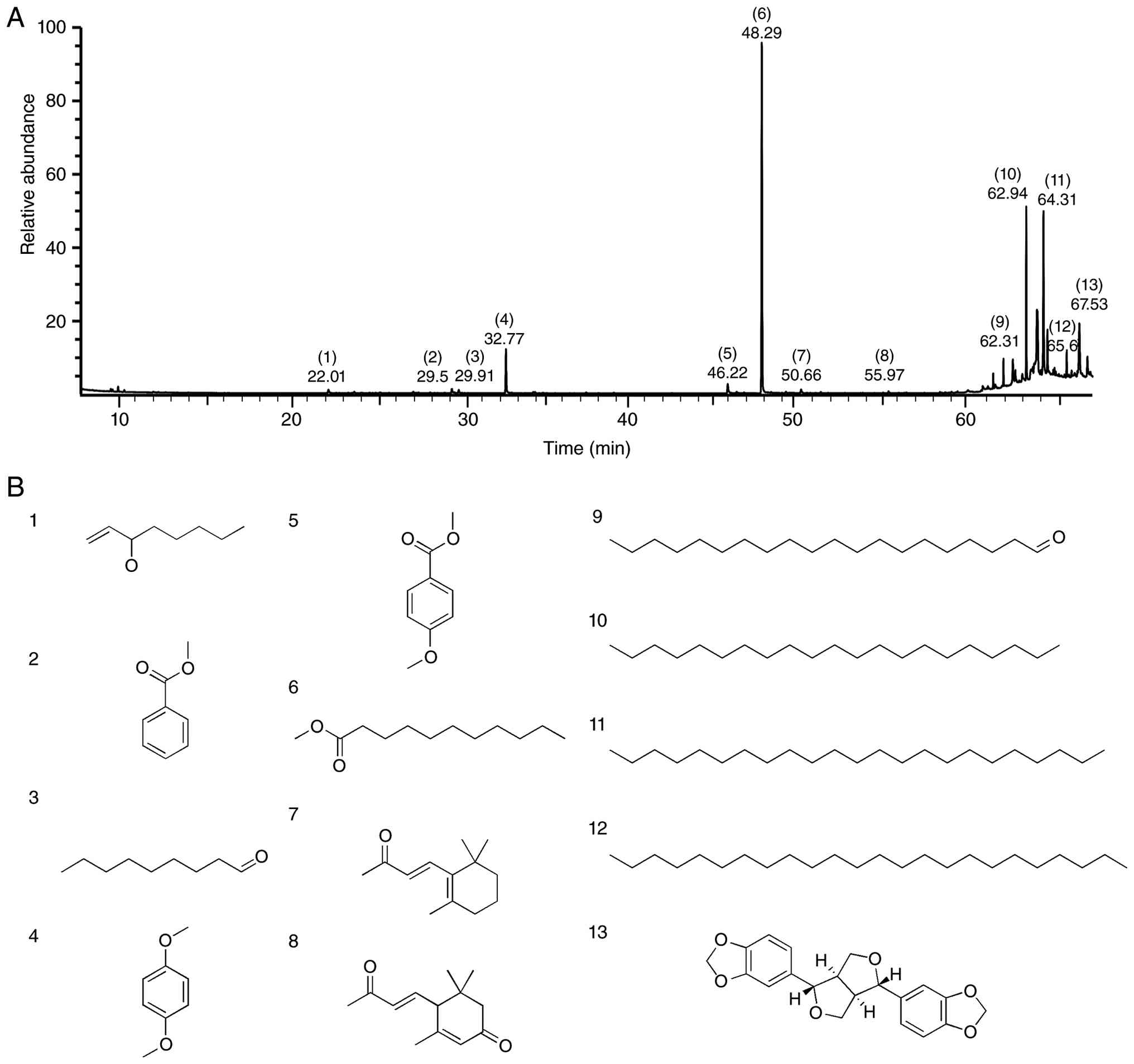
Figure 1

Gas chromatography/mass spectrometry
analysis of Paulownia tomentosa flower absolute. (A) Total
ion chromatogram showing the peaks corresponding to the 13 major
compounds listed in Table I. The
bracketed numbers and the numbers below them indicate the compound
numbers and retention times, respectively. (B) Chemical structures
of the 13 major compounds shown in panel A.

Figure 2

Effects of PTFAb on the expression of
SNARE proteins in RBL-2H3 cells. (A) RBL-2H3 cell viability.
RBL-2H3 cells were incubated in the presence or absence of PTFAb
(1–200 µg/ml) for 24 h. The final DMSO concentration was maintained
at 0.1% in all experimental groups, and control cells were treated
with 0.1% DMSO as the vehicle control. The cell viability was
analyzed using a WST assay (n=5). *P<0.05 compared with the
vehicle control cells. (B) RBL-2H3 cells were incubated for 48 h in
the presence or absence of PTFAb (0.1–100 µg/ml). The final DMSO
concentration was maintained at 0.1% in all groups, including the
vehicle control group. The cell lysates were immunoblotted with the
indicated antibodies. (C and D) The graphs show the expression
levels of the VAMP7 (C) and VAMP8 (D) proteins shown in panel B.
The expression levels of VAMP7 and VAMP8 were quantified by
normalization to β-actin (loading control) and presented as a
percentage relative to the vehicle control group. *P<0.05 vs.
the vehicle control cells. DMSO, dimethyl sulfoxide; PTFAb,
Paulownia tomentosa flower absolute; SNARE, soluble
N-ethylmaleimide-sensitive factor attachment protein receptor;
VAMP, vesicle-associated membrane protein; WST, water-soluble
tetrazolium.

Figure 3

Effects of PTFAb on β-hexosaminidase
and histamine release in IgE/DNP-stimulated RBL-2H3 cells. RBL-2H3
cells were incubated for 48 h in the presence or absence of PTFAb
(0.1–100 µg/ml). The final DMSO concentration was maintained at
0.1% in all experimental groups. The cells were treated with
anti-DNP IgE (200 ng/ml) for 10 h and then stimulated with DNP-BSA
(20 ng/ml) for 1 h. The culture media were collected and
centrifuged, and the levels of (A) β-hexosaminidase (n=3) and (B)
histamine (n=3) in the supernatant (conditioned media) were
measured using an enzyme immunoassay. The response in cells treated
with anti-DNP IgE alone in the presence of 0.1% DMSO was considered
100%. *P<0.05 vs. anti-DNP IgE/DNP-BSA-stimulated cells in the
presence of 0.1% DMSO alone. DMSO, dimethyl sulfoxide; anti-DNP
IgE, anti-dinitrophenyl immunoglobulin E; DNP-BSA,
2,4-dinitrophenyl-labeled bovine serum albumin; PTFAb, Paulownia
tomentosa flower absolute.

Figure 4

Effects of PTFAb on Syk, PI3K, and
AKT phosphorylation in RBL-2H3 cells. (A and B) Syk
phosphorylation. (A) Representative image. RBL-2H3 cells were
sensitized with anti-DNP IgE and subsequently stimulated with
DNP-BSA to induce Syk phosphorylation. The cells were treated with
PTFAb (0.1–100 µg/ml), and the expression levels of phosphorylated
Syk were analyzed by Western blotting (n=3). The final DMSO
concentration was maintained at 0.1% in all experimental groups.
(B) Quantification of the p-Syk levels normalized to the total Syk,
presented as a percentage relative to anti-DNP IgE-sensitized cells
treated with 0.1% DMSO alone. β-actin was used as a loading control
to verify equal protein loading. *P<0.05 vs. DNP-BSA-stimulated
cells in the presence of anti-DNP IgE alone. (C-E) Phosphorylation
of PI3K and AKT. (C) RBL-2H3 cells were sensitized with anti-DNP
IgE and then stimulated with DNP-BSA to induce PI3K and AKT
phosphorylation. The cells were treated with PTFAb (0.1–100 µg/ml),
and the expression levels of p-PI3K and p-AKT were analyzed by
Western blotting (n=3 for each protein). The final DMSO
concentration was maintained at 0.1% in all experimental groups.
Quantification of the (D) p-PI3K and (E) p-AKT levels normalized to
the respective total protein levels (total PI3K and total AKT), and
presented as percentages relative to anti-DNP-IgE-sensitized cells
treated with 0.1% DMSO alone. β-actin was used as a loading control
to verify equal protein loading. *P<0.05 vs. anti-DNP
IgE/DNP-BSA-stimulated cells in the presence of 0.1% DMSO alone.
DMSO, dimethyl sulfoxide; anti-DNP IgE, anti-dinitrophenyl
immunoglobulin E; DNP-BSA, 2,4-dinitrophenyl-labeled bovine serum
albumin; p-AKT, phosphorylated protein kinase B; p-PI3K,
phosphorylated phosphoinositide 3-kinase; PTFAb, Paulownia
tomentosa flower absolute; p-Syk, phosphorylated spleen
tyrosine kinase.

Figure 5

Effects of PTFAb on the proliferation
and migration of HaCaT cells. (A) Cell viability. HaCaT cells were
incubated in the presence or absence of PTFAb (1–500 µg/ml) for 24
h. The final DMSO concentration was maintained at 0.5% in all
experimental groups, and control cells were treated with 0.5% DMSO
as the vehicle control (−). The cell viability was analyzed using a
WST assay (n=5). *P<0.05 compared with the untreated cells. (B)
Proliferation. HaCaT cells were incubated in the presence or
absence of PTFAb (1–500 µg/ml) for 48 h. The final DMSO
concentration was maintained at 0.5% in all groups, including the
vehicle control group (−). Cell proliferation was analyzed using
the BrdU incorporation assay (n=3). rhEGF (50 ng/ml)-induced
proliferation was used as a positive control. The percentages
represent the levels compared to that of the vehicle control cells,
which are considered 100%. The results are presented as the means ±
SEMs. *P<0.05 compared to the quiescent state. (C and D)
Migration. HaCaT cells were incubated in the presence or absence of
PTFAb (1–500 µg/ml) for 210 min. The final DMSO concentration was
maintained at 0.5% in all experimental groups, including the
vehicle control group. The cell migration levels were assessed
using the Boyden chamber assay. (C) Representative images. The blue
spots indicate migrating cells. (D) Statistical graph obtained from
panel C. rhEGF (1 ng/ml)-induced migration was used as a positive
control. The percentages represent the levels compared to the
vehicle control, which are considered 100% (n=3). The results are
presented as the means ± SEMs. Scale bar=50 µm. *P<0.05 compared
with the vehicle control group. Con, vehicle control; BrdU,
5-bromo-2′-deoxyuridine; DMSO, dimethyl sulfoxide; PTFAb,
Paulownia tomentosa flower absolute; rhEGF, recombinant
human epidermal growth factor; SEMs, standard errors of the means;
WST, water-soluble tetrazolium.

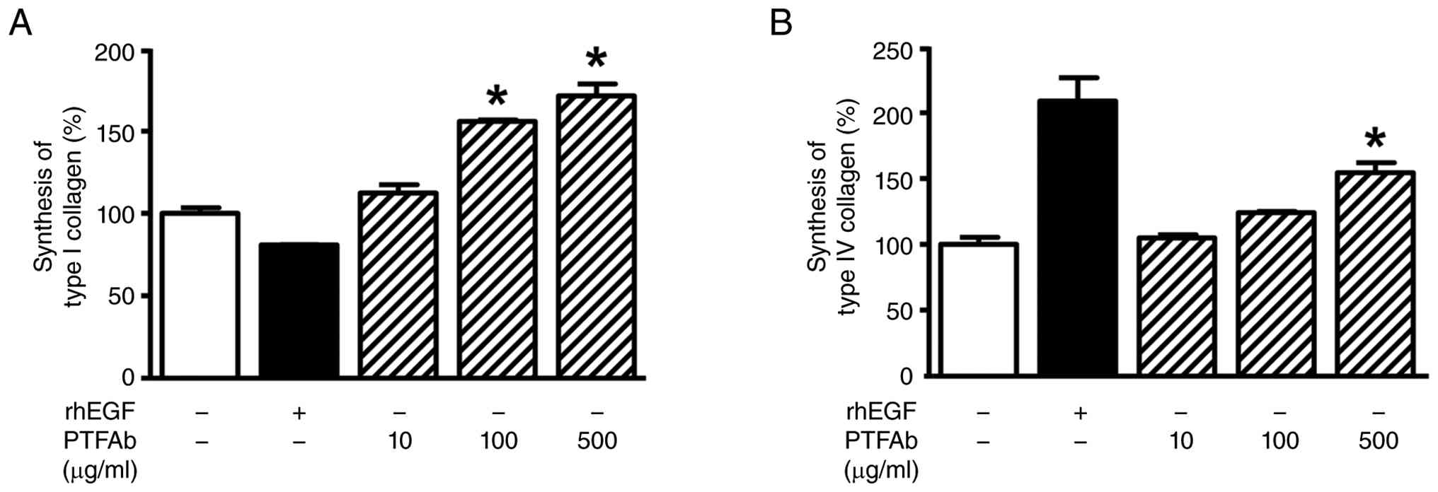
Figure 6

Effects of PTFAb on the synthesis of
type I and IV collagen in HaCaT cells. HaCaT cells were incubated
in the presence or absence of PTFAb (10, 100, and 500 µg/ml) for 48
h. The final DMSO concentration was maintained at 0.5% in all
experimental groups, and control cells were treated with 0.5% DMSO
as the vehicle control. The levels of (A) type I (n=3) and (B) IV
collagen (n=3) in the conditioned media were quantified using a
sandwich ELISA with the specific antibodies. The collagen synthesis
levels are expressed as percentages of the levels in the
conditioned media of vehicle control cells (−) considered for 100%
(n=3). rhEGF (50 ng/ml)-induced response was used as a positive
control. The results are presented as means ± SEMs. *P<0.05
compared to the vehicle control cells. DMSO, dimethyl sulfoxide;
ELISA, enzyme-linked immunosorbent assay; PTFAb, Paulownia
tomentosa flower absolute; rhEGF, recombinant human epidermal
growth factor; SEMs, standard errors of the means.

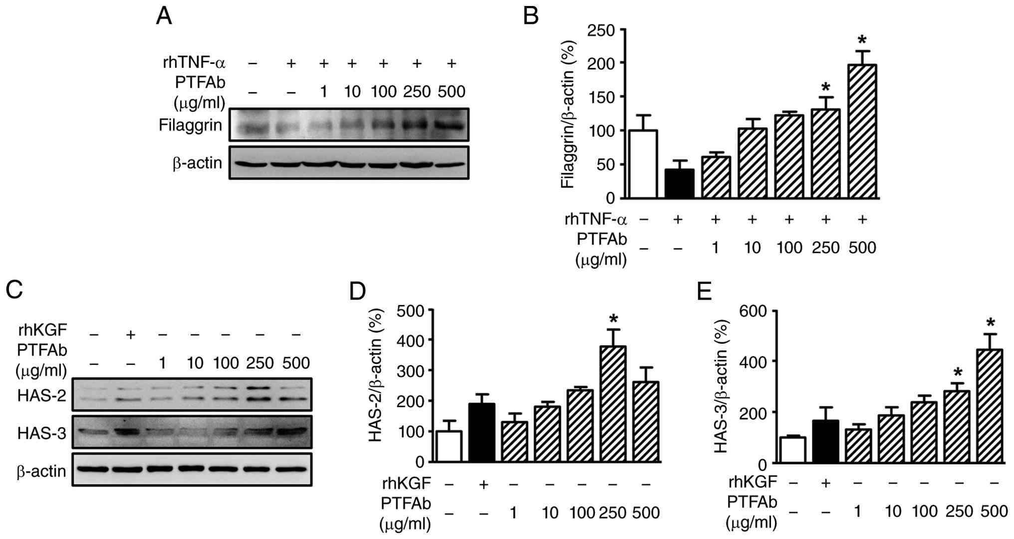
Figure 7

Effects of PTFAb on expression of
filaggrin and hyaluronan synthases in HaCaT cells. (A and B)
Filaggrin expression. (A) Representative image. HaCaT cells were
stimulated with TNF-α (5 ng/ml) to suppress filaggrin expression
and then treated with PTFAb (1–500 µg/ml). The final DMSO
concentration was maintained at 0.5% in all experimental groups.
The filaggrin protein levels were analyzed by immunoblotting (n=3).
(B) Statistical graph. The relative expression of filaggrin was
quantified by normalizing to β-actin (loading control) and
presented as a percentage of the 0.5% DMSO alone-treated control
group (vehicle control) (*P<0.05 vs. TNF-α-stimulated cells in
the presence of 0.5% DMSO alone). (C-E) Hyaluronan syntheses. (C)
Representative images. HaCaT cells were treated with PTFAb (1–500
µg/ml), and rhKGF (20 ng/ml) was applied to induce HAS expression.
The final DMSO concentration was maintained at 0.5% in all
experimental groups. The protein levels of HAS-2 and HAS-3 were
analyzed by Western blotting (n=3 for each protein). (D and E)
Statistical graph. The relative expression levels of (D) HAS-2 and
(E) HAS-3 were quantified by normalizing to β-actin (loading
control) and presented as percentages relative to the 0.5% DMSO
alone-treated vehicle control cells. rhKGF was used as a positive
control. *P<0.05 compared to 0.5% DMSO alone-treated control
group. DMSO, dimethyl sulfoxide; HAS, hyaluronan synthase; PTFAb,
Paulownia tomentosa flower absolute; rhKGF, recombinant
human keratinocyte growth factor; TNF-α, tumor necrosis
factor-α.

Figure 8

Effects of PTFAb on the expression of
ICAM-1 protein in HaCaT cells. (A) HaCaT cells were stimulated with
TNF-α (10 ng/ml) to induce ICAM-1 expression and then treated with
PTFAb (1–500 µg/ml). The final DMSO concentration was maintained at
0.5% in all experimental groups. The levels of ICAM-1 protein were
analyzed by immunoblotting (n=3). (B) The relative expression of
ICAM-1 was quantified by normalizing it to β-actin (loading
control) and expressed as a percentage relative to the 0.5% DMSO
alone-treated control group (vehicle control) (*P<0.05 vs.
TNF-α-stimulated cells in the presence of 0.5% DMSO alone). DMSO,
dimethyl sulfoxide; ICAM-1, intercellular adhesion molecule 1;
PTFAb, Paulownia tomentosa flower absolute; TNF-α, tumor
necrosis factor-α.
View References

1 

Sroka-Tomaszewska J and Trzeciak M: Molecular mechanisms of atopic dermatitis pathogenesis. Int J Mol Sci. 22:41302021. View Article : Google Scholar : PubMed/NCBI

2 

Mohd Kasim VNK, Noble SM, Liew KY, Tan JW, Israf DA and Tham CL: Management of atopic dermatitis via oral and topical administration of herbs in murine model: A systematic review. Front Pharmacol. 13:7857822022. View Article : Google Scholar : PubMed/NCBI

3 

Matsuda H, Nakamura S and Yoshikawa M: Degranulation inhibitors from medicinal plants in antigen-stimulated rat basophilic leukemia (RBL-2H3) cells. Chem Pharm Bull (Tokyo). 64:96–103. 2016. View Article : Google Scholar : PubMed/NCBI

4 

Woska JR Jr and Gillespie ME: SNARE complex-mediated degranulation in mast cells. J Cell Mol Med. 16:649–656. 2012. View Article : Google Scholar : PubMed/NCBI

5 

Hada M, Nishi K, Ishida M, Onda H, Nishimoto S and Sugahara T: Inhibitory effect of aqueous extract of Cuminum cyminum L. seed on degranulation of RBL-2H3 cells and passive cutaneous anaphylaxis reaction in mice. Cytotechnology. 71:599–609. 2019. View Article : Google Scholar : PubMed/NCBI

6 

McCarty MF, Lerner A, DiNicolantonio JJ and Benzvi C: Nutraceutical aid for allergies-strategies for down-regulating mast cell degranulation. J Asthma Allergy. 14:1257–1266. 2021. View Article : Google Scholar : PubMed/NCBI

7 

Furue M: Regulation of filaggrin, loricrin, and involucrin by IL-4, IL-13, IL-17A, IL-22, AHR, and NRF2: Pathogenic implications in atopic dermatitis. Int J Mol Sci. 21:53822020. View Article : Google Scholar : PubMed/NCBI

8 

Proksch E, Brandner JM and Jensen JM: The skin: An indispensable barrier. Exp Dermatol. 17:1063–1072. 2008. View Article : Google Scholar : PubMed/NCBI

9 

Potekaev NN, Borzykh OB, Medvedev GV, Pushkin DV, Petrova MM, Petrov AV, Dmitrenko DV, Karpova EI, Demina OM and Shnayder NA: The role of extracellular matrix in skin wound healing. J Clin Med. 10:59472021. View Article : Google Scholar : PubMed/NCBI

10 

Papakonstantinou E, Roth M and Karakiulakis G: Hyaluronic acid: A key molecule in skin aging. Dermatoendocrinol. 4:253–258. 2012. View Article : Google Scholar : PubMed/NCBI

11 

Evrard C, Lambert de Rouvroit C and Poumay Y: Epidermal hyaluronan in barrier alteration-related disease. Cells. 10:30962021. View Article : Google Scholar : PubMed/NCBI

12 

Diller RB and Tabor AJ: The role of the extracellular matrix (ECM) in wound healing: A review. Biomimetics (Basel). 7:872022. View Article : Google Scholar : PubMed/NCBI

13 

Choudhary V, Choudhary M and Bollag WB: Exploring skin wound healing models and the impact of natural lipids on the healing process. Int J Mol Sci. 25:37902024. View Article : Google Scholar : PubMed/NCBI

14 

Rerknimitr P, Otsuka A, Nakashima C and Kabashima K: The etiopathogenesis of atopic dermatitis: Barrier disruption, immunological derangement, and pruritus. Inflamm Regen. 37:142017. View Article : Google Scholar : PubMed/NCBI

15 

Singer KH, Tuck DT, Sampson HA and Hall RP: Epidermal keratinocytes express the adhesion molecule intercellular adhesion molecule-1 in inflammatory dermatoses. J Invest Dermatol. 92:746–750. 1989. View Article : Google Scholar : PubMed/NCBI

16 

Matsunaga T, Katayama I, Yokozeki H and Nishioka K: ICAM-1 expression on keratinocytes in mechanically-injured skin of a patient with atopic dermatitis. J Dermatol Sci. 12:219–226. 1996. View Article : Google Scholar : PubMed/NCBI

17 

Man G, Hu LZ, Elias PM and Man MQ: Therapeutic benefits of natural ingredients for atopic dermatitis. Chin J Integr Med. 24:308–314. 2018. View Article : Google Scholar : PubMed/NCBI

18 

Valdivieso-Ugarte M, Gomez-Llorente C, Plaza-Díaz J and Gil Á: Antimicrobial, antioxidant, and immunomodulatory properties of essential oils: A systematic review. Nutrients. 11:27862019. View Article : Google Scholar : PubMed/NCBI

19 

Kim DY, Won KJ, Kim YY, Yoo DY and Lee HM: Potential wound healing and anti-melanogenic activities in skin cells of Aralia elata (Miq.) Seem. flower essential oil and its chemical composition. Pharmaceutics. 16:10082024. View Article : Google Scholar : PubMed/NCBI

20 

Chung IM, Kim EH, Jeon HS and Moon HI: Protective effects of isoatriplicolide tiglate from Paulownia coreana against glutamate-induced neurotoxicity in primary cultured rat cortical cells. Nat Prod Commun. 5:851–852. 2010.PubMed/NCBI

21 

Si CL, Kim JK, Kwon DJ and Bae YS: Phenylpropanoid glycosides of Paulownia coreana Uyeki leaves. J Korean Wood Sci Technol. 34:78–82. 2006.

22 

Kim TJ: Korean resources plants (IV). Seoul National University Press; Seoul: pp. 76–77. 1996

23 

Kim NK, Kim MH, Yoon CS and Choi SW: Studies on the anti-inflammatory activity of Paulownia coreana Uyeki leaf extract. J Soc Cosmet Sci Korea. 32:241–247. 2006.

24 

Jin Q, Lee C, Lee JW, Lee D, Kim Y, Hong JT, Kim JS, Kim JH, Lee MK and Hwang BY: Geranylated flavanones from Paulownia coreana and their inhibitory effects on nitric oxide production. Chem Pharm Bull (Tokyo). 63:384–387. 2015. View Article : Google Scholar : PubMed/NCBI

25 

Teng Y, Zhong H, Yang X, Tao X and Fan Y: Current and emerging therapies for atopic dermatitis in the elderly. Clin Interv Aging. 18:1641–1652. 2023. View Article : Google Scholar : PubMed/NCBI

26 

International Union for Conservation of Nature, . The IUCN red list of threatened species. https://www.iucnredlist.orgMarch. 2026

27 

National Institute of Biological Resources (NIBR), . Korean Red Data Book of Vascular Plants (2021). https://species.nibr.go.krMarch. 2026

28 

Kim MJ, Won KJ, Kim DY, Won YR, Kim NY, Lee DK, Hong BS and Lee HM: Skin wound healing and anti-wrinkle-promoting in vitro biological activities of Caragana sinica flower absolute and its chemical composition. Pharmaceuticals (Basel). 16:2352023. View Article : Google Scholar : PubMed/NCBI

29 

Yang Y, Kong B, Jung Y, Park JB, Oh JM, Hwang J, Cho JY and Kweon DH: Soluble N-ethylmaleimide-sensitive factor attachment protein receptor-derived peptides for regulation of mast cell degranulation. Front Immunol. 9:7252018. View Article : Google Scholar : PubMed/NCBI

30 

Lee HP, Choi W, Kwon KW, You L, Rahmawati L, Luong VD, Kim W, Lee BH, Lee S, Kim JH and Cho JY: Inhibitory effects of grewia tomentosa Juss. on IgE-mediated allergic reaction and DNCB-induced atopic dermatitis. Plants (Basel). 11:25402022.PubMed/NCBI

31 

Bairoch A: The cellosaurus, a cell-line knowledge resource. J Biomol Tech. 29:25–38. 2018. View Article : Google Scholar : PubMed/NCBI

32 

Bae WB, Kim EH, Kim MJ and Yang SA: Inhibitory effects of Broussonetia kazinoki twig extract on allergic inflammatory reactions in TNF-α/IFN-γ-stimulated HaCaT and IgE-sensitized RBL-2H3 cells. Food Sci Preserv. 31:307–314. 2024. View Article : Google Scholar

33 

Bao Y, Wang S, Gao Y, Zhang W, Jin H, Yang Y and Li J: MicroRNA-126 accelerates IgE-mediated mast cell degranulation associated with the PI3K/Akt signaling pathway by promoting Ca2+ influx. Exp Ther Med. 16:2763–2769. 2018.PubMed/NCBI

34 

Siraganian RP, de Castro RO, Barbu EA and Zhang J: Mast cell signaling: The role of protein tyrosine kinase Syk, its activation and screening methods for new pathway participants. FEBS Lett. 584:4933–4940. 2010. View Article : Google Scholar : PubMed/NCBI

35 

Rivera J and Gilfillan AM: Molecular regulation of mast cell activation. J Allergy Clin Immunol. 117:1214–1226. 2006. View Article : Google Scholar : PubMed/NCBI

36 

Mathew-Steiner SS, Roy S and Sen CK: Collagen in wound healing. Bioengineering (Basel). 8:632021. View Article : Google Scholar : PubMed/NCBI

37 

Levin J, Friedlander SF and Del Rosso JQ: Atopic dermatitis and the stratum corneum: Part 1: The role of filaggrin in the stratum corneum barrier and atopic skin. J Clin Aesthet Dermatol. 6:16–22. 2013.PubMed/NCBI

38 

Malaisse J, Bourguignon V, De Vuyst E, Lambert de Rouvroit C, Nikkels AF, Flamion B and Poumay Y: Hyaluronan metabolism in human keratinocytes and atopic dermatitis skin is driven by a balance of hyaluronan synthases 1 and 3. J Invest Dermatol. 134:2174–2182. 2014. View Article : Google Scholar : PubMed/NCBI

39 

Kim DY, Won KJ, Hwang DI, Lee SY, Choi IH, Kim B and Lee HM: Essential oil from Chrysanthemum boreale flowers modulates SNARE protein-linked mast cell response and skin barrier proteins and ameliorates atopic dermatitis-like lesions in mice. Hortic Environ Biotechnol. 63:287–298. 2022. View Article : Google Scholar

40 

Yun Y, Kim K, Choi I and Ko SG: Topical herbal application in the management of atopic dermatitis: A review of animal studies. Mediators Inflamm. 2014:7521032014. View Article : Google Scholar : PubMed/NCBI

41 

Liu FT, Goodarzi H and Chen HY: IgE, mast cells, and eosinophils in atopic dermatitis. Clin Rev Allergy Immunol. 41:298–310. 2011. View Article : Google Scholar : PubMed/NCBI

42 

Li LC, Piao HM, Zheng MY, Lin ZH, Li G and Yan GH: Sesamin attenuates mast cell-mediated allergic responses by suppressing the activation of p38 and nuclear factor-κB. Mol Med Rep. 13:536–542. 2016. View Article : Google Scholar : PubMed/NCBI

43 

Wikramanayake TC, Stojadinovic O and Tomic-Canic M: Epidermal differentiation in barrier maintenance and wound healing. Adv Wound Care (New Rochelle). 3:272–280. 2014. View Article : Google Scholar : PubMed/NCBI

44 

Park S, Kang W, Choi D, Son B and Park T: Nonanal stimulates growth factors via cyclic adenosine monophosphate (cAMP) signaling in human hair follicle dermal papilla cells. Int J Mol Sci. 21:80542020. View Article : Google Scholar : PubMed/NCBI

45 

Putnins EE, Firth JD, Lohachitranont A, Uitto VJ and Larjava H: Keratinocyte growth factor (KGF) promotes keratinocyte cell attachment and migration on collagen and fibronectin. Cell Adhes Commun. 7:211–221. 1999. View Article : Google Scholar : PubMed/NCBI

46 

Brett D: A review of collagen and collagen-based wound dressings. Wounds. 20:347–356. 2008.PubMed/NCBI

47 

O'Toole EA: Extracellular matrix and keratinocyte migration. Clin Exp Dermatol. 26:525–530. 2001. View Article : Google Scholar : PubMed/NCBI

48 

Boudko SP, Danylevych N, Hudson BG and Pedchenko VK: Basement membrane collagen IV: Isolation of functional domains. Methods Cell Biol. 143:171–185. 2018. View Article : Google Scholar : PubMed/NCBI

49 

Motter Catarino C, Kaiser K, Baltazar T, Motter Catarino L, Brewer JR and Karande P: Evaluation of native and non-native biomaterials for engineering human skin tissue. Bioeng Transl Med. 7:e102972022. View Article : Google Scholar : PubMed/NCBI

50 

Szalus K and Trzeciak M: The role of collagens in atopic dermatitis. Int J Mol Sci. 25:76472024. View Article : Google Scholar : PubMed/NCBI

51 

Moosbrugger-Martinz V, Leprince C, Méchin MC, Simon M, Blunder S, Gruber R and Dubrac S: Revisiting the roles of filaggrin in atopic dermatitis. Int J Mol Sci. 23:53182022. View Article : Google Scholar : PubMed/NCBI

52 

Dang NN, Pang SG, Song HY, An LG and Ma XL: Filaggrin silencing by shRNA directly impairs the skin barrier function of normal human epidermal keratinocytes and then induces an immune response. Braz J Med Biol Res. 48:39–45. 2015. View Article : Google Scholar : PubMed/NCBI

53 

Hsu CY, Lecland N, Pendaries V, Viodé C, Redoulès D, Paul C, Merdes A, Simon M and Bierkamp C: Stabilization of microtubules restores barrier function after cytokine-induced defects in reconstructed human epidermis. J Dermatol Sci. 91:87–96. 2018. View Article : Google Scholar : PubMed/NCBI

54 

Bonté F: Skin moisturization mechanisms: New data. Ann Pharm Fr. 69:135–141. 2011. View Article : Google Scholar : PubMed/NCBI

55 

Zheng BW, Wang BY, Xiao WL, Sun YJ, Yang C and Zhao BT: Different molecular weight hyaluronic acid alleviates inflammation response in DNFB-induced mice atopic dermatitis and LPS-induced RAW 264.7 cells. Life Sci. 301:1205912022. View Article : Google Scholar : PubMed/NCBI

56 

Lee YI, Lee SG, Kim J, Choi S, Jung I and Lee JH: Proteoglycan combined with hyaluronic acid and hydrolyzed collagen restores the skin barrier in mild atopic dermatitis and dry, eczema-prone skin: A pilot study. Int J Mol Sci. 22:101892021. View Article : Google Scholar : PubMed/NCBI

57 

Griffiths CE and Nickoloff BJ: Keratinocyte intercellular adhesion molecule-1 (ICAM-1) expression precedes dermal T lymphocytic infiltration in allergic contact dermatitis (Rhus dermatitis). Am J Pathol. 135:1045–1053. 1989.PubMed/NCBI

58 

Griffiths CE, Voorhees JJ and Nickoloff BJ: Characterization of intercellular adhesion molecule-1 and HLA-DR expression in normal and inflamed skin: Modulation by recombinant gamma interferon and tumor necrosis factor. J Am Acad Dermatol. 20:617–629. 1989. View Article : Google Scholar : PubMed/NCBI

59 

Klesk K, Qian M and Martin RR: Aroma extract dilution analysis of cv. meeker (Rubus idaeus L.) red raspberries from Oregon and Washington. J Agric Food Chem. 52:5155–5161. 2004. View Article : Google Scholar : PubMed/NCBI

60 

Wang Q, Yang Y, Zhao X, Zhu B, Nan P, Zhao J, Wang L, Chen F, Liu Z and Zhong Y: Chemical variation in the essential oil of Ephedra sinica from Northeastern China. Food Chem. 98:52–58. 2006. View Article : Google Scholar

61 

Bozin B, Mimicá-Dukic N, Simin N and Anackov G: Characterization of the volatile composition of essential oils of some lamiaceae spices and the antimicrobial and antioxidant activities of the entire oils. J Agric Food Chem. 54:1822–1828. 2006. View Article : Google Scholar : PubMed/NCBI

62 

Tsuge S, Ohtan H and Watanabe C: Pyrolysis-GC/MS data book of synthetic polymers. Elsevier; pp. pp4202011

63 

Pino JA, Mesa J, Muñoz Y, Martí MP and Marbot R: Volatile components from mango (Mangifera indica L.) cultivars. J Agric Food Chem. 53:2213–2223. 2005. View Article : Google Scholar : PubMed/NCBI

64 

Wang Y, Finn C and Qian MC: Impact of growing environment on chickasaw blackberry (Rubus L.) aroma evaluated by gas chromatography olfactometry dilution analysis. J Agric Food Chem. 53:3563–3571. 2005. View Article : Google Scholar : PubMed/NCBI

65 

Jerković I, Tuberso CIG, Gugić M and Bubalo D: Composition of sulla (Hedysarum coronarium L.) honey solvent extractives determined by GC/MS: Norisoprenoids and other volatile organic compounds. Molecules. 15:6375–6385. 2010. View Article : Google Scholar : PubMed/NCBI

66 

Naka H, van Vang L, Inomata SI, Ando T, Kimura T, Honda H, Tsuchida K and Sakurai H: Sex pheromone of the persimmon fruit moth, Stathmopoda masinissa: Identification and laboratory bioassay of (4E, 6Z)-4,6-hexadecadien-1-ol derivatives. J Chem Ecol. 29:2447–2459. 2003. View Article : Google Scholar : PubMed/NCBI

67 

National Institute of Standards and Technology (NIST), . NIST Mass Spectrometry Data Center. NIST Gaithersburg, MD: 2014

Related Articles

  • Abstract
  • View
  • Download
  • Twitter
Copy and paste a formatted citation
Spandidos Publications style
Yoo DY, Won KJ, Kim DY, Kim YY, Bae JH, Yun JS and Lee HM: Ameliorative effects of <em>Paulownia tomentosa</em> flower absolute on atopic dermatitis‑related responses in mast cells and keratinocytes and its chemical composition. Mol Med Rep 34: 304, 2026.
APA
Yoo, D.Y., Won, K.J., Kim, D.Y., Kim, Y.Y., Bae, J.H., Yun, J.S., & Lee, H.M. (2026). Ameliorative effects of <em>Paulownia tomentosa</em> flower absolute on atopic dermatitis‑related responses in mast cells and keratinocytes and its chemical composition. Molecular Medicine Reports, 34, 304. https://doi.org/10.3892/mmr.2026.14015
MLA
Yoo, D. Y., Won, K. J., Kim, D. Y., Kim, Y. Y., Bae, J. H., Yun, J. S., Lee, H. M."Ameliorative effects of <em>Paulownia tomentosa</em> flower absolute on atopic dermatitis‑related responses in mast cells and keratinocytes and its chemical composition". Molecular Medicine Reports 34.5 (2026): 304.
Chicago
Yoo, D. Y., Won, K. J., Kim, D. Y., Kim, Y. Y., Bae, J. H., Yun, J. S., Lee, H. M."Ameliorative effects of <em>Paulownia tomentosa</em> flower absolute on atopic dermatitis‑related responses in mast cells and keratinocytes and its chemical composition". Molecular Medicine Reports 34, no. 5 (2026): 304. https://doi.org/10.3892/mmr.2026.14015
Copy and paste a formatted citation
x
Spandidos Publications style
Yoo DY, Won KJ, Kim DY, Kim YY, Bae JH, Yun JS and Lee HM: Ameliorative effects of <em>Paulownia tomentosa</em> flower absolute on atopic dermatitis‑related responses in mast cells and keratinocytes and its chemical composition. Mol Med Rep 34: 304, 2026.
APA
Yoo, D.Y., Won, K.J., Kim, D.Y., Kim, Y.Y., Bae, J.H., Yun, J.S., & Lee, H.M. (2026). Ameliorative effects of <em>Paulownia tomentosa</em> flower absolute on atopic dermatitis‑related responses in mast cells and keratinocytes and its chemical composition. Molecular Medicine Reports, 34, 304. https://doi.org/10.3892/mmr.2026.14015
MLA
Yoo, D. Y., Won, K. J., Kim, D. Y., Kim, Y. Y., Bae, J. H., Yun, J. S., Lee, H. M."Ameliorative effects of <em>Paulownia tomentosa</em> flower absolute on atopic dermatitis‑related responses in mast cells and keratinocytes and its chemical composition". Molecular Medicine Reports 34.5 (2026): 304.
Chicago
Yoo, D. Y., Won, K. J., Kim, D. Y., Kim, Y. Y., Bae, J. H., Yun, J. S., Lee, H. M."Ameliorative effects of <em>Paulownia tomentosa</em> flower absolute on atopic dermatitis‑related responses in mast cells and keratinocytes and its chemical composition". Molecular Medicine Reports 34, no. 5 (2026): 304. https://doi.org/10.3892/mmr.2026.14015
Follow us
  • Twitter
  • LinkedIn
  • Facebook
About
  • Spandidos Publications
  • Careers
  • Cookie Policy
  • Privacy Policy
How can we help?
  • Help
  • Live Chat
  • Contact
  • Email to our Support Team