Introduction
Atopic dermatitis (AD) is a representative chronic
inflammatory skin disease caused by a combination of immune
dysfunction and impaired skin barrier function. Its prevalence is
steadily increasing worldwide (1).
The pathogenesis of AD is shaped by a complex interplay of genetic
predisposition, environmental stimuli, immunological abnormalities,
and impaired skin barrier function (1). In particular, the excessive
activation of the T helper type 2 (Th2) cell immune response acts
as a key factor in exacerbating the inflammatory response of AD by
promoting the overproduction of immunoglobulin E (IgE) antibodies
in B cells and the sensitization and activation of mast cells
(2). The binding of IgE and its
receptors [high affinity IgE receptors (FcεRI)] activates mast
cells and triggers mast cell degranulation, releasing various
allergic or inflammatory mediators stored within the cells, such as
cytokines, histamine, and β-hexosaminidase, contributing directly
to the induction of itching and inflammatory skin lesions (2,3).
Mast cell degranulation is regulated by membrane fusion between
intracellular vesicles and the cell membrane. In addition, SNARE
(soluble N-ethylmaleimide-sensitive factor attachment protein
receptor) protein complexes play a key role in this process
(4). In particular, vesicle
membrane-located v(vesicle)-SNARE proteins, such as
vesicle-associated membrane protein (VAMP)7 and VAMP8, bind to
membrane-located t(target)-SNARE proteins, synaptosome-associated
protein (SNAP) 23, and syntaxin family proteins, forming a
quadruple-helix SNARE complex (4).
The bound SNARE complex induces membrane fusion and degranulation,
subsequently releasing allergic and inflammatory mediators from the
cells (4). This degranulation
response in activated mast cells is closely linked to the
activation of spleen tyrosine kinase (Syk) and its downstream
molecules, such as phosphatidylinositol 3-kinase (PI3K)/protein
kinase B (AKT) and mitogen-activated protein kinases (MAPKs)
signaling pathways (5,6). Therefore, SNARE protein complexes and
related signaling molecules are becoming important molecular
targets for modulating mast cell-mediated allergic and inflammatory
responses.
In addition to these immunological abnormalities,
structural impairment of the skin barrier function is another key
pathogenic factor determining the onset and exacerbation of AD
(7). The normal skin barrier
maintains homeostasis through the harmonious interaction of
keratinocyte differentiation-linked structural proteins [filaggrin
(FLG), loricrin (LOR), and involucrin (IVL)], a ceramide-centered
lipid layer, and extracellular matrix (ECM) components (e.g.,
collagen), protecting the body from external harmful environments
(8,9). FLG, LOR, and IVL are key structural
proteins of the outer layer of the epidermis (stratum corneum),
which is formed through the terminal differentiation of
keratinocytes and is essential for the skin barrier function
(7). These proteins regulate the
skin barrier integrity and hydration, and deficiencies in them
contribute to epidermal barrier dysfunction, a central feature of
AD (7). In addition to structural
proteins, proper hydration of the stratum corneum is critical for
barrier homeostasis. Hyaluronan (also called hyaluronic acid)
maintains skin moisture through its high water-binding capacity and
is synthesized by hyaluronan synthase (HAS) isoforms (HAS-1, −2,
and −3) (10,11). Alterations in hyaluronic acid
synthesis and degradation have been linked to the development and
progression of AD (11).
When the skin barrier is continuously damaged, the
tissue promotes the proliferation and migration of keratinocytes
and induces collagen synthesis, a key component of the ECM, to
re-epithelialize and structurally repair the damaged area (12,13).
On the other hand, the persistent inflammatory environment in AD
disrupts this normal repair process, and incomplete tissue
regeneration further exacerbates the barrier vulnerability
(14). In a cytokine-rich
inflammatory environment characteristic of AD, keratinocytes can
show enhanced intercellular adhesion molecule-1 (ICAM-1) expression
in response to inflammatory stimuli (15). This increased ICAM-1 expression may
promote the infiltration of immune cells into the skin and enhance
their interaction with ICAM-1, thereby triggering localized
inflammatory processes (16).
Therefore, controlling ICAM-1 expression regulation may help treat
skin inflammatory diseases such as AD.
The adverse effects of long-term use of synthetic
steroids and immunosuppressants have increased interest in safe and
effective natural ingredients that can modulate the immune
responses and restore the skin barrier (17). Plant essential oils contain a
variety of secondary metabolites with the potential to exert
diverse biological effects, including antibacterial,
anti-inflammatory, and wound healing properties (18,19).
Among these, Paulownia tomentosa (also called Paulownia
coreana Uyeki at the National Institute of Biological Resources
in Korea) is a deciduous tree belonging to the Paulowniaceae
family, native to Korea, China, and Japan (20). This plant is known for its timber
and ornamental value and is used widely in construction, furniture
making, musical instruments, and handicrafts (21). Traditionally, it has been used to
treat bronchial diseases such as cough, asthma, and bronchitis
(22). In addition,
pharmacological studies have reported that extracts and bioactive
compounds derived from various parts of the plant, including the
flowers, leaves, and fruit, have diverse biological activities,
such as anti-inflammatory, antioxidant, neuroprotective, and nitric
oxide inhibitory effects (20,23,24).
Nevertheless, little research has been conducted on
the effects of Paulownia tomentosa (PT) flower essential oil
on mast cell degranulation and skin barrier function, which are
related to the pathogenesis of AD. Although many topical and
systemic treatments for AD have been developed (25), their clinical utility has been
frequently limited because of their side effects and the complex
etiology of the disease. Therefore, novel strategies are essential
to overcome these existing limitations. In the present study, PT
flower absolute (PTFAb) was isolated, and its chemical composition
was analyzed by gas chromatography/mass spectrometry (GC/MS). The
effects of PTFAb on AD-related pathological aspects were also
investigated comprehensively, particularly mast cell degranulation,
its modulatory signaling, and skin barrier function-related
responses, in vitro. The following cells were used in this
in vitro study: rat basophilic leukemia cells (RBL-2H3
cells) as a mast cell model and human keratinocytes (HaCaT cells)
as an epithelial cell model.
Materials and methods
Materials
Fetal bovine serum (FBS; cat. no. SH30919.03) and
penicillin/streptomycin (P/S; cat. no. SH40003.01) were purchased
from Hyclone (Logan, UT, USA). Trypsin-ethylenediaminetetraacetic
acid (cat. no. 15400-054) was purchased from Gibco BRL
(Gaithersburg, MD, USA). Phosphate-buffered saline (PBS; cat. no.
LB 001-02) and Dulbecco's Modified Eagle Medium (DMEM; high
glucose, cat. no. LM 001-05; low glucose, cat. no. LM 001-11) were
supplied by Welgene (Daegu, Korea). Anti-β-actin antibody (cat. no.
A5441), anti-DNP IgE antibody (cat. no. D8406),
p-nitrophenyl-N-acetyl-β-D-glucosaminide (cat.
no. N9376), Tween-20 (cat. no. P2287), sucrose (cat. no. S1888),
polyvinylidene fluoride (PVDF) membrane (cat. no. IPVH00010),
dimethyl sulfoxide (DMSO; cat. no. D2650), LY294002 (cat. no.
440202), SB203580 (cat. no. S8307), and PD98059 (cat. no. P215)
were obtained from MilliporeSigma (St. Louis, MO, USA). Recombinant
human keratinocyte growth factor (rhKGF; cat. no. 251-KG),
recombinant human epidermal growth factor (rhEGF; cat. no. 236-EG)
(purity >97%), Recombinant human tumor necrosis factor-α
(rhTNF-α; cat. no. 210-TA), and anti-ICAM-1 antibody (cat. no. BAF
796) were acquired from R&D Systems (Minneapolis, MN, USA). The
EZ-CyTox kit (cat. no. EZ-3000) and bovine serum albumin (BSA; cat.
No. A0100-010) were supplied by DoGenBio (Seoul, Korea) and
GenDEPOT (Katy, TX, USA), respectively. Type I collagen (rat tail;
cat. no. 354236) was sourced from Corning Life Sciences (Corning,
NY, USA). The 5-bromo-2′-deoxyuridine (BrdU) kit (cat. no.
11669915001) and streptavidin-peroxidase (POD) conjugate (cat. no.
11089153001) was obtained from Roche (Indianapolis, IN, USA).
DNP-BSA (cat. no. A23018), monoclonal anti-type I and IV collagen
antibodies (cat. no. MA1-26771 and cat. no. MA1-22148,
respectively) and chemiluminescent substrate (cat. no. 37069) were
obtained from Thermo Scientific (Waltham, MA, USA). The antibodies
for phospho Syk (cat. no. 2711), ERK1/2 (cat. no. 9102), phospho
ERK1/2 (cat. no. 9101), p38 MAPK (cat. no. 9212), phospho p38 MAPK
(cat. no. 9211), JNK (cat. no. 9258), phospho JNK (cat. no. 4668),
PI3K (cat. no. 4292), phospho PI3K (cat. no. 4228), AKT (cat. no.
9272), phospho AKT (cat. no. 9271), horseradish peroxidase
conjugated rabbit immunoglobulin G (cat. no. 7074), and horseradish
peroxidase conjugated mouse immunoglobulin G (cat. no. 7076) were
purchased from Cell Signaling Technology, Inc. (Beverly, MA, USA).
RIPA buffer (cat. no. 9806) was also purchased from the same
company. The antibodies for Syk (cat. no. ab3993), VAMP7 (cat. no.
ab36195), VAMP8 (cat. no. ab76021), Syntaxin 1a (cat. no.
ab170890), Syntaxin 4 (cat. no. ab184545), SNAP 25 (cat. no.
ab109105), and polyclonal anti-type I and IV collagens (cat. no.
ab6577 and cat. no. ab6581, respectively) were obtained from Abcam
(Cambridge, UK). The antibodies for SNAP 23 (cat. no. sc-166244)
and filaggrin (cat. no. sc-66192) were supplied by Santa Cruz
Biotechnology (Dallas, TX, USA). Anti-HAS-2 (cat. no. NBP2-37446)
and anti-HAS-3 (cat. no. NBP1-86328) antibodies were purchased from
Novus Biologicals (Littleton, CO, USA). n-Hexane (cat. no.
4081-4110) and ethanol (cat. no. 4204-4410) were obtained from
DAEJUNG (Siheung, Korea) and piceatannol was from TCI (Tokyo,
Japan).
Extraction and preparation of
Paulownia tomentosa flower absolute
The flowers of Paulownia tomentosa were
harvested in Daepyeong-ri, Gwangdeok-myeon, Cheonan, Korea
(36.678200°N, 127.161120°E). Plant collection was conducted in
accordance with the guidelines of the Korea Forest Service
(Daejeon, Korea). Paulownia tomentosa is not classified as a
protected or endangered species. Its conservation status is listed
as ‘Least Concern’ in the IUCN Red List (26). It is classified not included in any
protected or threatened category in national databases, including
the Korean Red Data Book of Vascular Plants (27). The plant material was taxonomically
authenticated by Dr. Hyun-Jun Kim at the Forest Medicinal Resources
Research Center (Seoul, Korea). The voucher specimen (No. PCU-0001)
has been deposited in publicly accessible herbaria at the Korea
Essential Oil Resource Research Institute, Hoseo University (Asan,
Korea), and in the Korea Forest Plants Essential Oil Bank of the
National Institute of Forest Science (Seoul, Korea). The flower
essential oil of Paulownia tomentosa was obtained as an
absolute by organic solvent extraction. Briefly, 4 kg of
Paulownia tomentosa flowers were subjected to extraction
with n-hexane to obtain a concrete, which was then dissolved in
ethanol. The solvent was evaporated, leaving 5 g of the absolute
type of essential oil. The resulting absolute type of essential oil
was a yellow, viscous liquid, with a final yield of 0.125%
(w/w). The extracted absolute type of essential oil was
stored at −80°C until further use. For subsequent cell-based
experiments, the PTFAb sample was dissolved in DMSO, to prepare a
stock solution before treatment. The final concentration of DMSO
was adjusted to be identical in all experimental groups, including
the PTFAb-treated and control groups. For RBL-2H3 cell experiments,
the final DMSO concentration was maintained at 0.1%, whereas for
HaCaT cell experiments, it was maintained at 0.5%. The
corresponding DMSO concentrations were used in the control groups
as vehicle controls.
Identification of compounds in
PTFAb
PTFAb analysis was conducted by NICEM (the National
Instrumentation Center for Environmental Management, Seoul National
University, Seoul, Korea). Its components were identified by GC/MS
analysis using a TRACE 1310 GC unit coupled to an ISQ LT
single-quadrupole mass spectrometer (Thermo Scientific, Waltham,
MA, USA), as reported elsewhere (28). Briefly, the derivatized samples
were subjected to separation on a DB-5MS column (60 m × 0.25 mm,
0.25 µm; Agilent Technologies, Santa Clara, CA, USA) at a constant
flow rate of 1 ml/min using the following program: 50°C for 5 min,
50 to 65°C at 10°C/min, 65 to 210°C at 5°C/min, 210 to 310°C at
20°C/min, and 310°C for 10 min. The mass spectra were acquired in
the range of m/z 35 to m/z 550 at 0.2 scans/sec. The transfer line
and ion source temperatures were 300°C and 270°C, respectively. The
detected compounds were identified by comparing the mass spectra
and retention indices (RIs) with reference standards in the
NIST/NIH/EPA mass spectral library (NIST 11, version 2.0 g) and by
matching the retention times and spectra with those of commercially
available standards. A solution of C7-C30
n-alkanes was used as the standard to calculate the RIs.
Cell culture
RBL-2H3 cells (rat basophilic leukemia; cat. no.
22256), which were commonly used as a mast cell model (29,30),
were obtained from the Korea Cell Line Bank (Seoul, Korea) and the
HaCaT human keratinocyte cell line (Cellosaurus CVCL_0038) was
obtained from the National Institute for Korean Medicine
Development (Gyeongsan, Korea). According to the institute, the
cells were originally sourced from Keimyung University (Daegu,
Korea), which had purchased them from CLS Cell Lines Service
(Cytion; cat. no. 300493, Eppelheim, Germany), the official
DKFZ-approved supplier (31,32).
Cells were maintained under standard culture conditions. Their
identity and integrity were routinely confirmed by morphological
assessment and monitoring of growth characteristics. All cultures
were regularly tested for mycoplasma contamination, and cells
between passages 28 and 35 were used in experiments. The cells were
cultured in DMEM (Welgene Inc., Daegu, Korea) supplemented with 10%
FBS (HyClone, Logan, UT, USA) and 1% P/S (HyClone) at 37°C in a
humidified incubator containing 5% CO2.
Cell viability assay
The RBL-2H3 and HaCaT cells viability was assessed
using a water-soluble tetrazolium salt (WST) assay with the
EZ-CyTox kit (cat. no. EZ-3000; DoGenBio, Seoul, Korea). The cells
were seeded at 5×10³ cells per well in 96-well
microtiter plates and incubated overnight in a humidified
atmosphere containing 5% CO2 and 95% air at 37°C. Cells
were treated with various concentrations of PTFAb for 24 h, while
vehicle control cells received the corresponding concentration of
DMSO. Finally, 10 µl of EZ-Cytox reagent (cat. no. EZ-3000;
DoGenBio, Seoul, Korea) was added, and the plate was incubated at
37°C for 30 min. The absorbance at 450 nm was determined using a
multi-well plate reader (Synergy 2; BioTek Instruments, Winooski,
VT, USA).
Proliferation assay
The HaCaT cell proliferation level was evaluated
using a DNA synthesis-based BrdU incorporation assay with a BrdU
kit (cat. no. 11669915001; Roche, Indianapolis, IN, USA). Briefly,
96-well black flat-bottom plates were coated with type I collagen
(0.1 mg/ml), sourced from Corning Life Sciences (Corning, NY, USA),
for 30 min, and HaCaT cells were seeded at 2×10³
cells/well and incubated for 12 h. The cells were treated with in
PTFAb at the same concentrations described above, with rhEGF (50
ng/ml) as a positive control, and incubated for 36 h. Subsequently,
BrdU labeling solution (1:200 dilution with serum-free medium) was
added, and the cells were incubated for an additional 12 h at 37°C.
After incubation, the cells were fixed at room temperature for 30
min and incubated with peroxidase-conjugated anti-BrdU antibody
(1:100 dilution with diluent included in the BrdU kit) for 90 min
at room temperature. After the substrate reaction, the luminescence
was measured using a luminometer (Synergy 2; BioTek Instruments,
USA).
Migration assay
The HaCaT cell migration was evaluated using a
48-well microchemotaxis chamber (cat. no. 866-417-0014; Neuro
Probe, Gaithersburg, MD, USA). A polycarbonate membrane with an 8
µm pore size (cat. no. 1220686; GVS Life Science, Bologna, Italy)
and pre-coated with type I collagen (0.1 mg/ml) was used. The lower
chambers were filled with DMEM containing 0.1% BSA, supplemented
with rhEGF (1 ng/ml) as a positive control or PTFAb at various
concentrations. After assembling the collagen-coated membrane and
the upper chamber, the HaCaT cells were seeded into the upper
chamber at a density of 5×10⁴ cells/well in 50 µl of medium. The
chamber was incubated at 37°C in a humidified atmosphere containing
5% CO2 for 3 h and 30 min. After incubation, the
membrane was removed from the chamber, and the migrated cells were
fixed and stained using a Diff-Quick solution (cat. no. 38721;
Sysmex Corporation, Kobe, Japan). The migrated cells were
visualized under an optical microscope at ×200 magnification, and
their number was quantified by manual counting.
Collagen synthesis assay
HaCaT cells were seeded at a density of
5×10⁵ cells per 100-mm dish and incubated for 12 h at
37°C in a humidified atmosphere containing 5% CO2. After
incubation, the culture medium was removed, and the cells were
washed with PBS. The cells were then treated with PTFAb at the
indicated concentrations and further incubated for 48 h under the
same conditions. The conditioned medium (CM; supernatants) was
collected and centrifuged at 500, 800, and 1,000 × g for 10 min
each to obtain the supernatant. In parallel, the cells were washed
with PBS, lysed using a lysis buffer, and the total protein
concentration was determined using a DC protein assay kit (cat. no.
500-0116; Bio-Rad Laboratories, Hercules, CA, USA). For collagen
quantification, 96-well black flat-bottom plates (cat. no. 655090;
Greiner Bio-One, Kremsmünster, Austria) were coated with monoclonal
antibodies against type I and type IV collagen (cat. no. MA1-26771
and cat. no. MA1-22148, respectively; Thermo Scientific, Waltham,
MA, USA) at 2 µg/well in 100 µl and incubated at 4°C for 12 h.
After washing with PBS, the plates were blocked with PBS containing
1% BSA and 5% sucrose for 1 h at room temperature. The collected CM
was then added to each well and incubated for 90 min at room
temperature. After washing with PBS, biotinylated polyclonal
antibodies against type I and type IV collagen (cat. no. ab6577 and
cat. no. ab6581, respectively; Abcam, Cambridge, UK), diluted
1:2,000, were added and incubated for 90 min at room temperature.
The wells were then washed and incubated with POD conjugate (Roche,
Indianapolis, IN, USA) diluted 1:5,000 for 1 h. After a final wash
with PBS, chemiluminescent substrate solution (Thermo Scientific)
was added, and the luminescence was measured using a luminometer
(Synergy 2; Bio-Tek Instruments).
SNARE protein expression
The RBL-2H3 cells were seeded at a density of
3×10⁵ cells per 100-mm culture dish and incubated at
37°C in a humidified atmosphere containing 5% CO2 for 12
h. After incubation, the culture medium was removed, and the cells
were washed with PBS. The cells were then treated with various
concentrations of PTFAb and incubated at 37°C in an atmosphere
containing 5% CO2 for 48 h. The expression of SNARE
proteins, including VAMP7, VAMP8, syntaxin 1a, syntaxin 4, SNAP 23
and SNAP 25, was analyzed by immunoblotting.
β-Hexosaminidase and histamine release
assays
The RBL-2H3 cells were seeded in 24-well plates at a
density of 1.5×10⁴ cells per well and incubated at 37°C
in a humidified atmosphere containing 5% CO2 for 12 h.
After incubation, the culture medium was removed, and the cells
were washed with PBS. The cells were then treated with the
indicated PTFAb and incubated for 48 h at 37°C with 5%
CO2. Anti-DNP IgE (cat. no. D8406; MilliporeSigma, St.
Louis, MO, USA) was then diluted in DMEM containing 10% FBS and
added to the cells at a final concentration of 200 ng/ml, followed
by incubation for 10 h at 37°C in a humidified atmosphere
containing 5% CO2. After sensitization, the cells were
washed with Siraganian buffer (cat. no. BS067; Biosolution, Seoul,
Korea). Degranulation was induced by stimulation with DNP-BSA,
diluted in Siraganian buffer to a final concentration of 20 ng/ml,
and incubated in a humidified atmosphere containing 5%
CO2 for 1 h at 37°C. The culture supernatants were
collected and centrifuged sequentially at 500, 1,000, and 10,000 ×
g for 10 min each to obtain CM. In parallel, cells were washed with
PBS and lysed with a lysis buffer to extract the intracellular
proteins, which were quantified using a DC protein assay.
For the β-hexosaminidase release assay, 50 µl of the
collected supernatant was mixed with 100 µl of substrate buffer
containing 2 mM
4-p-nitrophenyl-N-acetyl-β-D-glucosaminide in
0.05 M sodium citrate buffer (pH 4.5) and incubated at 37°C for 3
h. The reaction was quenched by adding 100 µl of a stop solution
(0.2 M glycine-NaOH, pH 10.0), and the absorbance was measured at
405 nm using a multi-well plate reader (Synergy2; Bio-Tek
Instruments).
Histamine release was quantified from the same
conditioned medium using a histamine enzyme-linked immunoassay
(EIA) kit (cat. no. A05890; Cayman Chemical, Ann Arbor, MI, USA),
according to the manufacturer's instructions.
Activation analysis of Syk, MAPKs, and
PI3K/AKT
The RBL-2H3 cells were seeded at a density of
8×10⁵ cells per 60-mm culture dish and incubated at 37°C
in a humidified atmosphere containing 5% CO2 for 12 h.
The culture medium was removed, and the cells were washed with PBS.
The cells were then sensitized with Anti-DNP IgE (cat. no. D8406;
MilliporeSigma) diluted in DMEM containing 10% FBS to a final
concentration of 100 ng/ml and incubated for 12 h at 37°C with 5%
CO2. After sensitization, the IgE-containing medium was
removed, and the cells were washed with PBS. The cells were then
treated with various concentrations of PTFAb and incubated for 1 h
at 37°C. The medium was then removed, and the cells were washed
with Siraganian buffer. Degranulation was induced by a treatment
with DNP-BSA diluted in Siraganian buffer to a final concentration
of 20 ng/ml for 10 min. After stimulation, the cells were lysed
using RIPA buffer (Cell Signaling). The lysates were centrifuged at
17,000 × g for 15 min at 4°C to obtain the cell lysates. The
protein concentrations were determined using a DC protein assay,
and the levels of Syk, MAPKs, and PI3K/AKT phosphorylation were
analyzed by immunoblotting.
Filaggrin expression analysis
HaCaT cells were seeded at a density of
1×10⁶ cells per 100-mm dish and incubated for 12 h at
37°C in a humidified atmosphere containing 5% CO2. After
incubation, the culture medium was removed, and the cells were
washed with PBS. Except for the negative control group, the cells
were treated with different PTFAb concentrations dissolved in
culture medium containing TNF-α (5 ng/ml). The control group was
treated with the culture medium without TNF-α. The cells were then
incubated for an additional 24 h at 37°C in a 5% CO2
incubator. Filaggrin protein expression was analyzed by
immunoblotting.
Hyaluronan synthase-related protein
analysis
HaCaT cells were seeded at 1×10⁶ cells
per 100-mm dish and cultured for 12 h at 37°C in a humidified
incubator containing 5% CO2. After incubation, the
culture medium was removed, and the cells were washed with PBS,
followed by serum starvation in serum-free DMEM for 6 h. The cells
were treated with rhKGF (R&D Systems, Minneapolis, MN, USA) at
20 ng/ml as a positive control, or with the indicated
concentrations of PTFAb, and incubated for an additional 12 h at
37°C under 5% CO2. Expression of hyaluronan
synthesis-related proteins was analyzed by immunoblotting.
Immunoblotting
Immunoblotting analysis was performed as described
elsewhere (28). The cells were
lysed using RIPA buffer (Cell Signaling) and centrifuged at 17,000
× g for 15 min at 4°C. The total protein concentration in the
supernatant was determined using a DC protein assay kit. The
proteins (30–60 µg per lane) were fractionated by 8–12% sodium
dodecyl sulfate-polyacrylamide gel electrophoresis (SDS-PAGE) and
transferred electrophoretically to polyvinylidene fluoride (PVDF)
membranes at 4°C. The membranes were blocked with 3% skim milk or
BSA at room temperature for 2 h. Subsequently, the membranes were
washed with PBS containing 0.05% Tween-20 and incubated with the
primary antibodies against VAMP7 (dilution, 1:2,000), VAMP8
(dilution, 1:2,000), Syntaxin 1a (dilution, 1:1,000), Syntaxin 4
(dilution, 1:2,000), SNAP 23 (dilution, 1:1000), SNAP 25 (dilution,
1:2,000), phospho p38 MAPK (dilution, 1:1,000), p38 MAPK (dilution,
1:1,000), phospho ERK1/2 (dilution, 1:2,000), ERK 1/2 (dilution,
1:1,000), phospho JNK (dilution, 1:1,000), JNK (dilution, 1:1,000),
phospho Syk (dilution, 1:1,000), Syk (dilution, 1:500), phospho
PI3K (dilution, 1:1,000), PI3K (dilution, 1:1,000), phospho AKT
(dilution, 1:1,000), AKT (dilution, 1:1,000), filaggrin (dilution,
1:500), HAS-2 (dilution, 1:2,000), HAS-3 (dilution, 1:1,000), and
ICAM-1 (dilution, 1:1,000) at 4°C overnight. β-actin (dilution,
1:5,000) was incubated at room temperature for 30 min. The
membranes were then exposed to horseradish peroxidase conjugated
rabbit immunoglobulin G (dilution, 1:2,000) or mouse immunoglobulin
G (dilution, 1:2,000) at room temperature for 1 h. β-actin was used
as a loading control to confirm equal protein loading and for
normalization of protein expression levels. Specifically, the
levels of phosphorylated proteins were quantified as the ratio to
their corresponding total protein levels. Normalization to β-actin
was applied only for the quantification of total protein
expression. The protein bands were visualized using a
chemiluminescence substrate (cat. no. 2332632; ATTO, Tokyo, Japan)
and detected with a chemiluminescence imaging system (LuminoGraph,
ATTO, Tokyo, Japan).
Statistical analysis
All statistical analyses were performed using
GraphPad Prism version 5.0 (GraphPad Software, Inc., La Jolla, CA,
USA). The data are expressed as the mean ± standard error of the
mean (SEM). One-way ANOVA followed by Tukey's post hoc test was
used for multiple group comparisons. Results in the present study
are based on at least three independent experiments. P<0.05 was
considered to indicate a statistically significant difference.
Results
Chemical composition of PTFAb
The chemical composition of PTFAb was analyzed by
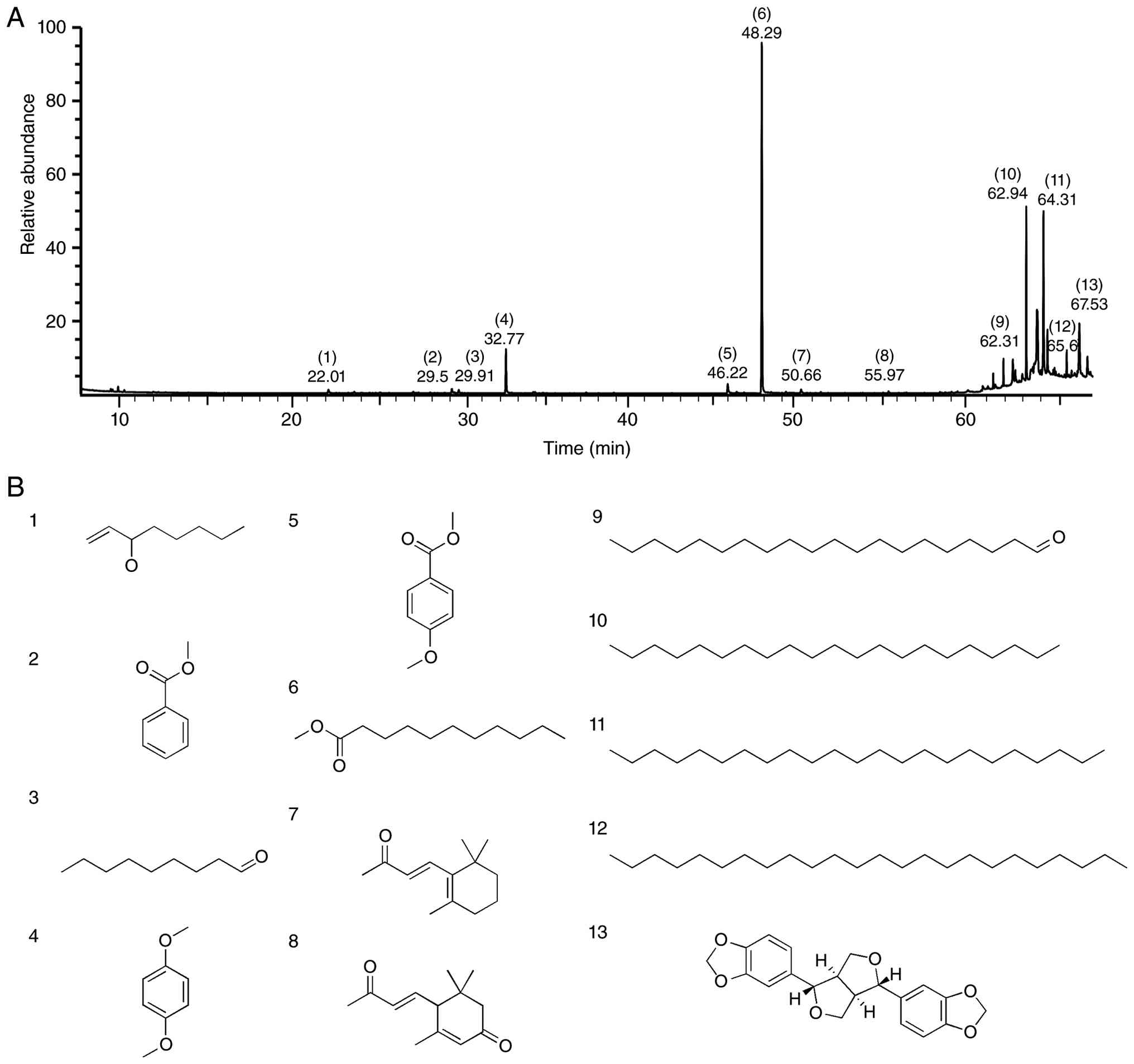
GC/MS, which identified 13 components (Table I and Fig. 1). Among the identified components,
methyl undecanoate (45.63%) had the highest content, followed in
order by heneicosane (13.62%), tricosane (13.62%), sesamin
(10.33%), 1,4-dimethoxybenzene (5.98%), tetracosane (4.79%), methyl
anisate (1.79%), eicosanal (1.36%), 1-octen-3-ol (0.73%), methyl
benzoate (0.71%), β-ionone (0.68%), nonanal (0.41%), and
3-oxo-α-ionone (0.35%) (Table
I).
 | Table I.Chemical composition of Paulownia
tomentosa flower absolute. |
Table I.
Chemical composition of Paulownia
tomentosa flower absolute.
|
|
| RI |
|
|
|
|---|
|
|
|
|
|
|
|
|---|
| Component name | RT, min | Observed | Literature | Area, % | CAS no. | (Refs.) |
|---|
| 1.
1-Octen-3-ol | 22.01 | 983 | 983 | 0.73 | 3391-86-4 | (59) |
| 2.
Methyl benzoate | 29.50 | 1,098 | 1,098 | 0.71 | 93-58-3 | (60) |
| 3.
Nonanal | 29.91 | 1,106 | 1,106 | 0.41 | 124-19-6 | (61) |
| 4.
1,4-Dimethoxybenzene | 32.77 | 1,164 | 1,165 | 5.98 | 150-78-7 | (62) |
| 5.
Methyl anisate | 46.22 | 1,383 | 1,386 | 1.79 | 121-98-2 | (62) |
| 6.
Methyl undecanoate | 48.29 | 1,427 | 1,427 | 45.63 | 1731-86-8 | (63) |
| 7.
β-Ionone | 50.66 | 1,486 | 1,486 | 0.68 | 14901-07-6 | (64) |
| 8.
3-Oxo-α-ionone | 55.97 | 1,658 | 1,665 | 0.35 | 79734-43-3 | (65) |
| 9.
Eicosanal | 62.31 | 2,029 | 2,211 | 1.36 | 2400-66-0 | (66) |
| 10.
Heneicosane | 62.94 | 2,101 | - | 13.62 | 629-94-7 |
|
| 11. Tricosane | 64.31 | 2,300 | - | 13.62 | 638-67-5 |
|
| 12.
Tetracosane | 65.60 | 2,500 | - | 4.79 | 646-31-1 |
|
| 13. Sesamin | 67.53 | 2,753 | 3,150 | 10.33 | 607-80-7 | (67) |
Alteration in SNARE proteins linked to
mast cell degranulation by PTFAb
The PTFAb test concentration for biological analysis
was first determined by observing the changes in cell viability in
RBL-2H3 mast cells treated with PTFAb. Treatment with PTFAb (1–200
µg/ml) showed a statistically significant decrease in cell
viability compared to the control group at 200 µg/ml, but no
significant decrease was observed at 1–150 µg/ml (Fig. 2A). Although there was no
significant decrease in cell viability at 150 µg/ml, the observed
cell morphology appeared less uniformly rounded and slightly more
irregular (Fig. S1). Therefore,
the PTFAb test concentration for subsequent biological analysis was
≤100 µg/ml, which did not affect the viability of RBL-2H3 cells.
SNARE proteins, such as v-SNAREs (VAMP7 and VAMP8) and t-SNAREs
(SNAP 23 and syntaxin 4), form trans-SNARE complexes to promote
membrane fusion. These proteins help regulate the mast cell
degranulation process and the release of inflammatory mediators,
such as histamine and cytokines (4). The influence of PTFAb on the
expression of SNARE proteins in RBL-2H3 mast cells was investigated
by immunoblotting. PTFAb (0.1–100 µg/ml) concentration-dependently
inhibited the expression of the v-SNARE proteins VAMP7 (Fig. 2B and C) and VAMP8 in RBL-2H3 cells
(Fig. 2B and D). The expression of
VAMP7 and VAMP8 in RBL-2H3 cells decreased significantly after the
treatment with PTFAb at 50–100 µg/ml. The reduced expression in
VAMP7 and VAMP8 showed a maximum at 100 µg/ml of PTFAb 22.02±8.61%
(Fig. 2C) and 20.61±7.40%
(Fig. 2D) of the untreated
control, respectively. On the other hand, the expression of the
t-SNARE proteins (syntaxin 1a, syntaxin 4, SNAP 23, and SNAP 25)
did not show significant changes after the treatment with PTFAb at
0.1–100 µg/ml (Fig. S2).
Effects of PTFAb on β-hexosaminidase
and histamine release in RBL-2H3 mast cells
Degranulation of activated mast cells results in the
release of β-hexosaminidase and histamine. These mediators are mast
cell degranulation markers and have been implicated in the
pathogenesis of AD (1,33). The effects of PTFAb (0.1–100 µg/ml)
on DNP-BSA (20 ng/ml)-induced β-hexosaminidase and histamine
release in anti-DNP IgE (200 ng/ml)-sensitized RBL-2H3 cells were
examined to determine if PTFAb inhibits mast cell degranulation.
DNP-BSA stimulation increased the levels of β-hexosaminidase
(329.09±4.77%) and histamine (193.02±6.64%) compared to the
anti-DNP IgE control (Fig. 3A and
B, respectively). By contrast, the PTFAb treatment at 1–100
µg/ml significantly reduced these levels in a
concentration-dependent manner. In particular, PTFAb exhibited
maximum inhibitory activity at 100 µg/ml, reducing β-hexosaminidase
and histamine to 146.06±4.34% (Fig.
3A) and 111.63±8.31% (Fig. 3B)
of the anti-DNP IgE control, respectively.
Effects of PTFAb on the
degranulation-related signaling molecules in RBL-2H3 mast
cells
During mast cell degranulation and the release of
inflammatory mediators, Syk phosphorylation, which accompanies the
activation of the IgE receptor FcεRI (Fc epsilon receptor I), plays
a key role in the initial signaling (34). PI3K/AKT and MAPK signaling pathways
are also involved in this process (35). Thus, the effects of PTFAb on these
key signaling molecules in mast cells were examined by observing
the phosphorylation of Syk, PI3K/AKT, and MAPKs (p38, ERK1/2, and
JNK) induced by DNP-BSA (20 ng/ml) in anti-DNP IgE antibody (100
ng/ml)-sensitized RBL-2H3 cells using immunoblotting to determine
how PTFAb affects these key signaling molecules in mast cells.
DNP-BSA significantly increased Syk phosphorylation (197.77±13.46%)
compared to the anti-DNP IgE control (Fig. 4A and B). On the other hand,
treatment with PTFAb at 75 and 100 µg/ml attenuated this level, and
the maximum inhibitory effect was observed at 100 µg/ml, reducing
Syk phosphorylation to 91.13±5.05% of the anti-DNP IgE control
(Fig. 4B). In addition, the
DNP-BSA treatment enhanced the phosphorylation levels of PI3K
(143.67±17.84%) and AKT (157.02±5.79%) compared to the anti-DNP IgE
control (Fig. 4C-E). The PTFAb
treatments at 75 and 100 µg/ml and at 50–100 µg/ml significantly
attenuated the increased levels of PI3K and AKT phosphorylation,
respectively. The maximum inhibitory effects on the phosphorylation
of these proteins were observed at 100 and 75 µg/ml, with the PI3K
and AKT phosphorylation levels reduced to 60.80±6.24% (Fig. 4D) and 59.42±9.37% of the anti-DNP
IgE control group, respectively (Fig.
4E). The total protein levels of Syk, PI3K, and AKT normalized
to β-actin are shown in Fig. S3,
indicating that the observed effects were not due to changes in
total protein expression. On the other hand, PTFAb (0.1–100 µg/ml)
did not affect the phosphorylation of MAPKs (p38, ERK1/2, and JNK)
induced by DNP-BSA (20 ng/ml) in anti-DNP IgE antibody (100
ng/ml)-activated RBL-2H3 cells (Fig.
S4).
 | Figure 4.Effects of PTFAb on Syk, PI3K, and
AKT phosphorylation in RBL-2H3 cells. (A and B) Syk
phosphorylation. (A) Representative image. RBL-2H3 cells were
sensitized with anti-DNP IgE and subsequently stimulated with
DNP-BSA to induce Syk phosphorylation. The cells were treated with
PTFAb (0.1–100 µg/ml), and the expression levels of phosphorylated
Syk were analyzed by Western blotting (n=3). The final DMSO
concentration was maintained at 0.1% in all experimental groups.
(B) Quantification of the p-Syk levels normalized to the total Syk,
presented as a percentage relative to anti-DNP IgE-sensitized cells
treated with 0.1% DMSO alone. β-actin was used as a loading control
to verify equal protein loading. *P<0.05 vs. DNP-BSA-stimulated
cells in the presence of anti-DNP IgE alone. (C-E) Phosphorylation
of PI3K and AKT. (C) RBL-2H3 cells were sensitized with anti-DNP
IgE and then stimulated with DNP-BSA to induce PI3K and AKT
phosphorylation. The cells were treated with PTFAb (0.1–100 µg/ml),
and the expression levels of p-PI3K and p-AKT were analyzed by
Western blotting (n=3 for each protein). The final DMSO
concentration was maintained at 0.1% in all experimental groups.
Quantification of the (D) p-PI3K and (E) p-AKT levels normalized to
the respective total protein levels (total PI3K and total AKT), and
presented as percentages relative to anti-DNP-IgE-sensitized cells
treated with 0.1% DMSO alone. β-actin was used as a loading control
to verify equal protein loading. *P<0.05 vs. anti-DNP
IgE/DNP-BSA-stimulated cells in the presence of 0.1% DMSO alone.
DMSO, dimethyl sulfoxide; anti-DNP IgE, anti-dinitrophenyl
immunoglobulin E; DNP-BSA, 2,4-dinitrophenyl-labeled bovine serum
albumin; p-AKT, phosphorylated protein kinase B; p-PI3K,
phosphorylated phosphoinositide 3-kinase; PTFAb, Paulownia
tomentosa flower absolute; p-Syk, phosphorylated spleen
tyrosine kinase. |
To further investigate the involvement of Syk/PI3K
and MAPK signaling pathways in the anti-degranulation activity of
PTFAb, pharmacological inhibitors targeting Syk, PI3K, p38, and
ERK1/2 were evaluated. PTFAb (50 µg/ml), piceatannol (a Syk
inhibitor; 30 µM), and LY294002 (a PI3K inhibitor; 0.1 µM)
significantly reduced anti-DNP IgE/DNP-induced β-hexosaminidase
release (Fig. S5A). Co-treatment
of PTFAb with either piceatannol or LY294002 further reduced
anti-DNP IgE/DNP-induced β-hexosaminidase release compared with
PTFAb treatment alone (Fig. S5B).
In contrast, treatment with SB203580 increased anti-DNP
IgE/DNP-induced β-hexosaminidase release (Fig. S5A), whereas co-treatment of PTFAb
with SB203580 partially attenuated the inhibitory effect of PTFAb
on anti-DNP IgE/DNP-induced β-hexosaminidase release (Fig. S5B). PD98059 alone or in
combination with PTFAb had little or no effect on anti-DNP
IgE/DNP-induced β-hexosaminidase release (Fig. S5A and B).
Effect of PTFAb on the proliferation
and migration of HaCaT cells
The cytotoxic effects of PTFAb on HaCaT cells were
first assessed using a WST assay to determine its regenerative
potential. The WST assay results showed that PTFAb significantly
enhanced cell viability at 250 and 500 µg/ml, while concentrations
at 1 to 100 µg/ml showed only a slight, non-significant increase
(Fig. 5A). Therefore, the entire
concentration range (1–500 µg/ml) of PTFAb was used for further
studies. BrdU assays showed that HaCaT cells proliferation was
increased significantly by PTFAb at 250 and 500 µg/ml, with the
maximum increase occurred at 500 µg/ml (143.98±12.34% compared to
the untreated control) (Fig. 5B).
Furthermore, the cell migration assay results showed that a PTFAb
treatment at 100–500 µg/ml significantly promoted the migration of
HaCaT cells, peaking at 840.00±52.46% at the 500 µg/ml (Fig. 5C and D).
 | Figure 5.Effects of PTFAb on the proliferation
and migration of HaCaT cells. (A) Cell viability. HaCaT cells were
incubated in the presence or absence of PTFAb (1–500 µg/ml) for 24
h. The final DMSO concentration was maintained at 0.5% in all
experimental groups, and control cells were treated with 0.5% DMSO
as the vehicle control (−). The cell viability was analyzed using a
WST assay (n=5). *P<0.05 compared with the untreated cells. (B)
Proliferation. HaCaT cells were incubated in the presence or
absence of PTFAb (1–500 µg/ml) for 48 h. The final DMSO
concentration was maintained at 0.5% in all groups, including the
vehicle control group (−). Cell proliferation was analyzed using
the BrdU incorporation assay (n=3). rhEGF (50 ng/ml)-induced
proliferation was used as a positive control. The percentages
represent the levels compared to that of the vehicle control cells,
which are considered 100%. The results are presented as the means ±
SEMs. *P<0.05 compared to the quiescent state. (C and D)
Migration. HaCaT cells were incubated in the presence or absence of
PTFAb (1–500 µg/ml) for 210 min. The final DMSO concentration was
maintained at 0.5% in all experimental groups, including the
vehicle control group. The cell migration levels were assessed
using the Boyden chamber assay. (C) Representative images. The blue
spots indicate migrating cells. (D) Statistical graph obtained from
panel C. rhEGF (1 ng/ml)-induced migration was used as a positive
control. The percentages represent the levels compared to the
vehicle control, which are considered 100% (n=3). The results are
presented as the means ± SEMs. Scale bar=50 µm. *P<0.05 compared
with the vehicle control group. Con, vehicle control; BrdU,
5-bromo-2′-deoxyuridine; DMSO, dimethyl sulfoxide; PTFAb,
Paulownia tomentosa flower absolute; rhEGF, recombinant
human epidermal growth factor; SEMs, standard errors of the means;
WST, water-soluble tetrazolium. |
Alteration of collagen synthesis in
HaCaT cells by PTFAb
Collagen synthesis strengthens the integrity of the
dermal and basement membranes, a key component of the skin barrier,
supporting keratinocyte migration and proliferation (36). The effects of PTFAb on collagen
synthesis in HaCaT cells were evaluated using a sandwich
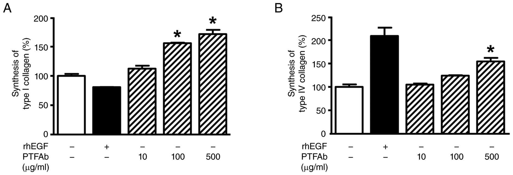
enzyme-linked immunosorbent assay (ELISA). Treatment with PTFAb
(10–500 µg/ml) significantly increased type I collagen synthesis at
100 and 500 µg/ml, and type IV collagen synthesis at 500 µg/ml.
Specifically, PTFAb at 500 µg/ml had the most prominent effect,
with synthesis levels reaching 171.45±7.27 and 154.42±7.53% for
type I (Fig. 6A) and IV (Fig. 6B), respectively.
Alteration of the skin barrier and
hyaluronan production-related proteins in HaCaT cells by PTFAb
A reduction in the filaggrin levels and HASs (HAS-1,
2, and 3)-mediated HA synthesis impairs the structural stability
and moisture retention of the skin barrier, contributing to skin
barrier compromise of AD (37,38).
Therefore, this study investigated the regulatory effects of PTFAb
on the expression of filaggrin and HAS in keratinocytes using
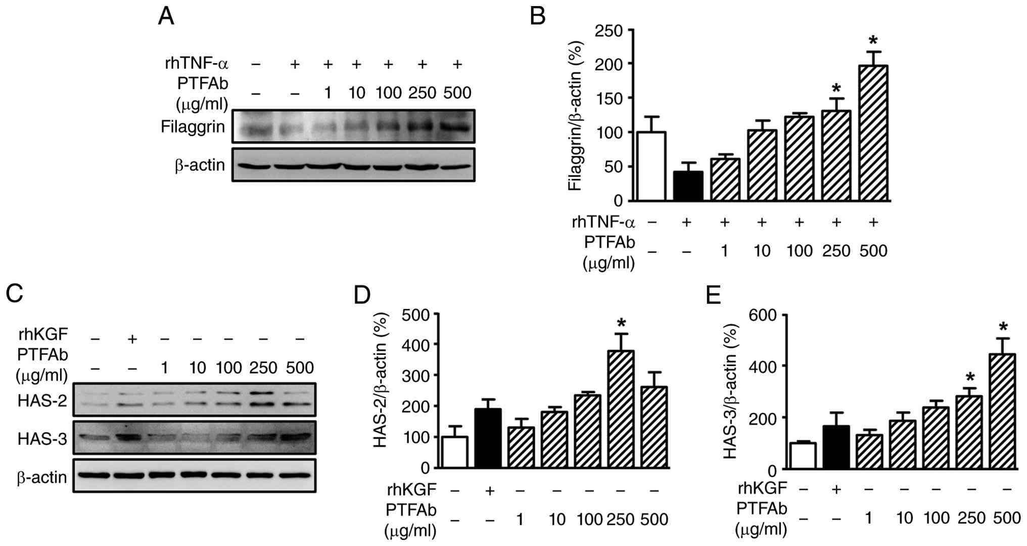
immunoblotting. FLG protein expression in HaCaT cells was
downregulated by a TNF-α treatment to 42.27±13.23% of the untreated
control. Nevertheless, treatment with PTFAb at 250 and 500 µg/ml
significantly restored FLG expression, which had been suppressed by
TNF-α. In particular, 500 µg/ml PTFAb had a maximum recovery effect
(196.08±20.47% of the untreated control) (Fig. 7A and B). In addition, this study
observed the influence of PTFAb on HAS expression in keratinocytes.
The effects of PTFAb on HAS-2 and HAS-3 expression were evaluated
at concentrations ranging from 1 to 500 µg/ml (Fig. 7C-E). The PTFAb treatment
significantly increased the HAS-2 levels by 377.11±55.21% compared
to the untreated control at 250 µg/ml (Fig. 7D). An increase (260.91±47.65% of
the untreated control) was also observed at 500 µg/ml, but did not
have statistical significance. In contrast, HAS-3 expression was
significantly upregulated at 250 and 500 µg/ml, with a maximum
effect of 443.78±61.56% at 500 µg/ml (Fig. 7E).
Effect of PTFAb on ICAM-1 expression
in HaCaT cells
Increased expression of ICAM-1 in keratinocytes by
inflammatory stimuli promotes the skin infiltration of immune cells
and local inflammatory responses, which may be associated with
abnormal skin barrier functions (15). Therefore, the changes in ICAM-1
protein expression were observed after a PTFAb treatment of
TNF-α-stimulated keratinocytes to determine if PTFAb can alleviate
skin inflammatory diseases through modulation of ICAM-1 expression.
As shown in Fig. 8A and B, the
TNF-α (10 ng/ml) treatment significantly increased ICAM-1
expression by 179.08±9.70% compared to the untreated control.
Nevertheless, the PTFAb treatment significantly reduced the
expression levels at 250 and 500 µg/ml. Specifically, the
expression was maximally suppressed, reaching 99.81±19.46% of the
untreated control (Fig. 8B).
Discussion
AD is a chronic inflammatory skin disease that
develops and progresses through the interaction of an imbalanced
immune response and impaired epidermal barrier function. These
complex pathological characteristics are associated with persistent
exacerbation and recurrent relapses (1). In particular, excessive secretion of
inflammatory mediators due to mast cell activation and dysfunction
of keratinocytes, which encompass the skin barrier, exacerbate the
inflammatory responses and impede the restoration of skin
homeostasis (1). Considering the
characteristics of AD, simultaneously modulating the inflammatory
responses and skin barrier function may be a practical therapeutic
approach. The present study evaluated the anti-atopic potential of
PTFAb, an absolute-type essential oil, using mast cell and
keratinocyte-based in vitro models. PTFAb suppressed the
mast cell responses associated with the secretion of inflammatory
mediators while simultaneously promoting the keratinocyte responses
involved in skin barrier formation and maintenance, suggesting that
PTFAb may be a promising functional material that can modulate
multiple AD-related cellular responses in mast cells and
keratinocytes.
Mast cell activation involves membrane fusion
between pre-existing granules or vesicles and the plasma membrane,
resulting in degranulation and the release of allergic mediators
stored within these granules (4).
The membrane fusion process in mast cells is regulated by the SNARE
complex, which is formed by the interaction between v-SNAREs and
t-SNAREs (4). VAMP7 and VAMP8 are
members of the v-SNARE proteins, and syntaxin 1a, syntaxin 4, SNAP
23, and SNAP 25 are members of the t-SNARE proteins. The
interaction between these v-SNARE and t-SNARE proteins promotes
mast cell degranulation. This mechanism was reported to be an
important molecular target for the treatment of AD inflammatory
lesions (29,39). Plant-derived substances inhibit
mast cell degranulation by downregulating SNARE proteins and
improve skin inflammatory symptoms in AD (39,40).
Therefore, attenuating mast cell hyper-degranulation might be an
important strategy for alleviating allergic inflammation. The
present study found that PTFAb reduced VAMP7 and VAMP8 expression
in RBL-2H3 cells, suggesting that PTFAb suppresses mast cell
membrane fusion by interfering with SNARE complex formation. On the
other hand, PTFAb did not suppress the expressions of syntaxin 1a,
syntaxin 4, SNAP 23, and SNAP 25 in RBL-2H3 cells. Therefore, PTFAb
may inhibit membrane fusion by selectively regulating the v-SNARE
proteins VAMP7 and VAMP8, thereby reducing mast cell
degranulation.
Excessive mast cell degranulation is a key
contributor to the allergic inflammation observed in AD (41). When activated by allergens and IgE,
mast cells release mediators such as histamine, β-hexosaminidase,
and cytokines via degranulation, triggering inflammatory reactions
(33,41). Among these, β-hexosaminidase and
histamine are widely recognized as reliable indicators of mast cell
degranulation because their release levels reflect the extent of
mediator secretion (33). A
literature survey showed that among 13 components identified from
PTFAb, sesamin was reported to reduce histamine release and the
production and secretion of pro-inflammatory cytokines in mast
cells stimulated with IgE (42),
suggesting that this constituent may contribute to the inhibitory
effect of PTFAb on mast cell-derived inflammatory allergic
reactions. In the present study, treatment with PTFAb markedly
reduced β-hexosaminidase and histamine release in RBL-2H3 cells
sensitized with anti-DNP IgE upon DNP-BSA stimulation. Hence, PTFAb
might suppress β-hexosaminidase and histamine release, which may
contribute to the regulation of AD-related allergic responses.
Binding FcεRI and IgE activates mast cells and
initiates the early activation of Syk, which then propagates
signals to various downstream signaling pathways associated with
the FcεRI-mediated degranulation and the production of allergic and
inflammatory cytokines (6,34). Among these pathways, the PI3K/AKT
axis plays a key role in inducing degranulation (33). In addition, the MAPK signaling
pathway, represented by ERK1/2, JNK, and p38, was reported to
regulate late-phase responses in the mast cells, including the
regulation of inflammatory cytokine and chemokine expression, and
degranulation (6,34). Therefore, targeting these signaling
pathways may be a promising strategy for suppressing mast
cell-mediated allergic inflammatory responses. A previous study
reported that the inhibition of Syk-PI3K/AKT and MAPK (p38, JNK,
and ERK1/2) phosphorylation by the plant extract was associated
with the suppression of DNCB-IgE-mediated mast cell degranulation
and inflammatory cytokine expression, probably ameliorating
DNCB-induced AD-like lesions in animals (30). RBL-2H3 cells and DNCB-induced skin
lesions are widely used as in vitro and in vivo
models, respectively, for studying the mast cell-mediated allergic
inflammatory responses and AD lesions (39). In the present study, the PTFAb
treatment significantly reduced the levels of Syk, PI3K, and AKT
phosphorylation in RBL-2H3 cells sensitized with anti-DNP IgE upon
DNP-BSA stimulation, whereas no significant changes were observed
in the phosphorylation of the MAPK family members, ERK1/2, JNK, and
p38. Therefore, these findings suggest that PTFAb may be associated
with reduced activation of the Syk-PI3K/AKT signaling pathway,
rather than broad suppression of all downstream signaling pathways
examined in this study.
In addition, to further investigate the potential
contribution of Syk-PI3K/AKT and MAPK signaling to the
anti-degranulation activity of PTFAb, we evaluated the effects of
pharmacological inhibitors targeting Syk, PI3K, and MAPKs on
anti-DNP IgE/DNP-BSA-induced β-hexosaminidase release. Co-treatment
of PTFAb with the Syk inhibitor piceatannol or the PI3K inhibitor
LY294002 further reduced anti-DNP IgE/DNP-BSA-induced
β-hexosaminidase release compared with PTFAb treatment alone.
Although these additive effects do not establish that the activity
of PTFAb is dependent on the Syk-PI3K/AKT pathway, they provide
supportive, but not definitive, pharmacological evidence that this
signaling axis may contribute to the observed effects of PTFAb.
Interestingly, inhibition of p38 with SB203580 increased anti-DNP
IgE/DNP-BSA-induced β-hexosaminidase release and partially
attenuated the inhibitory effect of PTFAb, whereas inhibition of
ERK1/2 with PD98059 had little effect either alone or in
combination with PTFAb. Consistent with these observations, PTFAb
did not significantly alter the phosphorylation of p38, ERK1/2, or
JNK, suggesting that MAPK signaling may not represent a major
molecular signaling pathway modulated by PTFAb. Collectively, these
findings indicate that PTFAb is associated with reduced
phosphorylation of Syk, PI3K, and AKT, whereas the inhibitor
experiments provide only supportive, non-definitive evidence for
the possible involvement of the Syk-PI3K/AKT signaling axis in its
anti-degranulation activity of PTFAb. Further studies are needed to
determine whether, and to what extent, this signaling pathway
contributes to the anti-degranulation effects of PTFAb and to
clarify the potential crosstalk between the Syk-PI3K/AKT and MAPK
signaling pathways in the regulation of mast cell
degranulation.
In addition to immune dysfunction leading to
dysregulated allergic responses, AD is characterized by impaired
skin barrier function (1). This
impaired barrier function can be linked to abnormalities in the
behavior of keratinocytes, the main cells in the epidermal layer of
the skin, because these cells are essential for restoring the
functional epidermal barrier through proliferation and migration
(43). These processes may
contribute to impaired epidermal renewal and delayed barrier
restoration in AD. Interestingly, nonanal, a compound isolated from
PTFAb, induces the expression of keratinocyte growth factor (KGF)
and stimulates the proliferation and migration in dermal papilla
cells (44). KGF promotes
keratinocyte proliferation and migration (45). Based on these reports, it is
suggested that PTFAb may exert stimulatory effects on HaCaT cell
migration and proliferation. In the present study, PTFAb stimulated
the proliferation and migration of HaCaT cells at non-cytotoxic
concentrations (250–500 µg/ml for proliferation and 100–500 µg/ml
for migration). These effects of PTFAb were mainly observed at
relatively high concentrations. PTFAb is a crude botanical extract
containing multiple constituents, and relatively high
concentrations are often required to elicit measurable biological
responses in in vitro studies. In addition, these
concentrations of PTFAb showed no cytotoxicity, suggesting that the
observed effects were not attributable to nonspecific cytotoxic
responses. Furthermore, topical administration may allow relatively
high local concentrations to be achieved at the site of
application, but this possibility requires confirmation in
appropriate in vivo studies. Nevertheless, the
physiological/pharmacological relevance of these concentrations
remains to be established through appropriate in vivo
studies.
Taken together, these findings suggest that PTFAb
has the potential to modulate AD-related keratinocyte responses by
promoting re-epithelialization and enhancing skin barrier-related
functions.
Despite these promising biological activities, the
active constituents responsible for the observed effects remain
unclear. While GC/MS analysis identified 13 constituents in PTFAb,
the present study did not isolate or identify the specific
compounds responsible for the biological activities observed in the
present study. Although several of the identified constituents,
including sesamin and nonanal, have previously been reported to
possess biological activities relevant to the present findings,
their individual contributions were not experimentally verified.
Therefore, the relationship between the chemical composition of
PTFAb and its biological activities remains to be established.
Because PTFAb is a complex mixture, its biological activities may
result from the effects of individual constituents or from additive
or synergistic interactions among multiple components. Further
studies are required to examine fractionated extracts, isolate
individual constituents, and elucidate the mechanisms underlying
their activities to determine which constituents contribute to the
cellular responses associated with the anti-AD potential of
PTFAb.
The structural stability of skin tissue is provided
by collagen, the predominant protein in the ECM, and influences key
cellular functions such as adhesion, migration, and proliferation
(46). Type I collagen is abundant
in the interstitial layer and supports the structural integrity and
elasticity of the skin, whereas type IV collagen in the basement
membrane contributes to keratinocyte migration and proliferation,
maintains the basement membrane structure, and mediates cell
adhesion and signaling (47–50).
In AD, reduced collagen levels have been linked to delayed wound
healing, decreased elasticity, and increased skin dryness (50). Plant-derived extracts have the
potential to support skin repair by stimulating collagen synthesis
in keratinocytes (19). The
results showed that PTFAb upregulated the production of collagen
types I and IV in HaCaT cells. Hence, PTFAb may enhance ECM
integrity and help stabilize skin barrier-related functions by
promoting collagen production in keratinocytes.
FLG, IVL, and LOR, which are produced by
keratinocytes, maintain the integrity of the skin barrier (7). Changes in the expression of these
epidermal barrier-related proteins are widely known as a major
cause of skin barrier dysfunction in AD (7). In particular, filaggrin mutations or
deficiencies are reported to be a key contributor to the
pathogenesis of AD, leading to a barrier dysfunction (51). In this context, studies have shown
that FLG deficiency impairs barrier integrity, whereas restoring or
upregulating FLG promotes barrier recovery from atopic,
inflammatory, or chemically induced skin damage (51–53).
These reports highlight the importance of regulating the barrier
proteins in maintaining and restoring the epidermal barrier
function in AD. In the present study, PTFAb enhanced FLG expression
in HaCaT cells exposed to TNF-α, highlighting its potential to
promote epidermal barrier recovery in AD. Future studies into its
effects on IVL and LOR expression could further clarify the
therapeutic potential of PTFAb in restoring the barrier
function.
Hyaluronic acid (HA), a glycosaminoglycan component
of the ECM, helps sustain skin moisture and elasticity through its
strong affinity for water; hence, it is a key molecule in skin
hydration maintenance (10,11).
Its synthesis in keratinocytes is mediated by HAS enzymes (HAS-1,
−2, and −3) (10). Maintaining
proper skin moisture is essential for preserving the barrier
integrity and the balance of skin functions, associated with a
normal skin barrier function (54). HA ameliorated
2,4-dinitrofluorobenzene-induced AD-like lesions in mice (55). An emollient containing hydrolyzed
collagen and HA improved the skin barrier function and enhanced
hydration in AD, as shown in the clinical, in vitro, and
ex vivo studies (56).
These reports highlight its potential use in AD therapy. In the
present study, PTFAb increased HAS-2 and HAS-3 expression in HaCaT
cells. Hence, PTFAb may help enhance HAS (HAS-2 and HAS-3)
expression in keratinocytes, probably improving skin hydration by
increasing HA production. Overall, PTFAb may promote cellular
responses associated with wound healing, skin barrier function, and
hydration in keratinocytes.
The interaction between keratinocytes, primary skin
cells, and various immune cells is associated with recurrent and
persistent inflammation in AD (1).
Keratinocytes show increased expression of adhesion molecules like
ICAM-1 in response to inflammatory stimuli. This process
facilitates the infiltration and retention of T cells in the skin,
contributing to skin barrier dysfunction and sustaining local
inflammatory response (57). A
previous study reported that TNF-α induces ICAM-1 in keratinocytes
and ICAM-1 is upregulated in keratinocytes in inflammatory lesions
of AD skin (58). In the present
study, TNF-α, which induces an inflammatory condition, increased
ICAM-1 expression, which was inhibited after PTFAb stimulation.
These findings suggest that PTFAb may attenuate AD-linked
inflammation, probably by reducing immune cell adhesion and
infiltration at the keratinocyte level. In addition, PTFAb
inhibited histamine and β-hexosaminidase release from mast cells,
highlighting its potential to suppress excessive immune activation
during the early phase of allergic responses. Collectively, these
findings suggest that PTFAb may exert anti-inflammatory effects by
regulating adhesion molecule expression and the release of
inflammatory mediators. Therefore, PTFAb may simultaneously
regulate multiple mast cell- and keratinocyte-mediated pathways
associated with AD-related cellular responses.
In conclusion, 13 compounds were identified from
PTFAb, showing that PTFAb modulates the key cellular responses
associated with the pathogenesis of AD. PTFAb downregulated the
expression of the v-SNARE proteins VAMP7 and VAMP8 in RBL-2H3 mast
cells. PTFAb also inhibited β-hexosaminidase and histamine release,
and suppressed Syk-PI3K/AKT phosphorylation in RBL-2H3 cells under
stimulation with anti-DNP IgE and DNP-BSA. In addition, in
keratinocytes, PTFAb promoted barrier-restorative functions by
enhancing cell proliferation and migration, increasing type I and
type IV collagen synthesis, restoring filaggrin expression reduced
by TNF-α, upregulating HAS-2 and HAS-3 expression, and suppressing
TNF-α-induced ICAM-1 expression. These findings demonstrate that
PTFAb enhances keratinocyte-mediated barrier repair and hydration
while reducing inflammatory adhesion molecule expression.
Collectively, these results suggest that PTFAb may modulate
cellular processes involved in AD pathogenesis through multiple
mechanisms, including the inhibition of mast cell degranulation and
the enhancement of keratinocyte barrier recovery. Importantly, this
study provides mechanistic evidence that PTFAb simultaneously
regulates two interdependent processes associated with AD: mast
cell-mediated allergic inflammation and keratinocyte-driven
epidermal barrier dysfunction, supporting its potential as a
multi-target natural product candidate. These findings provide a
scientific basis for further investigation of PTFAb as a natural
product-based material for AD-related research. Nevertheless,
additional validation in animal models and clinical studies will be
needed to clarify its therapeutic potential.
Several limitations of the present study should be
considered when interpreting the findings. Although HaCaT cells
were obtained from a documented commercial source and routinely
tested for mycoplasma contamination, short tandem repeat (STR)
profiling was not performed in our laboratory to independently
confirm cell identity. Therefore, this limitation should be taken
into account when interpreting the present results.
In addition, standard positive controls commonly
used in mast cell- and AD-related models were not included in the
present study. The primary objective of this study was to
investigate the biological effects and underlying mechanisms of
PTFAb by comparing its effects with corresponding stimulated
control groups, rather than to directly compare its efficacy with
established anti-allergic or anti-AD agents. Therefore, the effects
of PTFAb were evaluated based on its ability to attenuate
stimulus-induced cellular responses. Although positive controls
(rhEGF or rhKGF) were included in selected HaCaT cell-based assays
to validate specific keratinocyte responses, these assay-specific
controls do not address the absence of standard positive controls
for the mast cell- and AD-related assays and were not intended to
serve as reference compounds for mast cell degranulation or
AD-related immune responses. Future studies including appropriate
positive controls for mast cell and AD models will further
strengthen the comparative evaluation and validation of the
efficacy of PTFAb.
Supplementary Material
Supporting Data
Acknowledgements
The authors gratefully acknowledge Dr Hyun-Jun Kim
(Forest Medicinal Resources Research Center, Seoul, Korea) for
taxonomic authentication of Paulownia tomentosa.
Funding
The present study was supported by a grant from the R&D
Program for Forestry Technology of the Korea Forest Service (Korea
Forestry Promotion Institute) (grant no. RS-2024-00403260).
Availability of data and materials
The data generated in the present study may be
requested from the corresponding author, and the raw GC/MS data
generated during the current study are available from the
corresponding author upon reasonable request.
Authors' contributions
HML conceptualized the study. HML, KJW and DYK
performed the formal analysis. DYY, YYK, DYK, JHB and JSY conducted
the investigation. HML, KJW and DYY organized, managed, verified
and prepared the experimental data for analysis. HML, KJW and DYY
prepared the original draft of the manuscript. DYY, DYK, YYK, JHB
and JSY reviewed the draft. HML and KJW reviewed and edited the
manuscript. HML supervised the study and acquired funding. HML, KJW
and DYY confirm the authenticity of all the raw data. All authors
reviewed, discussed, edited, read and approved the final
manuscript.
Ethics approval and consent to
participate
Not applicable.
Patient consent for publication
Not applicable.
Competing interests
The authors declare that they have no competing
interests.
References
|
1
|
Sroka-Tomaszewska J and Trzeciak M:
Molecular mechanisms of atopic dermatitis pathogenesis. Int J Mol
Sci. 22:41302021. View Article : Google Scholar : PubMed/NCBI
|
|
2
|
Mohd Kasim VNK, Noble SM, Liew KY, Tan JW,
Israf DA and Tham CL: Management of atopic dermatitis via oral and
topical administration of herbs in murine model: A systematic
review. Front Pharmacol. 13:7857822022. View Article : Google Scholar : PubMed/NCBI
|
|
3
|
Matsuda H, Nakamura S and Yoshikawa M:
Degranulation inhibitors from medicinal plants in
antigen-stimulated rat basophilic leukemia (RBL-2H3) cells. Chem
Pharm Bull (Tokyo). 64:96–103. 2016. View Article : Google Scholar : PubMed/NCBI
|
|
4
|
Woska JR Jr and Gillespie ME: SNARE
complex-mediated degranulation in mast cells. J Cell Mol Med.
16:649–656. 2012. View Article : Google Scholar : PubMed/NCBI
|
|
5
|
Hada M, Nishi K, Ishida M, Onda H,
Nishimoto S and Sugahara T: Inhibitory effect of aqueous extract of
Cuminum cyminum L. seed on degranulation of RBL-2H3 cells and
passive cutaneous anaphylaxis reaction in mice. Cytotechnology.
71:599–609. 2019. View Article : Google Scholar : PubMed/NCBI
|
|
6
|
McCarty MF, Lerner A, DiNicolantonio JJ
and Benzvi C: Nutraceutical aid for allergies-strategies for
down-regulating mast cell degranulation. J Asthma Allergy.
14:1257–1266. 2021. View Article : Google Scholar : PubMed/NCBI
|
|
7
|
Furue M: Regulation of filaggrin,
loricrin, and involucrin by IL-4, IL-13, IL-17A, IL-22, AHR, and
NRF2: Pathogenic implications in atopic dermatitis. Int J Mol Sci.
21:53822020. View Article : Google Scholar : PubMed/NCBI
|
|
8
|
Proksch E, Brandner JM and Jensen JM: The
skin: An indispensable barrier. Exp Dermatol. 17:1063–1072. 2008.
View Article : Google Scholar : PubMed/NCBI
|
|
9
|
Potekaev NN, Borzykh OB, Medvedev GV,
Pushkin DV, Petrova MM, Petrov AV, Dmitrenko DV, Karpova EI, Demina
OM and Shnayder NA: The role of extracellular matrix in skin wound
healing. J Clin Med. 10:59472021. View Article : Google Scholar : PubMed/NCBI
|
|
10
|
Papakonstantinou E, Roth M and
Karakiulakis G: Hyaluronic acid: A key molecule in skin aging.
Dermatoendocrinol. 4:253–258. 2012. View Article : Google Scholar : PubMed/NCBI
|
|
11
|
Evrard C, Lambert de Rouvroit C and Poumay
Y: Epidermal hyaluronan in barrier alteration-related disease.
Cells. 10:30962021. View Article : Google Scholar : PubMed/NCBI
|
|
12
|
Diller RB and Tabor AJ: The role of the
extracellular matrix (ECM) in wound healing: A review. Biomimetics
(Basel). 7:872022. View Article : Google Scholar : PubMed/NCBI
|
|
13
|
Choudhary V, Choudhary M and Bollag WB:
Exploring skin wound healing models and the impact of natural
lipids on the healing process. Int J Mol Sci. 25:37902024.
View Article : Google Scholar : PubMed/NCBI
|
|
14
|
Rerknimitr P, Otsuka A, Nakashima C and
Kabashima K: The etiopathogenesis of atopic dermatitis: Barrier
disruption, immunological derangement, and pruritus. Inflamm Regen.
37:142017. View Article : Google Scholar : PubMed/NCBI
|
|
15
|
Singer KH, Tuck DT, Sampson HA and Hall
RP: Epidermal keratinocytes express the adhesion molecule
intercellular adhesion molecule-1 in inflammatory dermatoses. J
Invest Dermatol. 92:746–750. 1989. View Article : Google Scholar : PubMed/NCBI
|
|
16
|
Matsunaga T, Katayama I, Yokozeki H and
Nishioka K: ICAM-1 expression on keratinocytes in
mechanically-injured skin of a patient with atopic dermatitis. J
Dermatol Sci. 12:219–226. 1996. View Article : Google Scholar : PubMed/NCBI
|
|
17
|
Man G, Hu LZ, Elias PM and Man MQ:
Therapeutic benefits of natural ingredients for atopic dermatitis.
Chin J Integr Med. 24:308–314. 2018. View Article : Google Scholar : PubMed/NCBI
|
|
18
|
Valdivieso-Ugarte M, Gomez-Llorente C,
Plaza-Díaz J and Gil Á: Antimicrobial, antioxidant, and
immunomodulatory properties of essential oils: A systematic review.
Nutrients. 11:27862019. View Article : Google Scholar : PubMed/NCBI
|
|
19
|
Kim DY, Won KJ, Kim YY, Yoo DY and Lee HM:
Potential wound healing and anti-melanogenic activities in skin
cells of Aralia elata (Miq.) Seem. flower essential oil and its
chemical composition. Pharmaceutics. 16:10082024. View Article : Google Scholar : PubMed/NCBI
|
|
20
|
Chung IM, Kim EH, Jeon HS and Moon HI:
Protective effects of isoatriplicolide tiglate from Paulownia
coreana against glutamate-induced neurotoxicity in primary
cultured rat cortical cells. Nat Prod Commun. 5:851–852.
2010.PubMed/NCBI
|
|
21
|
Si CL, Kim JK, Kwon DJ and Bae YS:
Phenylpropanoid glycosides of Paulownia coreana Uyeki
leaves. J Korean Wood Sci Technol. 34:78–82. 2006.
|
|
22
|
Kim TJ: Korean resources plants (IV).
Seoul National University Press; Seoul: pp. 76–77. 1996
|
|
23
|
Kim NK, Kim MH, Yoon CS and Choi SW:
Studies on the anti-inflammatory activity of Paulownia
coreana Uyeki leaf extract. J Soc Cosmet Sci Korea. 32:241–247.
2006.
|
|
24
|
Jin Q, Lee C, Lee JW, Lee D, Kim Y, Hong
JT, Kim JS, Kim JH, Lee MK and Hwang BY: Geranylated flavanones
from Paulownia coreana and their inhibitory effects on
nitric oxide production. Chem Pharm Bull (Tokyo). 63:384–387. 2015.
View Article : Google Scholar : PubMed/NCBI
|
|
25
|
Teng Y, Zhong H, Yang X, Tao X and Fan Y:
Current and emerging therapies for atopic dermatitis in the
elderly. Clin Interv Aging. 18:1641–1652. 2023. View Article : Google Scholar : PubMed/NCBI
|
|
26
|
International Union for Conservation of
Nature, . The IUCN red list of threatened species. https://www.iucnredlist.orgMarch. 2026
|
|
27
|
National Institute of Biological Resources
(NIBR), . Korean Red Data Book of Vascular Plants (2021).
https://species.nibr.go.krMarch.
2026
|
|
28
|
Kim MJ, Won KJ, Kim DY, Won YR, Kim NY,
Lee DK, Hong BS and Lee HM: Skin wound healing and
anti-wrinkle-promoting in vitro biological activities of Caragana
sinica flower absolute and its chemical composition.
Pharmaceuticals (Basel). 16:2352023. View Article : Google Scholar : PubMed/NCBI
|
|
29
|
Yang Y, Kong B, Jung Y, Park JB, Oh JM,
Hwang J, Cho JY and Kweon DH: Soluble N-ethylmaleimide-sensitive
factor attachment protein receptor-derived peptides for regulation
of mast cell degranulation. Front Immunol. 9:7252018. View Article : Google Scholar : PubMed/NCBI
|
|
30
|
Lee HP, Choi W, Kwon KW, You L, Rahmawati
L, Luong VD, Kim W, Lee BH, Lee S, Kim JH and Cho JY: Inhibitory
effects of grewia tomentosa Juss. on IgE-mediated allergic reaction
and DNCB-induced atopic dermatitis. Plants (Basel).
11:25402022.PubMed/NCBI
|
|
31
|
Bairoch A: The cellosaurus, a cell-line
knowledge resource. J Biomol Tech. 29:25–38. 2018. View Article : Google Scholar : PubMed/NCBI
|
|
32
|
Bae WB, Kim EH, Kim MJ and Yang SA:
Inhibitory effects of Broussonetia kazinoki twig extract on
allergic inflammatory reactions in TNF-α/IFN-γ-stimulated HaCaT and
IgE-sensitized RBL-2H3 cells. Food Sci Preserv. 31:307–314. 2024.
View Article : Google Scholar
|
|
33
|
Bao Y, Wang S, Gao Y, Zhang W, Jin H, Yang
Y and Li J: MicroRNA-126 accelerates IgE-mediated mast cell
degranulation associated with the PI3K/Akt signaling pathway by
promoting Ca2+ influx. Exp Ther Med. 16:2763–2769.
2018.PubMed/NCBI
|
|
34
|
Siraganian RP, de Castro RO, Barbu EA and
Zhang J: Mast cell signaling: The role of protein tyrosine kinase
Syk, its activation and screening methods for new pathway
participants. FEBS Lett. 584:4933–4940. 2010. View Article : Google Scholar : PubMed/NCBI
|
|
35
|
Rivera J and Gilfillan AM: Molecular
regulation of mast cell activation. J Allergy Clin Immunol.
117:1214–1226. 2006. View Article : Google Scholar : PubMed/NCBI
|
|
36
|
Mathew-Steiner SS, Roy S and Sen CK:
Collagen in wound healing. Bioengineering (Basel). 8:632021.
View Article : Google Scholar : PubMed/NCBI
|
|
37
|
Levin J, Friedlander SF and Del Rosso JQ:
Atopic dermatitis and the stratum corneum: Part 1: The role of
filaggrin in the stratum corneum barrier and atopic skin. J Clin
Aesthet Dermatol. 6:16–22. 2013.PubMed/NCBI
|
|
38
|
Malaisse J, Bourguignon V, De Vuyst E,
Lambert de Rouvroit C, Nikkels AF, Flamion B and Poumay Y:
Hyaluronan metabolism in human keratinocytes and atopic dermatitis
skin is driven by a balance of hyaluronan synthases 1 and 3. J
Invest Dermatol. 134:2174–2182. 2014. View Article : Google Scholar : PubMed/NCBI
|
|
39
|
Kim DY, Won KJ, Hwang DI, Lee SY, Choi IH,
Kim B and Lee HM: Essential oil from Chrysanthemum boreale
flowers modulates SNARE protein-linked mast cell response and skin
barrier proteins and ameliorates atopic dermatitis-like lesions in
mice. Hortic Environ Biotechnol. 63:287–298. 2022. View Article : Google Scholar
|
|
40
|
Yun Y, Kim K, Choi I and Ko SG: Topical
herbal application in the management of atopic dermatitis: A review
of animal studies. Mediators Inflamm. 2014:7521032014. View Article : Google Scholar : PubMed/NCBI
|
|
41
|
Liu FT, Goodarzi H and Chen HY: IgE, mast
cells, and eosinophils in atopic dermatitis. Clin Rev Allergy
Immunol. 41:298–310. 2011. View Article : Google Scholar : PubMed/NCBI
|
|
42
|
Li LC, Piao HM, Zheng MY, Lin ZH, Li G and
Yan GH: Sesamin attenuates mast cell-mediated allergic responses by
suppressing the activation of p38 and nuclear factor-κB. Mol Med
Rep. 13:536–542. 2016. View Article : Google Scholar : PubMed/NCBI
|
|
43
|
Wikramanayake TC, Stojadinovic O and
Tomic-Canic M: Epidermal differentiation in barrier maintenance and
wound healing. Adv Wound Care (New Rochelle). 3:272–280. 2014.
View Article : Google Scholar : PubMed/NCBI
|
|
44
|
Park S, Kang W, Choi D, Son B and Park T:
Nonanal stimulates growth factors via cyclic adenosine
monophosphate (cAMP) signaling in human hair follicle dermal
papilla cells. Int J Mol Sci. 21:80542020. View Article : Google Scholar : PubMed/NCBI
|
|
45
|
Putnins EE, Firth JD, Lohachitranont A,
Uitto VJ and Larjava H: Keratinocyte growth factor (KGF) promotes
keratinocyte cell attachment and migration on collagen and
fibronectin. Cell Adhes Commun. 7:211–221. 1999. View Article : Google Scholar : PubMed/NCBI
|
|
46
|
Brett D: A review of collagen and
collagen-based wound dressings. Wounds. 20:347–356. 2008.PubMed/NCBI
|
|
47
|
O'Toole EA: Extracellular matrix and
keratinocyte migration. Clin Exp Dermatol. 26:525–530. 2001.
View Article : Google Scholar : PubMed/NCBI
|
|
48
|
Boudko SP, Danylevych N, Hudson BG and
Pedchenko VK: Basement membrane collagen IV: Isolation of
functional domains. Methods Cell Biol. 143:171–185. 2018.
View Article : Google Scholar : PubMed/NCBI
|
|
49
|
Motter Catarino C, Kaiser K, Baltazar T,
Motter Catarino L, Brewer JR and Karande P: Evaluation of native
and non-native biomaterials for engineering human skin tissue.
Bioeng Transl Med. 7:e102972022. View Article : Google Scholar : PubMed/NCBI
|
|
50
|
Szalus K and Trzeciak M: The role of
collagens in atopic dermatitis. Int J Mol Sci. 25:76472024.
View Article : Google Scholar : PubMed/NCBI
|
|
51
|
Moosbrugger-Martinz V, Leprince C, Méchin
MC, Simon M, Blunder S, Gruber R and Dubrac S: Revisiting the roles
of filaggrin in atopic dermatitis. Int J Mol Sci. 23:53182022.
View Article : Google Scholar : PubMed/NCBI
|
|
52
|
Dang NN, Pang SG, Song HY, An LG and Ma
XL: Filaggrin silencing by shRNA directly impairs the skin barrier
function of normal human epidermal keratinocytes and then induces
an immune response. Braz J Med Biol Res. 48:39–45. 2015. View Article : Google Scholar : PubMed/NCBI
|
|
53
|
Hsu CY, Lecland N, Pendaries V, Viodé C,
Redoulès D, Paul C, Merdes A, Simon M and Bierkamp C: Stabilization
of microtubules restores barrier function after cytokine-induced
defects in reconstructed human epidermis. J Dermatol Sci. 91:87–96.
2018. View Article : Google Scholar : PubMed/NCBI
|
|
54
|
Bonté F: Skin moisturization mechanisms:
New data. Ann Pharm Fr. 69:135–141. 2011. View Article : Google Scholar : PubMed/NCBI
|
|
55
|
Zheng BW, Wang BY, Xiao WL, Sun YJ, Yang C
and Zhao BT: Different molecular weight hyaluronic acid alleviates
inflammation response in DNFB-induced mice atopic dermatitis and
LPS-induced RAW 264.7 cells. Life Sci. 301:1205912022. View Article : Google Scholar : PubMed/NCBI
|
|
56
|
Lee YI, Lee SG, Kim J, Choi S, Jung I and
Lee JH: Proteoglycan combined with hyaluronic acid and hydrolyzed
collagen restores the skin barrier in mild atopic dermatitis and
dry, eczema-prone skin: A pilot study. Int J Mol Sci. 22:101892021.
View Article : Google Scholar : PubMed/NCBI
|
|
57
|
Griffiths CE and Nickoloff BJ:
Keratinocyte intercellular adhesion molecule-1 (ICAM-1) expression
precedes dermal T lymphocytic infiltration in allergic contact
dermatitis (Rhus dermatitis). Am J Pathol. 135:1045–1053.
1989.PubMed/NCBI
|
|
58
|
Griffiths CE, Voorhees JJ and Nickoloff
BJ: Characterization of intercellular adhesion molecule-1 and
HLA-DR expression in normal and inflamed skin: Modulation by
recombinant gamma interferon and tumor necrosis factor. J Am Acad
Dermatol. 20:617–629. 1989. View Article : Google Scholar : PubMed/NCBI
|
|
59
|
Klesk K, Qian M and Martin RR: Aroma
extract dilution analysis of cv. meeker (Rubus idaeus L.) red
raspberries from Oregon and Washington. J Agric Food Chem.
52:5155–5161. 2004. View Article : Google Scholar : PubMed/NCBI
|
|
60
|
Wang Q, Yang Y, Zhao X, Zhu B, Nan P, Zhao
J, Wang L, Chen F, Liu Z and Zhong Y: Chemical variation in the
essential oil of Ephedra sinica from Northeastern China. Food Chem.
98:52–58. 2006. View Article : Google Scholar
|
|
61
|
Bozin B, Mimicá-Dukic N, Simin N and
Anackov G: Characterization of the volatile composition of
essential oils of some lamiaceae spices and the antimicrobial and
antioxidant activities of the entire oils. J Agric Food Chem.
54:1822–1828. 2006. View Article : Google Scholar : PubMed/NCBI
|
|
62
|
Tsuge S, Ohtan H and Watanabe C:
Pyrolysis-GC/MS data book of synthetic polymers. Elsevier; pp.
pp4202011
|
|
63
|
Pino JA, Mesa J, Muñoz Y, Martí MP and
Marbot R: Volatile components from mango (Mangifera indica L.)
cultivars. J Agric Food Chem. 53:2213–2223. 2005. View Article : Google Scholar : PubMed/NCBI
|
|
64
|
Wang Y, Finn C and Qian MC: Impact of
growing environment on chickasaw blackberry (Rubus L.) aroma
evaluated by gas chromatography olfactometry dilution analysis. J
Agric Food Chem. 53:3563–3571. 2005. View Article : Google Scholar : PubMed/NCBI
|
|
65
|
Jerković I, Tuberso CIG, Gugić M and
Bubalo D: Composition of sulla (Hedysarum coronarium L.) honey
solvent extractives determined by GC/MS: Norisoprenoids and other
volatile organic compounds. Molecules. 15:6375–6385. 2010.
View Article : Google Scholar : PubMed/NCBI
|
|
66
|
Naka H, van Vang L, Inomata SI, Ando T,
Kimura T, Honda H, Tsuchida K and Sakurai H: Sex pheromone of the
persimmon fruit moth, Stathmopoda masinissa: Identification and
laboratory bioassay of (4E, 6Z)-4,6-hexadecadien-1-ol derivatives.
J Chem Ecol. 29:2447–2459. 2003. View Article : Google Scholar : PubMed/NCBI
|
|
67
|
National Institute of Standards and
Technology (NIST), . NIST Mass Spectrometry Data Center. NIST
Gaithersburg, MD: 2014
|