Introduction
Diminished ovarian reserve (DOR) is a prevalent
condition that affects the female reproductive system and has
attracted notable attention (1,2).
This condition is characterized by a decrease in the quantity and
quality of oocytes in the ovaries, resulting in symptoms such as
irregular menstrual cycles, reduced menstrual flow and infertility
(3–5). DOR affects 10–30% of female patients
of reproductive age and has been increasingly observed in younger
individuals, thereby posing a notable challenge to fertility and
overall patient well-being (6,7).
Research has indicated that various inherent and
external factors contribute to DOR. Age is a primary determinant of
this condition because the number of oocytes in the ovaries
declines steadily with advancing age. Furthermore, the steady
decline in oocyte quantity together with the accelerated rate of
follicular atresia with advancing age jointly affect oocyte quality
(8). Genetic anomalies also play a
notable role in DOR by directly impeding follicular development and
ovarian function (9). Chromosomal
and genetic abnormalities have been shown to affect primordial germ
cell generation, meiosis, follicle development and ovulation.
Common abnormalities in DOR include structural defects in the X
chromosome, translocations and aneuploidy (10). For example, genetic abnormality or
mutations in fragile X mental retardation 1, growth differentiation
factor 9 (GDF9), follicle-stimulating hormone receptor (FSHR), and
estrogen receptor 1 (ESR1), along with dysregulation of their
downstream encoded proteins, have been shown to directly impair
granulosa cell proliferation, steroidogenesis, and follicle
development, thereby contributing to DOR (11–14).
Furthermore, immune factors have been implicated in
diminishing the ovarian reserve, with certain autoimmune
conditions, including rheumatoid arthritis, Sjögren's syndrome and
systemic lupus erythematosus, being closely associated with DOR
(15,16). DOR is also associated with various
environmental influences and the psychological well-being of
individuals (17). The elucidation
of the pathogenic mechanisms related to DOR has advanced over the
past two decades, along with marked progress in biological research
techniques. However, targeted prevention and management strategies
for DOR cases of unknown etiologies remain challenging (1,2).
Assisted reproductive technology is widely
recognized in clinical practice as a viable treatment for patients
with DOR seeking to conceive (18). Although ovulation induction methods
can increase the number of retrieved oocytes and improve clinical
pregnancy rates, the success rate tends to be lower in individuals
with subpar oocyte quality. As such, hormone replacement therapy
combined with oocyte donation and embryo transfer is viewed as a
practical option for patients with poor oocyte quality (19). Additionally, adjunctive therapeutic
agents, such as growth hormones and dehydroepiandrosterone, exhibit
considerable potential for enhancing fertility outcomes (20,21).
Stem cell (SC) therapy has garnered notable interest for use in DOR
therapy due to its potential to repair damaged cells and stimulate
cell proliferation, thereby demonstrating notable promise for
restoring ovarian function and fertility in patients with DOR
(22). Notably, mesenchymal SCs
(MSCs) can promote the proliferation of ovarian GCs and inhibit GC
apoptosis, thereby aiding the restoration of ovarian reserve
function (23). However, stem cell
therapy remains in the early stages of research; thus, further
clinical studies and long-term safety and symptom monitoring are
necessary to assess its safety and effectiveness.
Hyperin is a flavonol glycoside compound extracted
from natural plants, such as Cuscuta chinensis, that has
previously been investigated for its antioxidative, anti-apoptotic,
anti-aging and immune-regulatory properties (24). However, the potential therapeutic
effect of hyperin on ovarian reserve function or follicular
development remains largely unexplored (25–27).
The present study isolated exosomes from human umbilical
cord-derived MSCs (UCMSCs) and used them to encapsulate hyperin via
ultrasonic technology. The efficacy of hyperin treatment was then
investigated in a mouse model of cyclophosphamide (CP)-induced DOR,
and the potential mechanisms underlying hyperin-mediated
therapeutic effects were explored.
Materials and methods
Cell lines and culture conditions
Human UCMSCs were provided by S-Evans (Hangzhou
Yiwensai Biotechnology Co., Ltd.) as primary cells at passage 2.
Upon receipt, the cells were thawed, cultured and expanded for
three sequential passages in αMEM (Sigma-Aldrich; Merck KGaA)
supplemented with 10% fetal bovine serum (FBS; Gibco; Thermo Fisher
Scientific, Inc.) in a humidified atmosphere (95% relative
humidity) at 37°C with 5% CO2, as reported in a previous
study (28). The identification of
UCMSCs in passage 5 was accomplished by performing morphological
examinations, detecting the expression of surface markers and
evaluating mesenchymal trilineage differentiation potential
(osteogenic, adipogenic and chondrogenic) according to the minimal
criteria defined by the International Society for Cellular Therapy
(29). UCMSCs in passage 5 were
also used in subsequent studies. KGN cells, which represent a human
ovarian granulosa tumor cell line, were purchased from Applied
Biological Materials Inc. (abm) (cat. no. T9195) and were cultured
in DMEM (Sigma-Aldrich; Merck KGaA) supplemented with 10% FBS
(Gibco; Thermo Fisher Scientific, Inc.) in a humidified atmosphere
at 37°C with 5% CO2.
Antibodies and reagents
Antibodies against Beclin-1(cat. no. ab207612), LC3
II (ab192890) and P62 (ab109012) were purchased from Abcam, and
anti-β-actin (AF0003) antibodies were obtained from Beyotime
Biotechnology. Antibodies against CD63 (67605-1-Ig), CD9
(20597-1-AP), CD81(66866-1-Ig) and TSG101(28283-1-AP) were
purchased from Proteintech. Hyperin was purchased from
Sigma-Aldrich (cat. no. 83388, Merck KGaA). CP (PHR1404) was
purchased from Merck KGaA.
Flow cytometry detection of surface
markers
For surface marker detection, UCMSCs
(1×10⁵ cells/tube) were harvested, washed with ice-cold
PBS (0.01 M, PH 7.4). Cells were blocked with 1% bovine serum
albumin (BSA; Sigma-Aldrich; Merck KGaA) in PBS for 20 min at 4°C
and incubated with FITC- and PE (phycoerythrin-conjugated primary
antibodies against CD73 (561014), CD90 (PE-conjugated; 561970),
CD105 (568552), CD19 (PE-conjugated; 555413), CD45 (PE-conjugated;
555483) and human leukocyte antigen-DR (HLA-DR) (FITC-conjugated;
555811) (all 1:100; Waters Biosciences) for 30 min at 4°C in the
dark. Cells were washed twice and analyzed using a FACSCalibur™
flow cytometer (Waters Biosciences) and FlowJo software version
10.6.2 (FlowJo LLC).
Staining for cell differentiation
Primarily, multi-lineage differentiation was induced
by transitioning UCMSCs at 90–95% confluence to specific induction
media [HUXUC-90021, HUXUC-90031, HUXUC-90041 for osteogenic,
adipogenic and chondrogenic differentiation, respectively; Cyagen
Bioscience (Suzhou), Inc.] in a humidified atmosphere (95% relative
humidity at 37°C with 5% CO2. For osteogenic
differentiation, cells were cultured for 3 weeks and fixed with
ice-cold 4% paraformaldehyde for 15 min, followed by staining with
2% Alizarin Red S (OriCell) for 10 min at room temperature to
visualize calcium deposits. For adipogenic differentiation, cells
were induced for 2 weeks, fixed with 4% paraformaldehyde for 15 min
at room temperature, followed by staining with 0.3% Oil Red O
working solution (OriCell) for 15 min at room temperature to
identify lipid droplets. For chondrogenic differentiation,
high-density micromass cultures were maintained for 3 weeks, fixed
with 4% paraformaldehyde for 15 min at room temperature, followed
by staining with 1% Alcian blue (OriCell) for 30 min at room
temperature to detect sulfated proteoglycans. Images were captured
using an inverted light microscope (Olympus IX73; Olympus
Corporation).
Exosome isolation
The αMEM containing 10% FBS was centrifuged at
100,000 × g for 10 h at 4°C to eliminate any presence of bovine
exosomes. Subsequently, the processed media underwent filtration
using a 0.22 µm polyethersulfone syringe filter (cat. no.
SLGPR33RS, Millipore; Merck KGaA) before being used for UCMSC
culture. After UCMSCs were cultured in the exosome-depleted media
for 48 h at 37°C, the cell conditioned supernatants (containing
secreted exosomes) were gathered and initially subjected to
centrifugation at 800 × g for 10 min at a temperature of 4°C to
remove intact cells and cellular debris. The exosomes were isolated
exclusively using the standard ultracentrifugation procedure.
Briefly, the cell-conditioned media were sequentially centrifuged
at 2,000 × g for 20 min at 4°C and 10,000 × g for 30 min at 4°C to
eliminate cellular debris and large vesicles. The supernatants were
then subjected to ultracentrifugation at 100,000 × g for 70 min at
4°C using an Optima L-90K ultracentrifuge (Beckman Coulter, Inc.).
The exosome pellets were washed with sterile PBS and
ultracentrifuged again at 100,000 × g for 70 min at 4°C for
purification. The exosome pellets were suspended in PBS or saline
solution; subsequently, a 20 µl aliquot of exosomes were lysed in
RIPA buffer (P0013B, Beyotime Biotechnology) to determine their
protein content using a BCA protein assay kit.
Hyperin was encapsulated into UCMSC-derived
extracellular vesicles (EVs) using ultrasonication. Briefly, 200 µl
hyperin solution was mixed with 200 µl of exosome suspension in a
1.5-ml microcentrifuge tube. The mixture was subjected to five
cycles of ultrasonication on ice (each cycle consisting of 30 sec
on and 90 sec off). Following ultrasonication, the mixture was
incubated on a constant-temperature shaker at 37°C for 1 h to allow
membrane recovery. Unencapsulated free hyperin was removed by
ultracentrifugation, and the drug-loaded exosomes (designated as
EV-H) were resuspend in PBS and stored at 4°C for subsequent
assays. The drug encapsulation efficiency (EE%) of exosomes was
calculated following UPLC-Q-TOF using the following equation:
EE%=(amount of encapsulated hyperin/initial amount of hyperin
added) ×100%. The influence of drug loading on EV size was analyzed
using nanoparticle tracking analysis (NTA). Briefly, exosome
samples were diluted in particle-free PBS to achieve an optimal
concentration range (1×10⁸−1×10⁹
particles/ml). NTA was performed using a ZetaView® PMX
120 analyzer (MicroTrac) equipped with a 488 nm laser. Videos of 60
sec durations were recorded at 11 positions at room temperature,
and data were analyzed using ZetaView software version 8.05.12
(Microtrac MRB) to determine the particle size distribution
profile. For stability evaluation, EV-Hs were incubated in PBS, 10%
mouse serum [36118ES08; Yisheng Biotechnology (Shanghai) Co., Ltd.]
or 50% mouse serum at 37°C for 72 h. The retained ratio of hyperin
in EV-Hs was measured to assess vesicular stability.
For cellular uptake tracking, isolated exosomes were
labeled with the green fluorescent lipophilic dye PKH67
(Sigma-Aldrich; Merck KGaA) according to the manufacturer's
instructions. PKH67-labeled exosomes (20 µg/ml) were incubated with
KGN cells in three treatment groups [Control, control EV (EV-C) and
hyperin-loaded EV (EV-H) groups] for 12 h at 37°C. Following
incubation, cells were washed with PBS, fixed with 4%
paraformaldehyde for 15 min at room temperature, and counterstained
with DAPI for 5 min at room temperature in the dark to visualize
cell nuclei. Fluorescence signals were visualized and captured
using a fluorescence microscope (Olympus IX73; Olympus
Corporation).
Animal experiments
A total of 60 female specific pathogen-free-grade
C57BL/6 mice aged 12 weeks (weight, 18–22 g) were purchased from
the Laboratory Animal Center of Hangzhou Medical College. All mice
were housed in an air-conditioned, SPF-grade animal room at a
temperature of 22±2°C and a relative humidity of 50–60% with a 12-h
light/dark cycle and free access to water and chow. All
experimental procedures were performed according to the National
Institutes of Health Guidelines for the Care and Use of Laboratory
Animals (30) and were approved by
the Medical Animal Experiment Ethics Committee of the Hangzhou
Medical College Laboratory Animal Center (Hangzhou, China; approval
no. ZJCLA-IACUC-20050016).
Mice were randomly divided into five treatment
groups: i) A control group; ii) a CP-induced DOR model group; iii)
oral administration (OA) group, comprising DOR model mice that were
orally administered hyperin; iv) a control EV (EV-C) group,
comprising DOR model mice injected with empty exosomes; and v) a
hyperin-loaded EV (EV-H) group, comprising DOR model mice injected
with hyperin-loaded exosomes (n=12 per group). The OA group
received intragastric administration of hyperin once every other
day at 4 mg/kg/d. The EV-C and EV-H groups received intraperitoneal
injections of 100 µl exosomes once every other day. A total of 1
week after hyperin treatment was initiated, all DOR model groups
received intraperitoneal injections of CP solution at a weekly dose
of 100 mg/kg; mice in the control group received an equal volume of
saline via intraperitoneal injection. After 3 weeks of CP
treatment, six mice in each group underwent euthanasia via
prolonged exposure to 5% isoflurane for 3–5 min, which was
administered via inhalation until cardiac arrest was confirmed.
Following blood sampling and sacrifice, the bilateral ovaries of
each mouse were harvested, trimmed of fat, and weighed. The ovarian
index was calculated using the following formula: Ovarian
index=[ovarian wet weight (mg)/body weight (g)] ×100%, as
previously described (31). The
remaining mice were kept on a normal diet for 2 weeks, after which
these mice were paired with fertile male C57BL/6 mice (age, 12
weeks; body weight, 22–28 g; housed under the aforementioned
conditions) at a female-to-male ratio of 2:1 for another 2 weeks to
assess pregnancy status. Successful mating and pregnancy status
were assessed by checking daily at 7:00 am for the presence of
vaginal plugs (designated as embryonic day 0.5), and confirmed by
subsequent gestational weight monitoring, conception time, and
litter delivery. Following the completion of the breeding and
fertility assessment, all mice were humanely euthanized using the
identical protocol. None of the animals were reused for other
subsequent experiments. Humane endpoints were pre-established to
minimize animal suffering. All mice were monitored daily for
clinical signs of distress, and mice were scheduled for immediate
mandatory early euthanasia upon meeting the following criteria: i)
Severe weight loss (>20% of baseline); ii) prolonged and
unrecoverable lethargy or unresponsiveness to stimulation; iii)
intractable hunched posture with inability to obtain food or water;
or iv) acute pain. Although mild, transient clinical signs of
CP-induced toxicity, such as dull coat and mild hunching, were
observed in the model groups, no mice reached the threshold for
severe, irreversible distress requiring early humane endpoint
intervention.
Serum preparation and enzyme-linked
immunosorbent assay (ELISA)
The mice underwent a 12-h fasting period before
being euthanized. Blood was obtained from the retrobulbar venous
plexus as a terminal procedure after inducing deep anesthesia using
5% isoflurane inhalation. An average volume of 0.4–0.5 ml whole
blood was collected from each mouse. A pyrogen and endotoxin-free
centrifuge tube was used for blood collection, and blood samples
were subsequently incubated at room temperature for 30 min.
Following incubation, blood was centrifuged at 1,000 × g for 10 min
at 4°C to separate the light-yellow serum layer from other blood
contents; this layer was then carefully aspirated, transferred into
a cryovial and stored at −80°C for future use. ELISA kits were
later used to detect the serum levels of anti-Müllerian hormone
(AMH) (ml037597), FSH (ml001910, mlbio) and estradiol (E2)
(ml001962; all Shanghai Enzyme-linked Biotechnology Co., Ltd.).
Analysis of mouse estrous cycle
To track estrous cycle dynamics, vaginal cytological
smears were collected once daily throughout the 28-day treatment
period across all experimental groups. Every morning at 8:00 am, a
sterilized cotton swab was moistened with physiological saline and
inserted into the vagina to a depth of ~0.5 cm. The swab was gently
rotated to collect secretions, which were then evenly applied onto
a glass slide and allowed to dry. An appropriate amount of ice-cold
4% paraformaldehyde was added in a dropwise manner to secretions
for post-fixation lasting 15 min. Subsequently, microscope slides
were stained with hematoxylin for 5 min, differentiated using
hydrochloric acid and alcohol for 10 sec, stained with eosin for 20
sec at room temperature and rinsed with running water thoroughly
before the slides were dried and sealed. The morphology of
exfoliated cells was examined under a light microscope to determine
the estrous-cycle stage of each sample. Estrous stages were
characterized as follows: i) Samples from mice in diestrus showed
numerous white blood cells and minimal mucus; ii) samples from mice
in proestrus displayed abundant nucleated epithelial cells
alongside few keratinized epithelial cells; iii) samples from mice
in estrus presented with predominantly keratinized epithelial
cells, and also contained a number of nucleated epithelial cells;
and iv) samples from mice in late estrus consisted primarily of
nucleated epithelial cells accompanied by leukocytes and small
amounts of keratinized epithelial cells as previously reported
(32,33).
Cell treatment and assessments of
mitochondrial membrane potential
KGN cells were harvested during the log-phase of
growth and seeded at a density of 3×10⁵ cells/ml. Cells
were subsequently randomly divided into four groups; for two of
these groups, cells were pre-treated with 100 µl EV-C and EV-H,
respectively for 48 h at 37°C. Subsequently, 10 µM
4-hydroperoxycyclophosphamide (4-HC) was added to each group of
cells for additional 24 h. Since 4-HC was dissolved in DMSO for
storage, an equivalent concentration of DMSO was added to the
control group to rule out solvent interference. Autophagy levels in
KGN cells were evaluated by measuring LC3 II, P62 and Beclin 1
protein expression via western blotting.
According to the manufacturer's instructions,
mitochondrial membrane potential was assessed using the JC-10
Mitochondrial Membrane Potential Assay Kit for flow cytometry
(Abcam) and microplate assays (Abcam). For flow cytometry analysis,
1×10⁶ cells were harvested and suspended in 1X JC-10
dye-loading solution at a volume of 500 µl, incubated at room
temperature for 30 min and subsequently analyzed via channels for
green-fluorescent monomeric and orange-fluorescent aggregate
signals using a FACSCalibur™ flow cytometer (Waters Biosciences)
and FlowJo software version 10.6.2 (FlowJo LLC). For microplate
assays, cells were placed in a 96-well plate and exposed to 50 µl
aliquots of JC-10 dye-loading solution at 37°C for 30 min.
Subsequently, the fluorescence intensities of aggregates and
monomeric forms of JC-10 were measured using a Synergy H1
microplate reader (BioTek; Agilent Technologies, Inc.) with
excitation/emission wavelengths of 490/530 and 490/590 nm after the
addition of assay buffer B (50 µl per well).
Hematoxylin and eosin (H&E)
staining
Mouse ovarian tissue samples (n=3) were harvested
and fixed in 4% paraformaldehyde at room temperature for 24 h prior
to paraffin embedding. Paraffin-embedded tissue blocks were sliced
into 5 µm-thick sections, with every 5th section collected for
analysis. The sections were dewaxed with xylene, rehydrated through
a descending ethanol series, stained with hematoxylin for 5 min at
room temperature, rinsed with tap water and differentiated using 1%
hydrochloric acid alcohol for 10 sec at room temperature.
Subsequently, sections were rinsed again with tap water, stained
with eosin for 20 sec at room temperature, dehydrated through
ascending ethanol and cleared with xylene. Neutral gum was added to
each section and the coverslips were sealed. Sections were observed
and images were captured under light microscope (Olympus BX53;
Olympus Corporation). Each section was evaluated and counted by the
same experienced observer; primary and secondary follicles were
classified as early-developing follicles, whereas primary,
secondary and antral follicles were classified as developing
follicles.
Western blot analysis
Total proteins were extracted from exosomes, mouse
ovarian tissue or KGN cells using RIPA lysis buffer containing 1%
PMSF (ST505, Beyotime Biotechnology), and protein concentration was
measured using BCA protein assay. A total of 20 µg protein/lane was
loaded into stacking gels and subsequently run through 10–12%
SDS-PAGE at a voltage of 60 V. Electrophoresis was performed on a
separation gel at 120 V. Proteins were transferred onto 0.45-µm
PVDF membranes in an ice-water mixture using a constant current of
330 mA for 90 min. Membranes were blocked with 5% non-fat dried
milk in TBST with 0.1%Tween-20 for 60 min at room temperature,
followed by overnight incubation at 4°C with the aforementioned
primary antibodies (all 1:1,000). Membranes were incubated with
horseradish peroxidase (HRP)-conjugated goat anti-rabbit (cat. no.
A0208, Beyotime Biotechnology) or HRP-conjugated goat anti-mouse
IgG (H+L) (both 1:5,000; A0216, Beyotime Biotechnology) secondary
antibodies on a shaker at room temperature for 1 h on the following
day. Immunoreactive proteins were visualized using an enhanced
chemiluminescence detection kit (P0018S, Beyotime Biotechnology)
and imaged with a chemiluminescent immunodetection system (ChemiDoc
XRS+; Bio-Rad Laboratories, Inc.). ImageJ (National Institutes of
Health, version 1.53) was used to analyze the grayscale values of
protein bands using anti-β-actin as a housekeeping control.
Transmission electron microscopy (TEM)
analysis
KGN cells were fixed in 2.5% glutaraldehyde at 4°C
for 4 h. Samples were then rinsed with PBS, dehydrated in solutions
of increasing ethanol and subsequently embedded in epoxy resin at
60°C for 48 h. Samples were sectioned at a thickness of 90 nm,
stained with 1% uranyl acetate for 2 min at room temperature and
lead citrate for 5 min at room temperature, and were subsequently
examined using an transmission electron microscope (H-7650;
Hitachi, Ltd.).
Ultra-performance liquid
chromatography-quadrupole-time-of-flight (UPLC-Q-TOF) mass
spectrometry analysis
Mouse serum samples were prepared for quantification
of hyperin, with quercetin (100 ng/ml) added as an internal
standard. Chromatographic separation was performed on an Agilent
1290 Infinity II LC system (Agilent Technologies, Inc.) using an
Agilent ZORBAX Eclipse Plus C18 column (2.1×100.0 mm; particle
size, 1.8 µm; Agilent Technologies, Inc.) maintained at 40°C, while
the autosampler was set at 4°C. The mobile phase consisted of 0.1%
(v/v) formic acid in water (solvent A) and acetonitrile (solvent
B). The gradient elution profile was optimized as follows: 0–2 min,
10% solvent B; 2–7 min, 10–40% solvent B; 7–10 min, 40–95% solvent
B; 10–12 min, 95% solvent B; and 12–15 min, 10% solvent B for
column re-equilibration. The flow rate was maintained at 0.3 µl/min
with an injection volume of 2 µl.
Mass spectrometry evaluation was conducted using an
Agilent 6545 Q-TOF mass spectrometer (Agilent Technologies, Inc.)
equipped with an electrospray ionization source operating in
positive ionization mode. The mass spectrometry parameters were
configured as follows: i) Capillary voltage, 3.5 kV; ii) gas
temperature, 320°C; iii) drying gas flow rate, 8 l/min; iv)
nebulizer pressure, 35 psi; and v) fragmentor voltage, 135 V.
Quantification was executed by monitoring the protonated molecular
ion [M+H]+ of hyperin at 465.10 m/z using Agilent
MassHunter software version B.08.00 (Agilent Technologies,
Inc).
Statistical analysis
All experiments were independently repeated ≥3
times. All data are presented as the mean ± standard deviation.
Statistical differences among multiple groups were evaluated using
one-way ANOVA followed by Tukey's post-hoc test in GraphPad 7.0
(Dotmatics). P<0.05 was considered to indicate a statistically
significant difference.
Results
Hyperin can be effectively loaded in
UCMSC-derived exosomes
UCMSCs were initially isolated and cultured up to
passage 5 and subjected to morphological assessment, lineage
differentiation and surface marker analysis. UCMSCs maintained
their typical spindle-shaped morphology (Fig. 1A) and exhibited robust tri-lineage
differentiation potential: osteogenic differentiation was confirmed
by mineralized calcium deposits (Fig.
1B), adipogenic differentiation by intracellular lipid droplet
accumulation (Fig. 1C) and
chondrogenic differentiation by matrix glycosaminoglycan synthesis
(Fig. 1D). Additionally, flow
cytometry analysis demonstrated high expression of classical MSC
positive surface markers CD73, CD90 and CD105, whereas
hematopoietic markers, such as CD19, CD45 and HLA-DR, were absent
(Fig. 1E), confirming the purity
and identity of UCMSCs. These results suggested that UCMSCs met the
criteria for standard MSC characteristics (26).
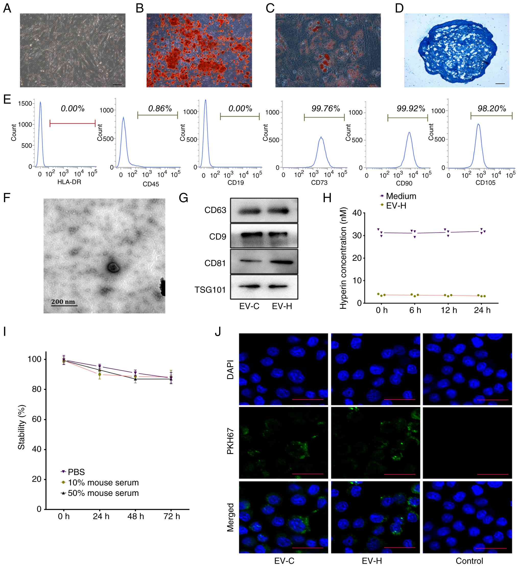 | Figure 1.UCMSC-derived exosomes achieve
effective hyperin drug delivery. (A) The morphology of UCMSCs in
passage 5 reached 90% confluence (scale bar, 50 µm). Then, the
culture medium was replaced with a specific lineage induction
medium. Representative inverted light microscopic images showing
cells 2–3 weeks post-lineage induction after staining with (B)
Alizarin Red S, (C) Oil Red O and (D) Alcian blue (scale bar, 50
µm). (E) Flow cytometry results indicated that UCMSCs were positive
for CD73, CD90 and CD105 and negative for CD19, HLA-DR and CD45.
(F) Representative transmission electron microscopy image of
hyperin-loaded exosomes (scale bar, 200 nm). (G) Western blot
assays revealed the positive expression of typical exosome markers
in UCMSC-derived exosomes. (H) Analysis of drug-loading capacity of
UCMSC-derived exosomes indicated that the exosomal loading rate of
hyperin was ~10% and remained stable within 24 h. (I) Analysis of
stability of hyperin-loaded exosomes in PBS, 10 and 50% mouse
serum. (J) Fluorescence microscopy was used to analyze the uptake
of PKH67-labeled exosomes (green signals) in KGN cells,
demonstrating that hyperin drug loading did not affect cellular
internalization (scale bar, 50 µm). EV-C, control extracellular
vesicle; EV-H, hyperin-loaded exosomes. |
Exosomes were isolated from culture medium that was
conditioned with UCMSCs for 48 h. NTA revealed comparable mean
diameters for EV-Cs (119 nm) and EV-Hs; 120 nm). Transmission
electron microscopy analysis provided evidence that the exosomes
maintained their characteristic double-layered membrane structure
(Fig. 1F), indicating that the
ultrasonication effectively encapsulated hyperin without destroying
the physical membrane integrity of the exosomes. Western blotting
was subsequently used to assess the expression levels of marker
proteins on the surface membrane of UCMSCs before and after
drug-loading. The results demonstrated that the exosomal markers
CD9, CD63, CD81 and TSG101 were positively expressed in both the
EV-C and EV-H groups (Fig.
1G).
Based on the results of UPLC-Q-TOF, the
concentration of hyperin in EV-Hs was 3.509±0.1923 nmol, whereas
the concentration of free hyperin in the supernatant was determined
to be 31.38±0.3413 nmol, indicating a drug-loading rate of ~10%.
Additionally, no significant decrease in the drug-loading capacity
of EV-Hs was observed within 24 h of drug loading at 4°C,
suggesting the feasibility of EV-H use in subsequent animal
experiments (Fig. 1H).
Furthermore, the stability of EV-Hs was evaluated in PBS, 10% mouse
serum and 50% mouse serum. Analysis revealed that after 72 h
incubation, the long-term stability of EV-Hs were 87.7±3.8% in PBS,
88.3±4.5% in 10% mouse serum, and 87.0±2.6% in 50% mouse serum
(Fig. 1I). Subsequently, the
uptake of exosomes by KGN cells was compared between the two groups
via staining of exosomes. As shown in Fig. 1J, green fluorescent signals were
observed in the cytoplasm of KGN cells in both EV groups,
indicating that hyperin drug loading did not notably affect the
uptake of exosomes. Thus, the results showed that EVs could be used
for drug-loading without their physical properties being altered
and could deliver hyperin efficiently and stably to affected
tissues for in vivo studies.
Exosome-based hyperin delivery reduces
CP-induced DOR symptoms in mice
The present study then assessed whether hyperin
could alleviate DOR symptoms in a murine model of the disease. As
illustrated in Fig. 2A, healthy
female mice were divided into five groups and administered
different treatments. No mortality was observed in any group
throughout the administration period prior to the experimental
endpoint. Starting on day 21, mice in the DOR group exhibited mild
clinical signs of CP-induced systemic discomfort, including a dull
coat, hunched backs and reduced activity levels. However, none
exhibited severe unresponsiveness or met the pre-established humane
endpoints for early euthanasia, as mice maintained active grooming,
preserved their righting reflex and maintained adequate food and
fluid intake. The body weights of mice from all groups showed no
significant differences during the first week (Fig. 2B). After receiving CP via
intraperitoneal injection on day 7, the body weights of mice in the
DOR, OA and EV-C groups increased slowly. Compared with the control
group, the DOR group showed a significant reduction in body weight
at all tested time points from day 12 onwards. The OA and EV-C
groups did not exhibit any statistically significant differences
when compared with each other or with the DOR group at any measured
time points. Although visual markers, such as coat color, showed
improvement in mice in the OA treatment group, mice exhibited
decreased food intake, which may have been attributed to the higher
dosage of hyperin administered via the intragastric method. Mice
treated with EV-H demonstrated markedly higher levels of weight
gain than those from the DOR, OA and EV-C treatment groups.
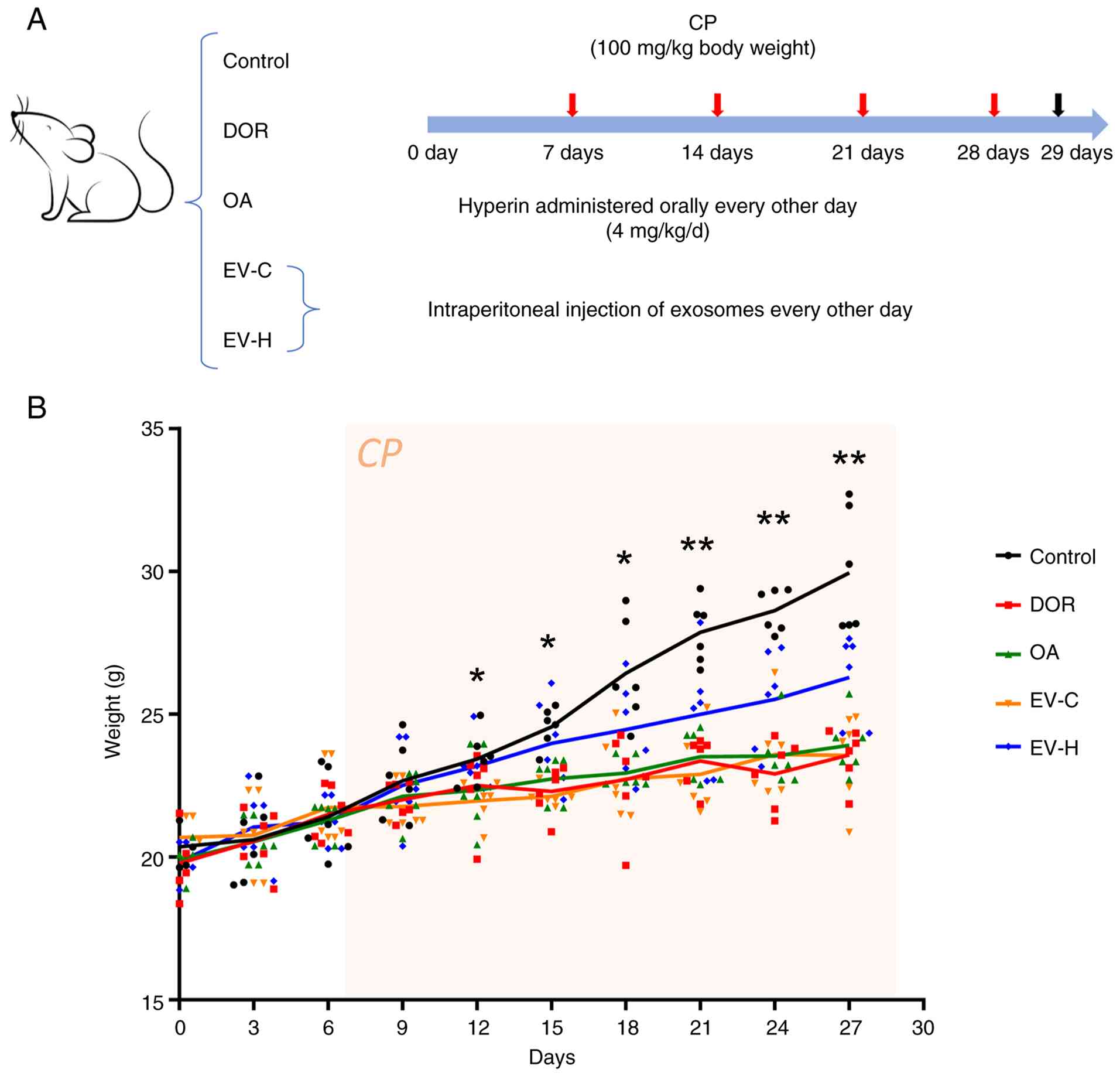 | Figure 2.Evaluation of the therapeutic effect
of hyperin on a CP-induced DOR mouse model. (A) Experimental
schematic flow diagram, showing how mice were randomly assigned to
five treatment groups: Control, DOR, OA, EV-C and EV-H groups.
Therapeutic administration of hyperin was initiated on day 1 and
performed once every other day. On day 7, a weekly dose of CP was
administered to mice all groups except the control group via
intraperitoneal injection to induce DOR-like pathology (red arrow).
Mice were systematically monitored and a subset of mice were
sacrificed on day 29 (black arrow). (B) Body weights of mice across
the five treatment groups were recorded longitudinally at
designated time points throughout the 4-week experimental period.
*P<0.05 and **P<0.01, vs control. DOR, diminished ovarian
reserve; CP, cyclophosphamide; OA, oral administration; EV-C,
control extracellular vesicle; EV-H, hyperin-loaded exosomes. |
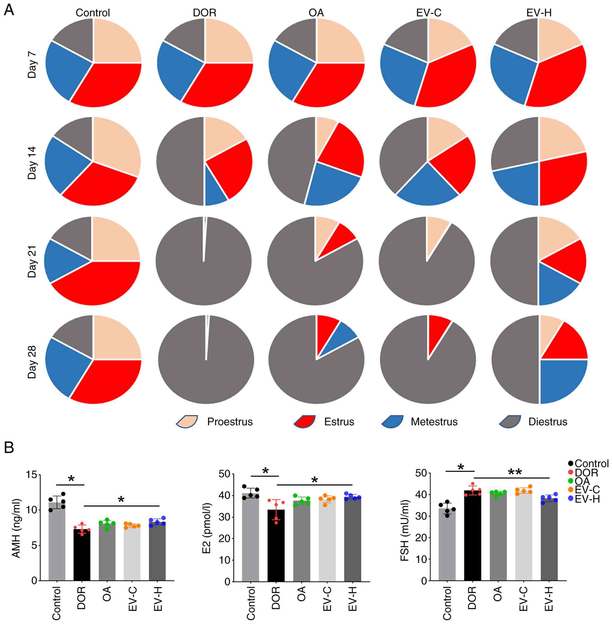
In addition, the present study simultaneously
assessed changes in the estrous cycle of female mice across
treatment groups, as shown in Fig.
3A. Before CP administration, all groups displayed a regular
estrous cycle with relatively even stage distribution (Fig. S1). Subsequently, the diestrus
phases gradually extended in the DOR group and these mice remained
stagnant in diestrus for an extended period after 2 weeks of
treatment. Mice from both the OA and EV-C groups showed notable
disruptions in their estrous cycles following CP injection, with a
notable prolongation of the estrus period; however, signs of estrus
were consistently present during treatment. Comparison among these
groups revealed a notable improvement in the estrous cycles of mice
within the EV-H group; even after 3 weeks of CP treatment,
EV-H-treated mice exhibited significant restoration of normal
estrous cyclicity and regular stage transitions. Mouse serum was
collected on day 29 following the final observation to assess the
AMH, FSH and E2 levels of mice in each group. As in Fig. 3B, analysis of mice in the DOR model
group revealed a significant decrease in AMH and E2 levels compared
with those in the control group, along with a significant increase
in serum FSH levels. No notable differences in AMH, FSH and E2
levels were observed between the DOR model group and the OA or EV-C
groups. However, compared with the DOR model group, a significant
increase in AMH levels was observed in mice within the EV-H group.
This increase was accompanied by a significant rise in E2 and a
significant decrease in FSH levels, indicating considerable
improvements in hormone levels in DOR model mice following EV-H
treatment.
As shown in Fig.
4A, ovarian follicles in all developmental stages were observed
in mice from the healthy control group, featuring plump oocytes and
abundant GCs in the antral follicles; furthermore, atretic
follicles were sporadically observed. By contrast, mice from the
DOR model group displayed a notable reduction in ovarian volume and
a less dense ovarian stroma. Fig.
4B showed that, compared with the control group, the quantity
of primordial, primary and secondary follicles decreased
significantly in the DOR group, whereas the number of atretic
follicles increased. No significant changes were observed in the
number of antral follicles between these groups. Mice from the OA
and EV-C groups showed notable increases in ovarian volume,
characterized by a less dense ovarian stroma and degenerated
oocytes with sparse, apoptotic GCs. Compared with the control, the
quantity of primordial, primary and secondary follicles decreased
significantly in the DOR group, whereas the number of atretic
follicles increased. No significant changes were observed in the
number of antral follicles between these groups. Comparatively,
mice in the EV-H group displayed marked histological protection,
featuring well-preserved and compact ovarian stroma, healthy plump
oocytes, and multiple layers of abundant, well-organized GCs.
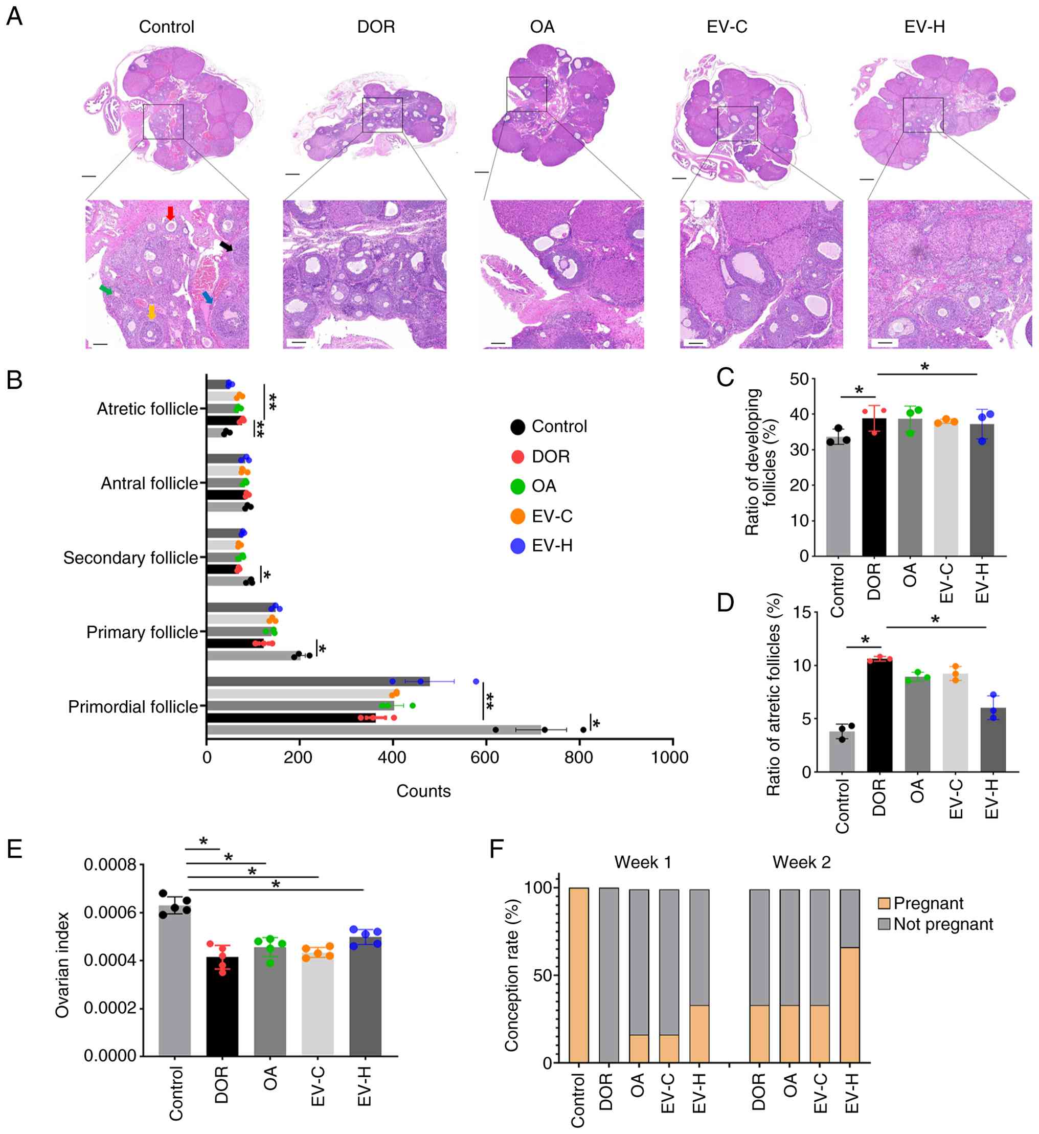 | Figure 4.Delivery of hyperin via exosomes
attenuates symptoms of cyclophosphamide-induced DOR in mice. (A)
Representative images of hematoxylin and eosin staining of ovaries
from mice in each treatment group (scale bar, 500 and 100 µm).
Black arrow, atretic follicle, blue arrow indicated antral
follicle, yellow arrow indicated secondary follicle, red arrow
indicated primary follicle, and green arrow indicated primordial
follicle. (B) Number of follicles of different grades in mice in
each treatment group. (C) The proportion of developing follicles in
each group. (D) The proportion of atretic follicles in each group.
(E) Ovarian index of mice in each group. n=5. (F) Comparison of
conception time of mice in each group. n=6. *P<0.05 and
**P<0.01. DOR, diminished ovarian reserve; OA, oral
administration; EV-C, control extracellular vesicle; EV-H,
hyperin-loaded exosomes. |
Furthermore, no notable improvements in the numbers
of other types of follicles or the number of atretic follicles in
these groups was observed compared with the DOR group.
Comparatively, mice in the EV-H group exhibited a significant
increase in the absolute number of primordial follicles compared
with those in the DOR group, whereas the absolute number of atretic
follicles significantly decreased, demonstrating that EV-H
effectively suppressed follicular atresia. CP treatment in DOR mice
induced severe depletion of the primordial pool, resulting in an
altered percentage distribution of developing follicles relative to
total remaining follicles (Fig.
4C). EV-H administration effectively normalized the proportion
of healthy developing follicles and significantly reduced the ratio
of atretic follicles (Fig. 4D),
demonstrating overall restoration of follicular pool composition.
On day 29, concurrent with serum collection, the ovarian index of
samples was evaluated (31). The
ovarian index values of treated mice in each group were
significantly decreased compared with that of the mice from the
control group (Fig. 4E).
Subsequently, to evaluate the fertility outcomes of each treatment,
the remaining mice in each group were maintained on a regular diet
for 2 weeks before being paired with fertile male C57BL/6 mice at a
ratio of 2:1; Fig. 4F depicts the
number of pregnancies in each group. A total of 7 days pf
cohabitation, all 6 control group mice became pregnant, whereas
none of the DOR group mice became pregnant. At this 1-week mark, 1
mouse each from the OA and EV-C groups was pregnant, whereas 2 EV-H
group mice became pregnant. By day 14 of cohabitation, mice from
the DOR, OA and EV-C groups demonstrated a pregnancy rate of 2 out
of 6, whereas mice in the EV-H group exhibited a pregnancy rate of
4 out of 6. These findings demonstrated that EV-H administration
notably improved ovarian follicle development and reduced
follicular atresia and improved functional fertility in mice with
CP-induced DOR.
Exosome-based hyperin delivery
suppresses CP-induced autophagy both in vivo and in vitro
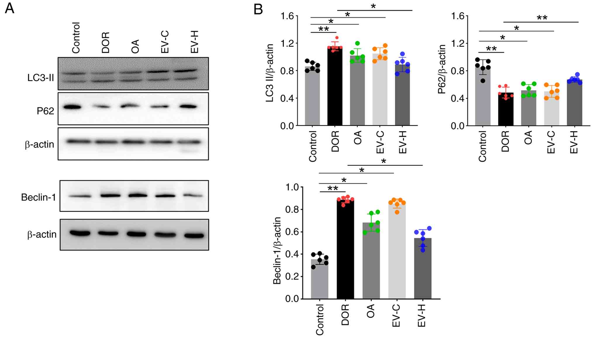
Subsequently, the present study investigated the
levels of autophagy within the ovarian tissues of each group of
mice. The differences in LC3-II, P62 and beclin-1 protein levels in
each group, as detected by western blot analysis, are presented in
Fig. 5. In the DOR model group,
the expression levels of LC3-II and beclin-1 increased
significantly compared with those in the control group, whereas the
P62 protein expression decreased significantly. Similarly, compared
with the control group, the OA and EV-C groups exhibited
significant increases in LC3-II and Beclin-1 expression, along with
a significant decrease in P62 expression. Notably, no significant
difference in LC3II, P62 and Beclin-1 expressions was observed
between the EV-H and control groups. Furthermore, compared with
mice in the DOR model group, mice in the EV-H group exhibited
significant decreases in the expression levels LC3-II and beclin-1
and a significant increase in P62 protein expression. These
findings indicated that EV-H effectively inhibited CP-induced
autophagy activation in mouse ovarian tissues.
Dysfunction of GCs, which represent the largest cell
group in the ovarian follicle and are responsible for secreting
steroid hormones and cytokines, has been shown to lead to
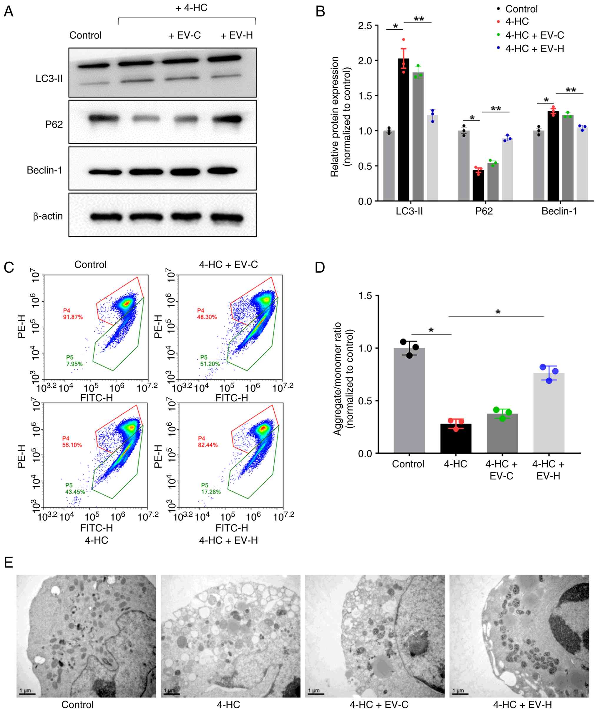
follicular atresia and may cause DOR (34). In the present study, the KGN
ovarian GC line, was utilized for in vitro experiments and
subject to treatment with 4-HC, the active metabolite of CP. As
shown in Fig. 6A and B, exposure
to 10 µM 4-HC for 24 h effectively triggered protein level
alterations indicative of autophagic activation in KGN cells. A
significant rise in LC3-II expression and beclin-1 protein levels
was observed in 4-HC treated cells compared with those in the
control group treated with DMSO. Cells treated with 4-HC also
exhibited a significant reduction in P62 protein levels.
Furthermore, pre-exposure to UCMSC-derived EV-C partially
attenuated the effects of 4-HC treatment on autophagy, but these
changes were not statistically significant. Notably, pre-exposure
to EV-H significantly mitigated the increase in the LC3-II and
beclin-1 protein expression levels induced by 4-HC treatment and
significantly increased P62 expression levels. These results
aligned with the present in vivo findings which established
that EV-H proficiently inhibits CP-induced autophagy
activation.
Following JC-10 staining flow cytometry and
microplate reader analyses were used to assess the mitochondrial
membrane potential of KGN cells subjected to different treatments
(Fig. 6C and D). Changes in the
red/green fluorescence intensity ratio of cell samples indicated
mitochondrial depolarization, with a significant decrease in
mitochondrial membrane potential being observed after 4-HC
treatment compared with controls. Pre-treatment with EV-H resulted
in a notable shift from primarily green- to red-fluorescence
emissions compared with 4-HC-treated cells, suggesting that
pre-treated cells exhibited greater mitochondrial membrane
potential. This was supported by the significant increase in
aggregate/monomer ratio observed in 4-HC + EV-H cells compared with
4-HC cell samples. To further support these observations, the
present study analyzed KGN cells via transmission electron
microscopy, which represents the benchmark method for investigating
autophagy (Fig. 6E). Compared with
the control group, the number of mitophagic vacuoles notably
increased in 4-HC-treated cells. However, compared with the 4-HC
group, the number of mitophagic vacuoles markedly decreased in the
EV-H pre-treatment group. These findings indicated that EV-H
pre-treatment effectively protected KGN cells against 4-HC-induced
damage.
Discussion
The clinical management of DOR remains a notable
challenge in reproductive medicine, particularly in cases involving
chemotherapy-induced premature ovarian insufficiency (35). Hyperin, a notable flavonol
glycoside used in traditional Chinese medicine (TCM), has long been
recognized for its potent antioxidant, anti-apoptotic and
reproductive-protective properties (36–38).
However, when administered via conventional OA, the therapeutic
efficacy of free hyperin was notably compromised; this was
potentially due to its rapid gastrointestinal degradation and
notable hepatic first-pass metabolism, and consequently, its low
bioavailability in target ovarian tissue (39). To circumvent these pharmacokinetic
limitations, the present study successfully engineered a novel
nanomedicine delivery system by encapsulating hyperin into human
UCMSC-derived exosomes via ultrasonication, thereby yielding a
stable and efficient hyperin formulation. The findings of the
present study demonstrated that EV-H markedly outperformed
therapies involving free hyperin in restoring hormone balance,
preserving follicular reserves and mitigating GC injury, thereby
offering a promising exosome-based strategy for DOR therapy.
The key to understanding the occurrence and
development of DOR lies in establishing animal models that mimic
its pathological mechanisms. Animal models of DOR established using
various DOR induction methods, such as chemotherapy and
drug-induced damage, have provided a solid experimental foundation
for research (39–41). The chemotherapy drug CP is commonly
used to induce DOR and establish DOR animal models due to its
ability to notably damage ovarian function in mice and replicate
the pathological characteristics of DOR (40). As such, CP-based models facilitate
a more comprehensive investigation of treatment efficacy in DOR and
further elucidate the corresponding mechanisms of action for such
treatments than other DOR models. In the present study, a DOR mouse
model was established via the intraperitoneal administration of CP
at a dose of 100 mg/kg weekly for 3 consecutive weeks. Over time,
DOR model mice exhibited progressively disrupted estrous cycles,
which eventually stagnated in the diestrus phase. Following CP
treatment, mice in the model group demonstrated markedly reduced
serum AMH levels, elevated FSH levels, decreased ovarian index
values, a depleted primordial follicle pool and a significantly
increased incidence of follicular atresia, as well as increased
conception time. All of these observations were consistent with the
clinical symptoms of DOR, indicating successful modeling (41,42).
Mechanistically, the maintenance of ovarian
homeostasis is intricately linked to the precise regulation of
autophagy in GCs. Autophagy fundamentally plays dual roles in
reproductive toxicology (43,44).
Under physiological baseline conditions or mild cellular stress,
autophagy acts as a pro-survival, homeostatic mechanism aimed at
recycling damaged organelles and clearing reactive oxygen species
(ROS) (45). However, chemotherapy
with alkylating agents, such as CP, triggers an influx of
intracellular ROS and results in notable amounts of double-stranded
DNA breaks within GCs (46). Under
such notable and chronic toxic stress, the autophagic flux shifts
from a beneficial, restorative response into an unchecked,
cytotoxic pathway that directly executes GC apoptosis and
accelerates follicular atresia (47,48).
Notably, treatment with EV-H significantly mitigated these
alterations, partially restoring the expression levels of LC3-II,
beclin-1 and P62 towards their respective baseline levels. Rather
than completely suppressing an important physiological process,
EV-H acted as a therapeutic modulator that mitigated the underlying
cellular stress, as evidenced by the restoration of the
mitochondrial membrane potential in KGN cells following EV-H
treatment, thereby rescuing GCs from entering the cytotoxic
autophagic cascade.
The present study highlighted the potential of
integrating TCM with exosome technology for the treatment of DOR,
utilizing exosomes derived from UCMSCs for hyperin delivery, and
observed a notable protective effect on ovarian-reserve function in
mice. Exosomes are natural extracellular vesicles that are widely
distributed in the body and possess intrinsic therapeutic
advantages, such as non-immunogenicity and easy cell membrane
penetration (49). In previous
years, research on exosome-based drug delivery has expanded,
demonstrating promising applications for treating various
challenging diseases and tumors by successfully loading
small-molecule chemical drugs and nucleic acid therapeutics into
exosomes (50,51). The difference in administration
routes between the OA group, which was subject to intragastric
administration, and the exosome groups, which received hyperin via
intraperitoneal injection, represents a notable pharmacokinetic
variable. However, because EV-H treatment displayed markedly
improved efficacy in restoring follicle counts and hormone profiles
than empty EV-C administered via the same administration route, the
present study demonstrated that the therapeutic benefits observed
in the EV-H group were driven by hyperin encapsulation rather than
route-dependent bioavailability alone. Compared with direct gavage
and other drug-administration methods reported in literature
(26,52), utilizing exosomes as drug carriers
notably enhanced the targeted delivery and therapeutic bioefficacy
of hyperin in vivo, thereby improving its therapeutic
efficacy and minimizing the adverse effects associated with direct
hyperin administration.
In conclusion, the present study demonstrated that
hyperin-loaded exosomes exhibited notable therapeutic potential for
the treatment of DOR. By integrating TCM principles with modern
scientific methodologies, the present study represented a
comprehensive investigation into the mechanisms by which TCM could
support DOR treatment, offering more effective and safer
therapeutic options for affected patients. These findings will
markedly contribute toward therapies supporting female reproductive
health. However, it is important to consider individual variations
in treatment response and assess the long-term effectiveness of the
treatment method established in the present study. Therefore, the
present study presented an integrated therapeutic approach that
combined TCM with modern biotechnology and offered innovative
prospects for future clinical DOR treatments.
Supplementary Material
Supporting Data
Acknowledgements
The authors would like to express their sincere
gratitude to Miss Xin Pan (Key Laboratory of Cell-Based Drug and
Applied Technology Development in Zhejiang Province (Hangzhou,
China) for administrative support and coordination in laboratory
animal management and compliance during the present study.
Funding
The present study was supported by the Zhejiang Provincial
Medical and Health Science and Technology Project (grant no.
2025KY476) and the Key Technologies R&D Program of Zhejiang
Province (grant nos. 2019C03041 and 2021C03077).
Availability of data and materials
The data generated in the present study may be
requested from the corresponding author.
Authors' contributions
YY and YWu were responsible for performing the
experiments and writing the original draft of the manuscript. JZ
and YWa assisted with the experiments and analyzed the data. HY
contributed toward data interpretation. GL performed animal
experiments. RP and LL contributed toward conceptualization,
reviewing and editing the manuscript, supervision and project
management. All authors read and approved the final version of the
manuscript. YY and YWu confirm the authenticity of all the raw
data.
Ethics approval and consent to
participate
All experimental procedures were performed according
to the National Institutes of Health Guidelines for the Care and
Use of Laboratory Animals and approved by the Medical Animal
Experiment Ethics Committee of the Hangzhou Medical College
Laboratory Animal Center, otherwise known as the Zhejiang Center of
Laboratory Animals (approval no. ZJCLA-IACUC-20050016).
Patient consent for publication
Not applicable.
Competing interests
The authors declare that they have no competing
interests.
References
|
1
|
Broekmans FJ, Soules MR and Fauser BC:
Ovarian aging: Mechanisms and clinical consequences. Endocr Rev.
30:465–493. 2009. View Article : Google Scholar : PubMed/NCBI
|
|
2
|
Park SU, Walsh L and Berkowitz KM:
Mechanisms of ovarian aging. Reproduction. 162:R19–R33. 2021.
View Article : Google Scholar : PubMed/NCBI
|
|
3
|
Chon SJ, Umair Z and Yoon MS: Premature
ovarian insufficiency: Past, present, and future. Front Cell Dev
Biol. 9:6728902021. View Article : Google Scholar : PubMed/NCBI
|
|
4
|
Scantamburlo VM, Linsingen RV, Centa LJR,
Toso KFD, Scaraboto D, Araujo Júnior E and Kulak Junior J:
Association between decreased ovarian reserve and poor oocyte
quality. Obstet Gynecol Sci. 64:532–539. 2021. View Article : Google Scholar : PubMed/NCBI
|
|
5
|
Yao LN, Lin WQ, Jiang N, Li C, Cao HF, Li
H and Qian JH: Comparative study of assisted reproductive outcomes
between young patients with occult premature ovarian insufficiency
and advanced-age patients. J Int Med Res. 48:3000605209346562020.
View Article : Google Scholar : PubMed/NCBI
|
|
6
|
Abrahami N, Izhaki I and Younis JS: Do
young women with unexplained infertility show manifestations of
decreased ovarian reserve? J Assist Reprod Genet. 36:1143–1152.
2019. View Article : Google Scholar : PubMed/NCBI
|
|
7
|
Liu T, Li Q, Wang S, Chen C and Zheng J:
Transplantation of ovarian granulosa-like cells derived from human
induced pluripotent stem cells for the treatment of murine
premature ovarian failure. Mol Med Rep. 13:5053–5058. 2016.
View Article : Google Scholar : PubMed/NCBI
|
|
8
|
Xie Z, Li Y, Chen Z, Cao Q, Zhang C, Fei
Y, Li K and Guan Y: Effects of Bushen Huoxue method for female with
decreased ovarian reserve: A protocol for systematic review and
meta-analysis. Medicine (Baltimore). 99:e229572020. View Article : Google Scholar : PubMed/NCBI
|
|
9
|
Lv Z, Lv Z, Song L, Zhang Q and Zhu S:
Role of lncRNAs in the pathogenic mechanism of human decreased
ovarian reserve. Front Genet. 14:10560612023. View Article : Google Scholar : PubMed/NCBI
|
|
10
|
Fusco F, Paciolla M, Chen E, Li X, Genesio
R, Conti A, Jones J, Poeta L, Lioi MB, Ursini MV and Miano MG:
Genetic and molecular analysis of a new unbalanced X;18
rearrangement: Localization of the diminished ovarian reserve
disease locus in the distal Xq POF1 region. Hum Reprod.
26:3186–3196. 2011. View Article : Google Scholar : PubMed/NCBI
|
|
11
|
Ferder IC, Espeche LD, Bruque CD,
Parborell F, Tesone M and Dain L: Expression and characterisation
of Fmr1 splice variants during folliculogenesis in the rat. Reprod
Fertil Dev. 34:1034–1042. 2022. View
Article : Google Scholar : PubMed/NCBI
|
|
12
|
Paulini F and Melo EO: The role of
oocyte-secreted factors GDF9 and BMP15 in follicular development
and oogenesis. Reprod Domest Anim. 46:354–361. 2011. View Article : Google Scholar : PubMed/NCBI
|
|
13
|
Wu W, Han J, Cao R, Zhang J, Li B, Liu Z,
Liu K, Li Q, Pan Z, Chen J and Liu H: Sequence and regulation of
the porcine FSHR gene promoter. Anim Reprod Sci. 154:95–104. 2015.
View Article : Google Scholar : PubMed/NCBI
|
|
14
|
Gogola J, Hoffmann M and Ptak A:
Persistent endocrine-disrupting chemicals found in human follicular
fluid stimulate the proliferation of granulosa tumor spheroids via
GPR30 and IGF1R but not via the classic estrogen receptors.
Chemosphere. 217:100–110. 2019. View Article : Google Scholar : PubMed/NCBI
|
|
15
|
Henes M, Froeschlin J, Taran FA, Brucker
S, Rall KK, Xenitidis T, Igney-Oertel A, Lawrenz B and Henes JC:
Ovarian reserve alterations in premenopausal women with chronic
inflammatory rheumatic diseases: Impact of rheumatoid arthritis,
Behcet's disease and spondyloarthritis on anti-Mullerian hormone
levels. Rheumatology (Oxford). 54:1709–1712. 2015. View Article : Google Scholar : PubMed/NCBI
|
|
16
|
Chen J, Wu S, Wang M, Zhang H and Cui M: A
review of autoimmunity and immune profiles in patients with primary
ovarian insufficiency. Medicine (Baltimore). 101:e325002022.
View Article : Google Scholar : PubMed/NCBI
|
|
17
|
Kim Y, Lee E, Song JY, Kim Y and Lee S:
Association between environmental pollutants and the FSH/AMH ratio
as a marker of ovarian reserve. Environ Anal Health Toxicol.
37:e2022029–0. 2022. View Article : Google Scholar : PubMed/NCBI
|
|
18
|
Osmanlioglu S, Berker B, Aslan B, Şükür
YE, Özmen B, Sönmezer M, Atabekoğlu CS and Aytaç R: Presence of
endometrioma does not impair embryo quality and assisted
reproductive technology (ART) cycle outcome in diminished ovarian
reserve (DOR) patients. Reprod Sci. 30:1540–1547. 2023. View Article : Google Scholar : PubMed/NCBI
|
|
19
|
Vigneswaran K and Hamoda H: Hormone
replacement therapy-Current recommendations. Best Pract Res Clin
Obstet Gynaecol. 81:8–21. 2022. View Article : Google Scholar : PubMed/NCBI
|
|
20
|
Gonda KJ, Domar AD, Gleicher N and Marrs
RP: Insights from clinical experience in treating IVF poor
responders. Reprod Biomed Online. 36:12–19. 2018. View Article : Google Scholar : PubMed/NCBI
|
|
21
|
Monteiro CS, Scheffer BB, Carvalho RF and
Scheffer JB: The impact of dehydroepiandrosterone in poor ovarian
responders on assisted reproduction technology treatment. JBRA
Assist Reprod. 23:414–417. 2019.PubMed/NCBI
|
|
22
|
Pellicer N, Cozzolino M, Diaz-Garcia C,
Galliano D, Cobo A, Pellicer A and Herraiz S: Ovarian rescue in
women with premature ovarian insufficiency: Facts and fiction.
Reprod Biomed Online. 46:543–565. 2023. View Article : Google Scholar : PubMed/NCBI
|
|
23
|
Chen H, Liu C, Zhu S, Li S, Zhang Q, Song
L and Liang X: The therapeutic effect of stem cells on
Chemotherapy-Induced premature ovarian failure. Curr Mol Med.
21:376–384. 2021. View Article : Google Scholar : PubMed/NCBI
|
|
24
|
Wu JJ, Zhang J, Xia CY, Ding K, Li XX, Pan
XG, Xu JK, He J and Zhang WK: Hypericin: A natural anthraquinone as
promising therapeutic agent. Phytomedicine. 111:1546542023.
View Article : Google Scholar : PubMed/NCBI
|
|
25
|
Wang X, Fan G, Wei F, Bu Y and Huang W:
Hyperoside protects rat ovarian granulosa cells against hydrogen
peroxide-induced injury by sonic hedgehog signaling pathway. Chem
Biol Interact. 310:1087592019. View Article : Google Scholar : PubMed/NCBI
|
|
26
|
Zhu F, Gao J, Zeng F, Lai Y, Ruan X and
Deng G: Hyperoside protects against cyclophosphamide induced
ovarian damage and reduced fertility by suppressing
HIF-1alpha/BNIP3-mediated autophagy. Biomed Pharmacother.
156:1137432022. View Article : Google Scholar : PubMed/NCBI
|
|
27
|
You F, Cao J, Cheng L, Liu X and Zeng L:
Hyperin alleviates Triptolide-induced ovarian granulosa cell injury
by regulating AKT/TSC1/mTORC1 signaling. Evid Based Complement
Alternat Med. 2021:93992612021. View Article : Google Scholar : PubMed/NCBI
|
|
28
|
Pan W, Gu L, Yang H, Xu C, Yang Z, Lu Q,
Shi Y, Zhang L, Shao J, Chen Y, et al: Repeat-dose toxicity study
of human umbilical cord mesenchymal stem cells in cynomolgus
monkeys by intravenous and subcutaneous injection. Front Cell Dev
Biol. 11:12737232023. View Article : Google Scholar : PubMed/NCBI
|
|
29
|
Dominici M, Le Blanc K, Mueller I,
Slaper-Cortenbach I, Marini F, Krause D, Deans R, Keating A,
Prockop DJ and Horwitz E: Minimal criteria for defining multipotent
mesenchymal stromal cells. The International Society for Cellular
Therapy position statement. Cytotherapy. 8:315–317. 2006.
View Article : Google Scholar : PubMed/NCBI
|
|
30
|
Council NR: Guide for the Care and Use of
Laboratory Animals: Eighth Edition. Washington, DC: The National
Academies Press; 2011
|
|
31
|
Huang C, Song K, Ma W, Ding J, Chen Z and
Zhang M: Immunomodulatory mechanism of Bushen Huoxue Recipe
alleviates cyclophosphamide-induced diminished ovarian reserve in
mouse model. J Ethnopharmacol. 208:44–56. 2017. View Article : Google Scholar : PubMed/NCBI
|
|
32
|
Byers SL, Wiles MV, Dunn SL and Taft RA:
Mouse estrous cycle identification tool and images. PLoS One.
7:e355382012. View Article : Google Scholar : PubMed/NCBI
|
|
33
|
Ajayi AF and Akhigbe RE: Staging of the
estrous cycle and induction of estrus in experimental rodents: An
update. Fertil Res Pract. 6:52020. View Article : Google Scholar : PubMed/NCBI
|
|
34
|
Chen Q, Chen Q, Song Y, Xiang Y, Li Q,
Sang Y, Zhang L, Bai L and Zhu Y: Downregulation of homeobox A1 in
human granulosa cells is involved in diminished ovarian reserve
through promoting cell apoptosis and mitochondrial dysfunction. Mol
Cell Endocrinol. 580:1120842024. View Article : Google Scholar : PubMed/NCBI
|
|
35
|
Cedars MI: Managing poor ovarian response
in the patient with diminished ovarian reserve. Fertil Steril.
117:655–656. 2022. View Article : Google Scholar : PubMed/NCBI
|
|
36
|
Wierman ME and Kiseljak-Vassiliades K:
Should dehydroepiandrosterone be administered to Women? J Clin
Endocrinol Metab. 107:1679–1685. 2022. View Article : Google Scholar : PubMed/NCBI
|
|
37
|
Somigliana E, Li Piani L, Paffoni A,
Salmeri N, Orsi M, Benaglia L, Vercellini P and Vigano' P:
Endometriosis and IVF treatment outcomes: Unpacking the process.
Reprod Biol Endocrinol. 21:1072023. View Article : Google Scholar : PubMed/NCBI
|
|
38
|
Zhang C and Xu X: Advancement in the
treatment of diminished ovarian reserve by traditional Chinese and
Western medicine. Exp Ther Med. 11:1173–1176. 2016. View Article : Google Scholar : PubMed/NCBI
|
|
39
|
Buigues A, Marchante M, Herraiz S and
Pellicer A: Diminished ovarian reserve Chemotherapy-induced mouse
model: A tool for the preclinical assessment of new therapies for
ovarian damage. Reprod Sci. 27:1609–1619. 2020. View Article : Google Scholar : PubMed/NCBI
|
|
40
|
He R, Fan Q, Li Y, Zhu Q, Hu D, Du J, Xing
Y, Li H, Liang X and Yang Y: Identification of common and specific
genes involved in mouse models of Age-related and
Cyclophosphamide-Induced diminished ovarian reserve. Reprod Sci.
30:1965–1978. 2023. View Article : Google Scholar : PubMed/NCBI
|
|
41
|
Bunnewell SJ, Honess ER, Karia AM, Keay
SD, Al Wattar BH and Quenby S: Diminished ovarian reserve in
recurrent pregnancy loss: A systematic review and meta-analysis.
Fertil Steril. 113:818–827.e3. 2020. View Article : Google Scholar : PubMed/NCBI
|
|
42
|
Kučera M, Štěpán Ml J and Štelcl M:
Possibilities and real meaning of assessment of ovarian reserve.
Ceska Gynekol. 83:307–311. 2018.PubMed/NCBI
|
|
43
|
Chhabra V, Meenakshi S, Maity S, Saini D,
Saini M, Murti K and Kumar N: Impact of fluoride exposure on
reproductive health: Insights into molecular mechanisms and health
implications. Reprod Toxicol. 135:1089072025. View Article : Google Scholar : PubMed/NCBI
|
|
44
|
Nazeerulla F, Banerjee I, Paul MK, Bisht
B, Basu D, Bhutia SK and Mukhopadhyay S: Autophagy as an architect
of embryogenesis: Understanding the new mechanisms, emerging
paradigms, and future directions. Life Sci. 391:1242772026.
View Article : Google Scholar : PubMed/NCBI
|
|
45
|
Debnath J, Gammoh N and Ryan KM: Autophagy
and autophagy-related pathways in cancer. Nat Rev Mol Cell Biol.
24:560–575. 2023. View Article : Google Scholar : PubMed/NCBI
|
|
46
|
Kawano M, McKey J, Batchvarov IS and Capel
B: Granulosa cell death is a significant contributor to
DNA-damaging chemotherapy-induced ovarian insufficiencydagger. Biol
Reprod. 112:906–915. 2025. View Article : Google Scholar : PubMed/NCBI
|
|
47
|
Zhu F, Hu C, Huang S, Cao X, Li X, Song Y,
Chen M and Xiao J: Qilin Pills protect against diminished ovarian
reserve by suppressing pathological overactivation of
PINK1/Parkin-mediated mitophagy in granulosa cells. J
Ethnopharmacol. 369:1218622026. View Article : Google Scholar : PubMed/NCBI
|
|
48
|
Chen L, Zeng L, Pan S, Zu L, Pan H and Fan
L: β-sitosterol in Yijing Hugui decoction prevents
cyclophosphamide-induced premature ovarian insufficiency via the
AKT1/Nrf2 pathway. Cytotechnology. 77:762025. View Article : Google Scholar : PubMed/NCBI
|
|
49
|
Kalluri R and LeBleu VS: The biology,
function, and biomedical applications of exosomes. Science.
367:eaau69772020. View Article : Google Scholar : PubMed/NCBI
|
|
50
|
Kimiz-Gebologlu I and Oncel SS: Exosomes:
Large-scale production, isolation, drug loading efficiency, and
biodistribution and uptake. J Control Release. 347:533–543. 2022.
View Article : Google Scholar : PubMed/NCBI
|
|
51
|
Liang Y, Duan L, Lu J and Xia J:
Engineering exosomes for targeted drug delivery. Theranostics.
11:3183–3195. 2021. View Article : Google Scholar : PubMed/NCBI
|
|
52
|
Zhou Q, Tang H, Wang Y, Hua Y, Ouyang X
and Li L: Hyperoside mitigates PCOS-associated adipogenesis and
insulin resistance by regulating NCOA2-mediated PPAR-gamma
ubiquitination and degradation. Life Sci. 364:1234172025.
View Article : Google Scholar : PubMed/NCBI
|