Introduction
Statistical projections indicated that 1,596,670 new
cases of cancer and 571,950 mortalities would occur in the United
States in 2011 (1). Oral cancer is
a serious health problem in many other parts of the world and the
eighth-leading cause of cancer-related death in men. Certain
studies suggest that the risk factors for oral cancer are tobacco,
alcohol, ultraviolet light and oral lesions (2,3).
Although the incidence of oral cancer is low, patients have a poor
prognosis, and the five-year survival rate has remained unchanged
at approximately 50%. Accordingly, the development of more
effective therapeutic strategies for the prevention and therapy of
oral cancer is imperative.
Mcl-1 is a Bcl-2-family protein that is essential in
apoptosis control, and it rapidly decreases during apoptosis
(4). In human malignancies, the
increased expression of Mcl-1 causes tumor progression and
chemoresistance (5). Natural
products derived from plant sources modulate apoptosis through the
downregulation of Mcl-1. Lycorine isolated from Amaryllidaceae
lycoris induces apoptosis and causes a rapid turnover of Mcl-1
expression in human leukemia cell lines (6). The apoptotic effects of Honokiol,
purified from magnolia, appear to be associated with the
downregulation of Mcl-1 in B-cell chronic lymphocytic leukemia
(7). Thus, the downregulation of
Mcl-1 may be an attractive therapeutic strategy for inducing
apoptosis. The pro-apoptotic protein Bak is constitutively
integrated in the mitochondrial outer membrane, but changes
conformation and forms oligomeric complexes in response to
apoptotic stimuli (8). Notably, the
downregulation of Mcl-1 by chemotherapeutic agents is associated
with the activation of Bak (9–11).
Therefore, the study of Bak in cancer cells expressing Mcl-1 may
provide a promising strategy.
Sanguisorba officinalis L. has
anti-inflammatory, anti-allergic and anxiolytic activities
(12–15). Moreover, several triterpenoids
isolated from the roots of S. officinalis L. have been shown
to inhibit the growth of tumor cell lines (16). However, its effects in oral cancer
and the mechanism of S. officinalis L.-induced apoptosis
remain poorly defined. In this study, we provide experimental
evidence that an extract of S. officinalis L. inhibits cell
growth and induces apoptosis in oral cancer cell lines.
Materials and methods
Reagents
Hot water extract of S. officinalis L. (HESO)
was kindly provided by Professor Ki-Han Kwon (Kwangju University,
Kwangju, Korea). PARP antibody was obtained from BD Pharmingen (San
Jose, CA, USA). Sp1 and actin antibodies were obtained from Santa
Cruz Biotechnology, Inc. (Santa Cruz, CA, USA). Antibodies against
Mcl-1, Bak and survivin were obtained from Cell Signaling
Technology, Inc. (Charlottesville, VA, USA).
Cell culture and chemical treatment
HSC4 cells were provided by Hokkaido University
(Hokkaido, Japan) and HN22 cells were provided by Dankook
University (Cheonan, Korea). Both cells were cultured in Dulbecco’s
modified Eagle’s medium (DMEM) supplemented with 10% fetal bovine
serum (FBS) and antibiotics at 37°C in a 5% CO2
incubator. Cells were treated with vehicle (DMSO) or HESO (200, 400
and 600 μg/ml for HSC4 cells and 100, 200 and 400 μg/ml for HN22
cells) for 48 h.
MTS assay
The effect of HESO on cell viability was tested
using the CellTiter 96 Aqueous One Solution Cell Proliferation
Assay kit (Promega, Madison, WI, USA) according to the
manufacturer’s instructions for
3-(4,5-dimethylthiazol-20yl)-(3-carboxymethoxyphenyl)-2-(4-sulphophenyl)-2H-tetrazolium
(MTS) assay. Briefly, cells were seeded in 96-well plates and
incubated for different times with different doses of HESO. The
absorbance was measured at 490 and 690 nm (background) using an
ELISA microplate reader (BioTek Instruments, Inc., Madison, WI,
USA). The data were expressed as the percentage of cell viability
compared to the control.
DAPI staining
Apoptotic cells with chromatin condensation and
nuclear fragmentation were confirmed morphologically using
fluorescent nuclear dye 4′-6-diamidino-2-phenylindole (DAPI; Sigma
Chemical Co, MO, USA). HSC4 and HN22 cells treated with HESO were
harvested by trypsinization and fixed in 100% methanol at room
temperature for 10 min. Both cells were deposited on slides and
stained with DAPI solution (2 μg/ml). The cell morphology was
observed under a fluorescence microscope.
Western blot analysis
Whole cell lysates were extracted using lysis buffer
and the protein concentration of lysates was quantified using the
DC Protein Assay (Bio-Rad Laboratories, Hercules, CA, USA). Samples
containing equal amounts of protein were separated by SDS-PAGE and
transferred to Immun-Blot PVDF membranes (Bio-Rad). The membranes
were blocked with 5% skimmed milk in TBST at room temperature for 2
h, and incubated overnight at 4°C with primary antibodies against
PARP, Sp1, Mcl-1, Bak, survivin or actin, followed by incubation
with HRP-conjugated secondary antibodies. Antibody-bound proteins
were detected using the ECL Western Blotting Luminol reagent (Santa
Cruz Biotechnology, Inc.).
Detection of Bak activation
HSC4 cells were harvested and whole-cell lysates
were extracted using lysis buffer. The extracted protein was
analyzed by western blot analysis. Bak activation was detected
using the primary antibody recognizing only the active form of Bak
(Ab-2).
Cross-linking
For Bak oligomerization, HSC4 cells were treated
with vehicle (DMSO) or HESO (200, 400 and 600 μg/ml) for 48 h.
Cells were harvested and suspended in conjugation buffer with 10 mM
EDTA. Lysates were incubated with 0.2 mM bismaleimide (BMH; Thermo
Scientific, Rockford, IL, USA) at room temperature for 1 h, and
then extracted with lysis buffer for western blot analysis.
Statistical analysis
Student’s t-test was used to determine the
significance of differences between the control and treatment
groups, and p<0.05 was considered to indicate a statistically
significant result.
Results
Growth inhibition effects of HESO on
human oral cancer cells
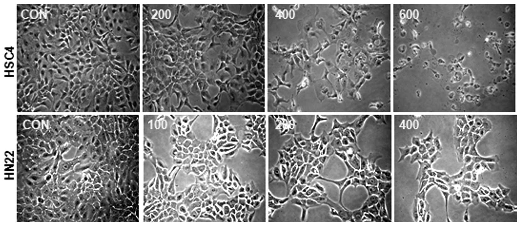
To investigate whether HESO inhibits the growth of
HSC4 and HN22 human oral cancer cells, cells were treated with DMSO
or various doses of HESO (200, 400 and 600 μg/ml for HSC4 cells and
100, 200 and 400 μg/ml for HN22 cells) for 24 or 48 h. The
morphological changes were observed using an optical microscope
after 48 h. Results showed that HESO-treated cells were detached in
a dose-dependent manner (Fig. 1).
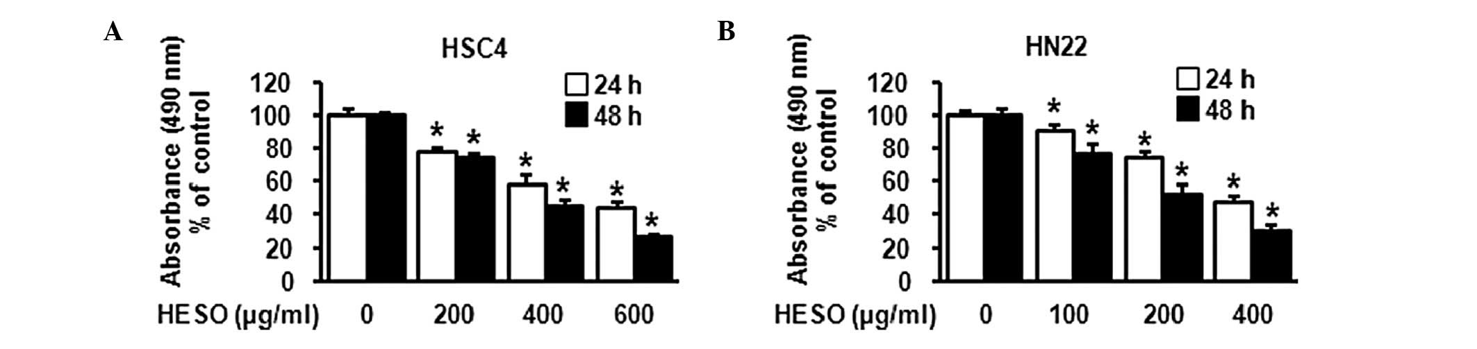
The effect of HESO on cell viability was examined using the MTS
assay. The cell viability of both cell lines decreased signficantly
and dose-dependently upon treatment with HESO (Fig. 2A and B). The IC50 value was 391.3
μg/ml for HSC4 cells and 259.9 μg/ml for HN22 cells at 48 h. These
results indicated that HESO inhibited growth of human oral cancer
cells.
Apoptotic effect of HESO on human oral
cancer cells
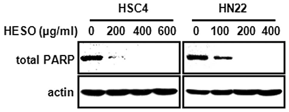
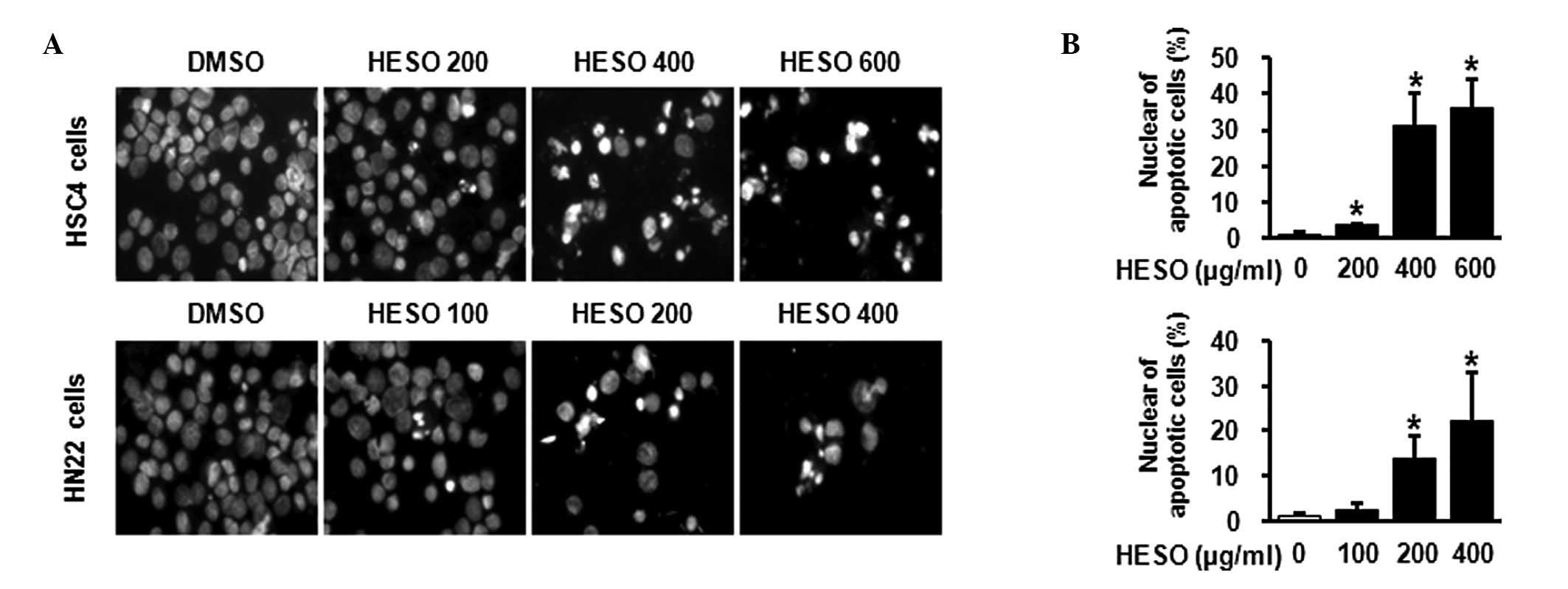
We then examined whether the growth inhibition by
HESO was associated with an apoptotic effect using western blot
analysis and DAPI staining. Fig. 3
demonstrates that there was a decrease in total PARP expression in
both cells lines following treatment with HESO for 48 h, indicating
that PARP might be cleaved to yield a 89-kDa fragment. In addition,
HSC4 and HN22 cells treated with various doses of HESO for 48 h
exhibited chromatin condensation and nuclear fragmentation,
characteristic features of cells undergoing apoptosis. The
percentage of cells with nuclear fragmentation in the HESO-treated
group compared with the DMSO-treated group is shown in Fig. 4A and B. These results demonstrate
that HESO induced apoptosis in human oral cancer cells.
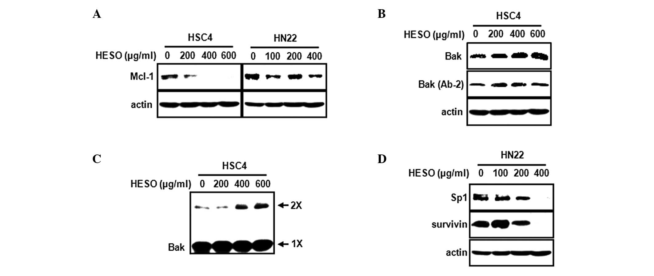
HESO induces downregulation of either
Mcl-1 or Sp1
An earlier study suggest that downregulation of
Mcl-1 protein may be required to initiate the apoptosis cascade
leading to cytochrome c release (17). Our own previous study found that the
inhibition of Mcl-1 by tolfenamic acid induces apoptosis in
mucoepidermoid carcinoma (18).
Thus, we investigated whether HESO affects Mcl-1 expression in HSC4
and HN22 oral cancer cells. Fig. 5A
shows that HESO decreased Mcl-1 expression in HSC4 cells but had no
significant effect on Mcl-1 in HN22 cells. To further investigate
whether the downregulation of Mcl-1 by HESO in HSC4 cells affects
the downstream targets Bak and Bax, HSC4 cells were treated with
various doses of HESO, and Bak and Bax protein levels were
measured. Fig. 5B shows that HESO
caused a marked increase in Bak expression, but failed to modulate
Bax expression. During the apoptotic cell death process, Bak
undergoes a conformational change and oligomerization on the
mitochondrial outer membrane. To evaluate whether Bak underwent a
conformational change from the inactive form to the active form
during HESO-induced apoptosis, we measured Bak using an antibody
recognizing only the active form. The results demonstrated that
HESO treatment resulted in an increase in the active form of Bak,
indicating that Bak underwent a conformational change. We next
investigated whether the Bak protein oligomerized on the
mitochondrial outer membrane. Following treatment of HSC4 cells
with HESO, BMH was used to crosslink oligomerized Bak. Fig. 5C shows that the level of
oligomerized Bak increased in HESO-treated HSC4 cells. Next, we
investigated whether HESO affected Sp1 in HN22 cells, since HESO
did not change Mcl-1 protein expression. HESO was found to
significantly decrease Sp1 and also reduce the expression of
survivin, its downstream target (Fig.
5D).
Discussion
The evasion of apoptosis is associated with tumor
progression, chemotherapy resistance and poor clinical outcome.
Cancer cells escape from apoptosis by increasing the expression of
anti-apoptotic proteins, including Bcl-2, Bcl-xl, Mcl-1 and
Bfl-1/A1 (19). In particular,
Mcl-1 is relatively highly expressed in malignant cell types,
suggesting that Mcl-1 is associated with the evasion of apoptosis.
Recently, several groups have suggested that downregulation of
Mcl-1 enhances apoptosis in diverse cancer cells. Targeting Mcl-1
by small interfering RNA markedly decreases its expression in
resistant melanoma cells and sensitizes them to Fas-induced
apoptosis (20). The use of
cyclin-dependent kinase (CDK) inhibitors, including flavopiridol
and roscovitine derivative seliciclib, potentiates Mcl-1
downregulation to induce apoptosis (21–23).
Moreover, the mitogen-activated protein kinase kinase 1 inhibitor
PD98059 and Raf inhibitor BAY43-9006 (sorafenib) synergistically
enhance apoptosis induced by the Bcl-2/Bcl-xL inhibitor ABT-737 by
downregulating Mcl-1 expression (5,24,25).
These findings suggest that downregulation of Mcl-1 is essential
for the regulation of apoptotic cell death.
In this study, we determined whether HESO was
capable of inhibiting cell growth and decreasing Mcl-1 expression
to induce apoptosis in oral cancer cells. We found that HESO
inhibited cell growth and induced apoptosis in HSC4 and HN22 oral
cancer cell lines. However, HESO-induced apoptosis in HSC4 cells
was associated with a decrease in Mcl-1 expression, whereas Mcl-1
expression did not change in HN22 cells.
Permeabilization of the mitochondria during
apoptosis is regulated by pro-apoptotic proteins, including
multidomain Bak and Bax, and BH3-only; Bim and tBid, which are
activators of Bax/Bak; and Bad, Bik, Noxa, Puma, Hrk and Bmf, which
are sensitizers of Bax/Bak. These interplay with anti-apoptotic
proteins, including Bcl-2, Bcl-xl, Mcl-1 and Bfl-1/A1. Upon the
induction of apoptosis, several apoptogenic factors (including
cytochrome c and AIF) are released from the mitochondria to the
cytosol (26,27). AIF is involved in initiating a
caspase-independent pathway of apoptosis, while cytochrome c binds
to Apaf-1 and caspase 9, leading to complex formation and the
activation of caspases such as caspase 3 and caspase 7 (28). Recent studies suggest that Bak
activation is required for cytochrome c to be released and for
apoptosis to occur (29–32). This process is followed by the
induction of a conformational change and oligomerization of Bak in
the mitochondrial outer membrane in response to multiple death
signals (33,34). Thus, we investigated whether Bak
underwent a conformational change and whether oligomerization
occurred on the mitochondrial outer membrane when HESO induced
apoptosis in HSC4 cells. We found that HESO induced the
conformational change and oligomerization of Bak. These results
showed that the downregulation of Mcl-1 by HESO in HSC4 cells may
be associated with the modulation of Bak protein.
In view of the observation that Mcl-1 expression was
not altered by HESO in HN22 cells, we hypothesized that the
mechanism of HESO on apoptotic activity might be different in each
cell line. Therefore, we focused on the molecular target for
HESO-induced apoptosis in HN22 cells. Previous studies reported
that Sp1 is overexpressed in many human tumor and cancer cell lines
(35,36). Furthermore, we reported that
downregulation of Sp1 induces apoptosis in cervical, prostate and
oral cancer cells (37–39). Thus, we examined whether
HESO-induced apoptosis was associated with the Sp1 protein. We
found that HESO decreased Sp1 expression in HN22 cells. Several
studies found that Sp1 regulates survivin as its downstream target
in various cancer cell lines (37,39–41).
We also found that HESO significantly decreased survivin,
suggesting that the downregulation of Sp1 by HESO in HN22 cells
resulted in a decrease of survivin expression.
In summary, HESO decreased cell growth and induced
apoptosis in HSC4 and HN22 human oral cancer cells. HESO reduced
Mcl-1 protein in HSC4 cells and caused the activation and
oligomerization of Bak, inducing apoptosis. In HN22 cells, HESO
decreased Sp1 and its downstream target, survivin. Although a study
of the antitumor effect of HESO in animal models is required, our
results clearly demonstrate that HESO may be a potential drug
candidate against oral cancer, targeting either Mcl-1 or Sp1.
Acknowledgements
This study was supported by the National Research
Foundation of Korea (NRF) and funded by the Ministry of Education,
Science and Technology (2011-0019173).
References
|
1
|
Siegel R, Ward E, Brawley O and Jemal A:
Cancer statistics: the impact of eliminating socioeconomic and
racial disparities on premature cancer deaths. CA Cancer J Clin.
61:212–236. 2011. View Article : Google Scholar : PubMed/NCBI
|
|
2
|
Mashberg A, Boffetta P, Winkelman R and
Garfinkel L: Tobacco smoking, alcohol drinking, and cancer of the
oral cavity and oropharynx among U.S. veterans. Cancer.
72:1369–1375. 1993. View Article : Google Scholar : PubMed/NCBI
|
|
3
|
Neville BW and Day TA: Oral cancer and
precancerous lesions. CA Cancer J Clin. 52:195–215. 2002.
View Article : Google Scholar : PubMed/NCBI
|
|
4
|
Michels J, Johnson PW and Packham G:
Mcl-1. Int J Biochem Cell Biol. 37:267–271. 2005. View Article : Google Scholar
|
|
5
|
Liu XS, Jiang J, Jiao XY, Wu YE, Lin JH
and Cai YM: Lycorine induces apoptosis and down-regulation of Mcl-1
in human leukemia cells. Cancer Lett. 274:16–24. 2009. View Article : Google Scholar : PubMed/NCBI
|
|
6
|
Battle TE, Arbiser J and Frank DA: The
natural product honokiol induces caspase-dependent apoptosis in
B-cell chronic lymphocytic leukemia (B-CLL) cells. Blood.
106:690–697. 2005. View Article : Google Scholar : PubMed/NCBI
|
|
7
|
Lee NH, Lee MY, Lee JA, Jung DY, Seo CS,
Kim JH and Shin HK: Anti-asthmatic effect of Sanguisorba
officinalis L. and potential role of heme oxygenase-1 in an
ovalbumin-induced murine asthma model. Int J Mol Med. 26:201–208.
2010.
|
|
8
|
Van Delft MF and Huang DC: How the Bcl-2
family of proteins interact to regulate apoptosis. Cell Res.
16:203–213. 2006.PubMed/NCBI
|
|
9
|
Dash R, Richards JE, Su ZZ, Bhutia SK,
Azab B, Rahmani M, Dasmahapatra G, Yacoub A, Dent P, Dmitriev IP,
et al: Mechanism by which Mcl-1 regulates cancer-specific apoptosis
triggered by mda-7/IL-24, an IL-10-related cytokine. Cancer Res.
70:5034–5045. 2010. View Article : Google Scholar : PubMed/NCBI
|
|
10
|
Okumura K, Huang S and Sinicrope FA:
Induction of Noxa sensitizes human colorectal cancer cells
expressing Mcl-1 to the small-molecule Bcl-2/Bcl-xL inhibitor,
ABT-737. Clin Cancer Res. 14:8132–8142. 2008. View Article : Google Scholar : PubMed/NCBI
|
|
11
|
Chen S, Dai Y, Harada H, Dent P and Grant
S: Mcl-1 down-regulation potentiates ABT-737 lethality by
cooperatively inducing Bak activation and Bax translocation. Cancer
Res. 67:782–791. 2007. View Article : Google Scholar : PubMed/NCBI
|
|
12
|
Lee NH, Lee MY, Lee JA, Jung DY, Seo CS,
Kim JH and Shin HK: Anti-asthmatic effect of Sanguisorba
officinalis L. and potential role of heme oxygenase-1 in an
ovalbumin-induced murine asthma model. Int J Mol Med. 26:201–208.
2010.
|
|
13
|
Shin TY, Lee KB and Kim SH: Anti-allergic
effects of Sanguisorba officinalis on animal models of
allergic reactions. Immunopharmacol Immunotoxicol. 24:455–468.
2002.PubMed/NCBI
|
|
14
|
Yu T, Lee YJ, Yang HM, Han S, Kim JH, Lee
Y, Kim C, Han MH, Kim MY, Lee J and Cho JY: Inhibitory effect of
Sanguisorba officinalis ethanol extract on NO and PGE
production is mediated by suppression of NF-kappaB and AP-1
activation signaling cascade. J Ethnopharmacol. 134:11–17.
2011.
|
|
15
|
Hachiya A, Kobayashi A, Ohuchi A, Kitahara
T and Takema Y: The inhibitory effect of an extract of
Sanguisorba officinalis L. on ultraviolet B-induced
pigmentation via the suppression of endothelin-converting
enzyme-1alpha. Biol Pharm Bull. 24:688–692. 2001.PubMed/NCBI
|
|
16
|
Liu X, Cui Y, Yu Q and Yu B: Triterpenoids
from Sanguisorba officinalis. Phytochemistry. 66:1671–1679.
2005. View Article : Google Scholar : PubMed/NCBI
|
|
17
|
Nijhawan D, Fang M, Traer E, Zhong Q, Gao
W, Du F and Wang X: Elimination of Mcl-1 is required for the
initiation of apoptosis following ultraviolet irradiation. Genes
Dev. 17:1475–1486. 2003. View Article : Google Scholar : PubMed/NCBI
|
|
18
|
Choi KH, Shim JH, Huong LD, Cho NP and Cho
SD: Inhibition of myeloid cell leukemia-1 by tolfenamic acid
induces apoptosis in mucoepidermoid carcinoma. Oral Dis.
17:469–475. 2011. View Article : Google Scholar : PubMed/NCBI
|
|
19
|
Gul O, Basaga H and Kutuk O: Apoptotic
blocks and chemotherapy resistance: strategies to identify Bcl-2
protein signatures. Brief Funct Genomic Proteomic. 7:27–34. 2008.
View Article : Google Scholar : PubMed/NCBI
|
|
20
|
Chetoui N, Sylla K, Gagnon-Houde JV,
Alcaide-Loridan C, Charron D, Al-Daccak R and Aoudjit F:
Down-regulation of mcl-1 by small interfering RNA sensitizes
resistant melanoma cells to fas-mediated apoptosis. Mol Cancer Res.
6:42–52. 2008. View Article : Google Scholar : PubMed/NCBI
|
|
21
|
Ma Y, Cress WD and Haura EB:
Flavopiridol-induced apoptosis is mediated through up-regulation of
E2F1 and repression of Mcl-1. Mol Cancer Ther. 2:73–81.
2003.PubMed/NCBI
|
|
22
|
Gojo I, Zhang B and Fenton RG: The
cyclin-dependent kinase inhibitor flavopiridol induces apoptosis in
multiple myeloma cells through transcriptional repression and
down-regulation of Mcl-1. Clin Cancer Res. 8:3527–3538. 2002.
|
|
23
|
MacCallum DE, Melville J, Frame S, Watt K,
Anderson S, Gianella-Borradori A, Lane DP and Green SR: Seliciclib
(CYC202, R-Roscovitine) induces cell death in multiple myeloma
cells by inhibition of RNA polymerase II-dependent transcription
and down-regulation of Mcl-1. Cancer Res. 65:5399–5407. 2005.
View Article : Google Scholar : PubMed/NCBI
|
|
24
|
Konopleva M, Contractor R, Tsao T, Samudio
I, Ruvolo PP, Kitada S, Deng X, Zhai D, Shi YX, Sneed T, et al:
Mechanisms of apoptosis sensitivity and resistance to the BH3
mimetic ABT-737 in acute myeloid leukemia. Cancer Cell. 10:375–388.
2006. View Article : Google Scholar : PubMed/NCBI
|
|
25
|
Lin X, Morgan-Lappe S, Huang X, Li L,
Zakula DM, Vernetti LA, Fesik SW and Shen Y: ‘Seed’ analysis of
off-target siRNAs reveals an essential role of Mcl-1 in resistance
to the small-molecule Bcl-2/Bcl-XL inhibitor ABT-737. Oncogene.
26:3972–3979. 2007.
|
|
26
|
Fulda S, Scaffidi C, Susin SA, Krammer PH,
Kroemer G, Peter ME and Debatin KM: Activation of mitochondria and
release of mitochondrial apoptogenic factors by betulinic acid. J
Biol Chem. 273:33942–33948. 1998. View Article : Google Scholar : PubMed/NCBI
|
|
27
|
Li Y, He K, Huang Y, Zheng D, Gao C, Cui L
and Jin YH: Betulin induces mitochondrial cytochrome c release
associated apoptosis in human cancer cells. Mol Carcinog.
49:630–640. 2010.PubMed/NCBI
|
|
28
|
Zong WX, Li C, Hatzivassiliou G, Lindsten
T, Yu QC, Yuan J and Thompson CB: Bax and Bak can localize to the
endoplasmic reticulum to initiate apoptosis. J Cell Biol.
162:59–69. 2003. View Article : Google Scholar : PubMed/NCBI
|
|
29
|
Germain M, Milburn J and Duronio V: MCL-1
inhibits BAX in the absence of MCL-1/BAX Interaction. J Biol Chem.
283:6384–6392. 2008. View Article : Google Scholar : PubMed/NCBI
|
|
30
|
Shankar S and Srivastava RK: Bax and Bak
genes are essential for maximum apoptotic response by curcumin, a
polyphenolic compound and cancer chemopreventive agent derived from
turmeric, Curcuma longa. Carcinogenesis. 28:1277–1286. 2007.
View Article : Google Scholar
|
|
31
|
Labi V, Erlacher M, Kiessling S and
Villunger A: BH3-only proteins in cell death initiation, malignant
disease and anticancer therapy. Cell Death Differ. 13:1325–1338.
2006. View Article : Google Scholar : PubMed/NCBI
|
|
32
|
Leber B, Lin J and Andrews DW: Embedded
together: the life and death consequences of interaction of the
Bcl-2 family with membranes. Apoptosis. 12:897–911. 2007.
View Article : Google Scholar : PubMed/NCBI
|
|
33
|
Upreti M, Chu R, Galitovskaya E, Smart SK
and Chambers TC: Key role for Bak activation and Bak-Bax
interaction in the apoptotic response to vinblastine. Mol Cancer
Ther. 7:2224–2232. 2008. View Article : Google Scholar : PubMed/NCBI
|
|
34
|
Heath-Engel HM and Shore GC: Regulated
targeting of Bax and Bak to intracellular membranes during
apoptosis. Cell Death Differ. 13:1277–1280. 2006. View Article : Google Scholar : PubMed/NCBI
|
|
35
|
Wang L, Wei D, Huang S, Peng Z, Le X, Wu
TT, Yao J, Ajani J and Xie K: Transcription factor Sp1 expression
is a significant predictor of survival in human gastric cancer.
Clin Cancer Res. 9:6371–6380. 2003.PubMed/NCBI
|
|
36
|
Shi Q, Le X, Abbruzzese JL, Peng Z, Qian
CN, Tang H, Xiong Q, Wang B, Li XC and Xie K: Constitutive Sp1
activity is essential for differential constitutive expression of
vascular endothelial growth factor in human pancreatic
adenocarcinoma. Cancer Res. 61:4143–4154. 2001.
|
|
37
|
Shim JH, Shin JA, Jung JY, Choi KH, Choi
ES, Cho NP, Kong G, Ryu MH, Chae JI and Cho SD: Chemopreventive
effect of tolfenamic acid on KB human cervical cancer cells and
tumor xenograft by downregulating specificity protein 1. Eur J
Cancer Prev. 20:102–111. 2011. View Article : Google Scholar : PubMed/NCBI
|
|
38
|
Choi ES, Shim JH, Jung JY, Kim HJ, Choi
KH, Shin JA, Nam JS, Cho NP and Cho SD: Apoptotic effect of
tolfenamic acid in androgen receptor-independent prostate cancer
cell and xenograft tumor through specificity protein 1. Cancer Sci.
102:742–748. 2011. View Article : Google Scholar : PubMed/NCBI
|
|
39
|
Shin JA, Shim JH, Jeon JG, Choi KH, Choi
ES, Cho NP and Cho SD: Apoptotic effect of Polygonum Cuspidatum in
oral cancer cells through the regulation of specificity protein 1.
Oral Dis. 17:162–170. 2011. View Article : Google Scholar : PubMed/NCBI
|
|
40
|
Li F and Altieri DC: Transcriptional
analysis of human survivin gene expression. Biochem J. 344(Pt 2):
305–311. 1999. View Article : Google Scholar
|
|
41
|
Chun JY, Hu Y, Pinder E, Wu J, Li F and
Gao AC: Selenium inhibition of survivin expression by preventing
Sp1 binding to its promoter. Mol Cancer Ther. 6:2572–2580. 2007.
View Article : Google Scholar : PubMed/NCBI
|