Introduction
Non-small cell lung cancer (NSCLC) accounts for
approximately 75–80% of all lung cancers and is the leading cause
of cancer-related mortality worldwide (1,2).
Twenty percent of stage I and 30% of stage II NSCLC patients
(3) who are treated with curative
intent experience recurrence of cancer, which is often incurable at
the time of discovery, and have a 5-year survival rate of less than
50% (1). Despite years of intense
efforts to control lung cancer mortality with surgical resection,
chemotherapy and radiotherapy, NSCLC remains the leading cause of
cancer-related mortality. This clearly indicates an urgent
requirement for elucidating the mechanisms of lung cancer
carcinogenesis, as well as discovering new approaches for its
prevention and treatment.
The transcription factor growth arrest and DNA
damage-inducible gene 153 (GADD153), also known as CHOP (4,5), is
considered to function as a proapoptotic molecule. GADD153 belongs
to the CCAAT/enhancer binding protein (C/EBP) family of
transcription factors. It forms heterodimers with other C/EBP
family proteins and changes their transcriptional activity
(5,6).
GADD153 is ubiquitously expressed at a low level in
several cell types, and its expression is induced by a variety of
stress factors (5,7), including genotoxic stress, endoplasmic
reticulum (ER) stress and nutrient depletion. Stress induces
GADD153 expression at transcriptional and post-transcriptional
levels (4,8–11). ER
stress is induced by environmental conditions that are frequently
encountered in cancer growth; therefore, the role of ER stress in
carcinogenesis and tumor progression is being actively researched
(12,13). Overexpression of GADD153 has been
reported to lead to cell cycle arrest and/or apoptosis (14,15).
Disruption of the GADD153 gene has been identified to render cells
more resistant to ER stress-induced apoptosis, while exogenous
GADD153 is capable of inducing growth arrest and/or apoptosis
(14–16). GADD153 is one of the important
factors in the death of cancer cells and in the anticarcinogenic
process, where it downregulates cell growth and survival rate.
Studies have suggested that GADD153 triggers the critical early
events leading to the initiation of apoptosis, which are considered
to be of significance in the prognosis of early NCSLC (17). In this study, we aimed to determine
whether the expression of GADD153 is an indicator of good prognosis
in stage I NSCLC patients. Consequently, we evaluated GADD153
expression in 76 stage I NSCLC tissue samples and investigated the
correlation between expression and clinical and pathological
findings.
Materials and methods
Study population and samples
Tissue specimens were obtained from 76 patients who
were diagnosed with stage I primary NSCLC and underwent curative
surgical removal of a primary lesion at the Yonsei Medical Center
(Seoul, Republic of Korea) between 1995 and 1998. Pathological
evaluation was established for histological classification and
staging in all patients. No patient underwent radiotherapy or
chemotherapy prior to or after surgery until the disease recurred.
Of the 76 patients, 56 (73.7%) were male and 20 (26.3%) were
female. The age of the patients ranged from 31 to 83 years, with a
mean age of 61.8±9.97 years, which was similar to the age
distribution in our institution’s large database of patients with
stage I NSCLC (data not shown). All the clinical and pathological
information and follow-up data were based on studies from our tumor
registry service. The study was reviewed and approved by the
institution’s Surveillance Committee and their permission was
obtained to use tissue blocks and other pertinent information from
patient files.
Immunohistochemical staining for
GADD153
Paraffin-embedded 4 μm-thick tissue sections from 76
primary stage I NSCLC samples were stained with mouse monoclonal
antibody against human GADD153 (Abcam, Ltd., Cambridge, UK). All
sections were deparaffinized in a series of xylene baths and
rehydrated using a graded alcohol series. Paraffin sections were
retrieved via microwave treatment and treated with 0.3% hydrogen
peroxidase to block endogenous peroxidase activity. The sections
were then incubated with the primary anti-GADD153 antibody.
Following incubation, the sections were processed
using standard avidin-biotin immunohistochemical techniques
according to the manufacturer’s instructions (Vector Laboratories,
Burlingame, CA, USA). Diaminobenzidine was used as a chromogen and
hematoxylin was used for counterstaining. Adjacent normal-appearing
bronchial epithelium within each tissue section served as an
internal reference. The intensity of GADD153 immunostaining was
evaluated within the cell membrane and cytoplasm of invasive cancer
components. Cell staining was regarded as positive or negative and
all slides were independently evaluated by two pathologists who
were unaware of the clinical and pathological information of the
subjects. Cancer cells in at least 4 fields were counted at ×200
magnification. The GADD153 immunoreactivity level was classified by
the proportion of positive cells: 0, <5% positive cells; 1+,
5–30% positive cells; 2+, 30–50% positive cells; 3+, >50%
positive cells. The intensity of GADD153 expression was also
scored; 0, negative to weak; 1, moderate; 2, strong. The score was
the sum of the intensity and the percentage of positive cells. A
score of ≤1 was applied as a cut-off point for loss of GADD153
expression.
Statistical analysis
Survival data of all patients were obtained from the
Korea National Statistical Office. A two-sample t-test for
independent samples and an χ2-test were used for
continuous and categorical variables, respectively. The
Kaplan-Meier estimator was used to compute survival rate
probability as a function of time and the log-rank test was used to
compare survival time between the groups. All statistical tests
were two-sided. SPSS software (version 15.0) was used throughout
and P<0.05 was considered to indicate a statistically
significant difference.
Results
NSCLC patient characteristics
The NSCLC cases included 56 (73.7%) male and 20
(26.3%) female patients (age range, 31–83 years; mean age,
61.8±9.97 years). The demographic characteristics of the patients
are shown in Table I.
 | Table IStage I NSCLC patient characteristics
relative to GADD153 expression. |
Table I
Stage I NSCLC patient characteristics
relative to GADD153 expression.
| GADD153 expression in
tumor tissues | |
|---|
|
| |
|---|
| Patient
characteristics | Positive (n=29) | Negative (n=47) | P-value |
|---|
| Gender | | | |
| Male | 18 | 38 | 0.07 |
| Female | 11 | 9 | |
| Smoking status | | | |
| Smoker; packs/year,
mean ± SD | 21; 45.0±28.73 | 33; 35.2±19.69 | 0.112; 0.311 |
| Non-smoker | 6 | 4 | |
| Unknown | 2 | 10 | |
| Pathology | | | 0.674 |
| Adenocarcinoma | 16 | 21 | |
| Squamous cell
carcinoma | 12 | 24 | |
| Other | 1 | 2 | |
| Histological
grade | | | 0.305 |
|
Well-differentiated | 12 | 10 | |
| Moderately
differentiated | 7 | 17 | |
| Poorly
differentiated | 6 | 12 | |
| Unclassified | 4 | 8 | 0.735 |
| TNM stage | | | |
| T1N0M0 | 7 | 13 | |
| T2N0M0 | 22 | 34 | |
| Distant
metastasis | 0 | 8 | 0.029 |
| Mean survival time
(months, 95% CI) | | | |
| Disease-free | 54.19
(50.736–57.652) | 46.97
(38.062–55.881) | 0.084 |
|
Disease-specific | 53.85
(50.702–57.002) | 28.80
(13.895–43.705) | 0.020 |
| Overall | 52.18
(46.735–57.624) | 53.29
(49.123–57.447) | 0.743 |
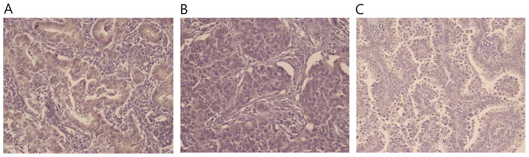
GADD153 immunohistochemistry
Immunohistochemical staining of GADD153 in normal
and cancer tissue sections was conducted (Fig. 1). Immunoreactivity for the GADD153
antibody was identified primarily in the cytoplasm of cancer cells.
However, in the normal lung tissue, bronchial epithelial cells
demonstrated weak immunoreactivity. The intensity of GADD153
staining in the cytoplasm was strong, granular and distinct in the
lung cancer tissue. The number of cases with GADD153 positive
staining was 29 (38.7%). There was heterogeneity of staining inside
the same tumor with sporadic, patchy, focal or diffused patterns.
Such intramural heterogeneity made scoring difficult in certain
cases, but immunohistochemical staining of GADD153 was stable and
reproducible.
Correlation between GADD153 expression
and various clinicopathological parameters
A total of 37 (48.7%) of the 76 stage I NSCLCs were
adenocarcinomas, 36 (47.3%) were squamous cell carcinomas and 3
(3.9%) were other histological types of NSCLC. GADD153 expression
was positive in 29 (38.2%) and negative in 47 (61.8%) cases. We
were able to classify 64 tissue samples for histological grade of
differentiation; 12 (54.5%) out of 22 were well-differentiated, 7
(29.2%) out of 24 were moderately differentiated and 6 (33.3%) out
of 18 were poorly differentiated. Analysis of GADD153 expression
revealed that expression was not influenced by histological type
(P=0.674) or differential grade (P=0.305). In a subgroup analysis
among subtypes of adenocarcinoma, no differences were observed
between bronchioloalveolar carcinomas and other subtypes of
adenocarcinoma. The smoking status of the patients did not
influence the expression of GADD153, and none of the patients with
GADD153 expression experienced distant metastasis of statistical
significance (P=0.029) (Table
I).
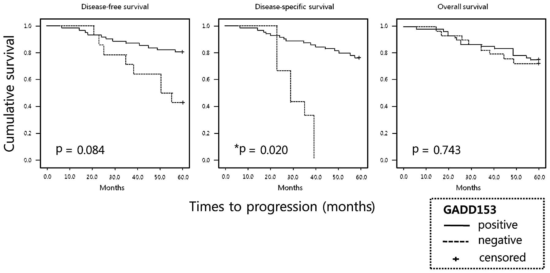
Survival analysis
The total follow-up period of the patients who were
alive at the time of analysis was 5 years. A total of 21 (27.6%) of
the 76 patients died during the follow-up period; 11 (14.5%)
succumbed to cancer-related events. The 5-year survival rate
probability of the patient population was 72.3%, which is similar
to previous results from a large-scale study (18). Overall survival and disease-free
survival rate curves did not demonstrate statistically significant
differences between NSCLC patients with and without GADD153
expression (Fig. 2). However, the
patients with GADD153 expression had a significantly improved
disease-specific survival rate compared to patients with negative
GADD153 expression (28.80 vs. 53.85 months; P=0.020) (Table I; Fig
2). Subgroup analyses classified according to pathological
diagnosis, grade of differentiation or TNM stages demonstrated that
GADD153 expression status did not influence disease-free,
disease-specific and overall survival rate in NSCLC patients (data
not shown).
Discussion
It has been well-established that mutational
activation of the Ras gene is a key factor in human cancer
development (19). Oncogenic Ras
proteins transform cells via multiple downstream signaling
cascades, which lead to the phosphorylation and activation of
proliferation-inducing transcription factors, including Elk-1,
Ets-2 and c-Myc (19,20). Oncogenic Ras has also been
identified to downregulate the expression of proapoptotic proteins,
including the Bcl-2 family protein Bak and the transcriptional
repressor Par-4 (21–24). One study also demonstrated that
oncogenic Ras downregulated GADD153 expression at protein and mRNA
levels (25).
GADD153, which belongs to the C/EBP family of
transcription factors, forms heterodimers with other members of the
C/EBP family, resulting in the inhibition of transcriptional
activities (5,17). The GADD153 gene is typically induced
in response to cellular stress (17). Previously, it has been reported that
GADD153 expression may be regulated through various MAP kinase
signaling pathways, and that the particular signaling pathway
involved is dependent upon the type of stimuli (16,26,27).
There is considerable evidence indicating that GADD153 is directly
involved in the apoptosis pathway. It has been demonstrated that
GADD153 upregulates the proapoptotic factor BH-3 (BIM) and
downregulates the antiapoptotic Bcl-2 (28,29).
Therefore, GADD153 increases cellular sensitivity to apoptosis by
suppressing the transcription of antiapoptotic Bcl-2 (29). Previous studies have demonstrated
that oncogenic Ras downregulates GADD153 expression and exogenous
GADD153 inhibits Ras-induced cellular transformation (25). Therefore, GADD153 is important not
only in killing cancer cells, but also in the anticarcinogenic
process, where it downregulates cell growth and survival rate. A
previous study has suggested that GADD153 may act as a critical
marker of early response to cell injury, and each molecule is known
to function in a different signal transduction pathway responsive
to cell injury (30). The main role
of the GADD153 gene is to block proliferation at G1 and 2
checkpoints in response to DNA damage. Transfection of the GADD153
gene into various cancer cell lines induces apoptosis without any
stress-inducing factors, indicating that GADD153 is directly
involved in the regulation of apoptosis (16). Furthermore, the GADD153 protein
plays a significant role in the induction of apoptosis of cancer
cells treated with N-(4-hydroxyphenyl)retinamide, a synthetic
retinoid, in a retinoic acid receptor-independent pathway (31,32).
These results strongly suggest that the expression of the GADD153
gene is a new molecular mechanism of antitumor activity. However,
studies on the clinical relevance of GADD153 expression in human
cancer are extremely limited. In previous studies, increased
expression of GADD153 correlated with a lower tumor stage in colon
cancer and with a higher survival rate in melanoma (33,34). A
further study on lung cancer demonstrated that GADD153 expression
correlated with a larger tumor size, higher pathological T stage,
higher TNM stage and shorter overall survival rate (35). Therefore, we focused on the
expression of GADD153 in patients with stage I (3) primary NSCLC and its association with
clinical outcome. The aim of this study was to examine the
expression of GADD153 in stage I NSCLC with respect to
prognosis.
We examined the prognostic significance of GADD153
expression in formalin-fixed paraffin-embedded tissues and revealed
that GADD153 expression was downregulated in a significant amount
of patients with stage I NSCLC. Overall, 29 of 76 tumors (38.7%)
expressed GADD153, and its expression was localized in the membrane
and cytoplasm rather than in the nucleus of NSCLC cells. Notably,
patients with GADD153 expression demonstrated a positive
correlation with improved disease-specific survival rate (P=0.020).
However, our data indicated that GADD153 expression was not
associated with an improved overall survival rate and was only
slightly associated with greater disease-free survival; no
statistically significant differences were identified. We
demonstrated that GADD153 expression was closely associated with
and may have a role in the prevention of stage I NSCLC distant
metastasis (P=0.029); thus, the greater disease-specific survival
rate in patients with GADD153 expression. Subsequently, we analyzed
the association of GADD153 expression with the clinicopathological
parameters of stage I NSCLC patients. GADD153 was associated with
distant metastasis, but none of the other clinicopathological
features of stage I NSCLC patients, suggesting that GADD153 may be
involved in apoptotic events preventing metastasis.
Since this study was a preliminary investigation,
the number of patients with stage I NSCLC was relatively small (76
patients) and the longest follow-up time was 60 months. This study
has revealed that GADD153 expression is a candidate marker that may
aid in the stratification of patients according to prognosis
following curative surgical removal of a primary lesion. Further
comprehensive studies involving the mechanisms that induce
expression of GADD153 in NSCLC are required to define the role of
GADD153 in lung carcinogenesis. The significant association with
survival rate observed in the present study is of particular
relevance and should be confirmed in additional cohorts of
patients.
Acknowledgements
This study was supported by the Institutional Grant
from Yonsei University College of Medicine (6-2008-0198) provided
to YS Chang through the Human Barrier Research Institute.
References
|
1
|
Jemal A, Siegel R, Xu J and Ward E: Cancer
statistics, 2010. CA Cancer J Clin. 60:277–300. 2010. View Article : Google Scholar
|
|
2
|
Greenlee RT, Hill-Harmon MB, Murray T and
Thun M: Cancer statistics, 2001. CA Cancer J Clin. 51:15–36. 2001.
View Article : Google Scholar
|
|
3
|
Sobin L and Wittekind Ch: TNM
Classification of Malignant Tumors. 6th edition. Wiley-Liss; New
York: pp. 99–103. 2002
|
|
4
|
Luethy JD, Fargnoli J, Park JS, Fornace AJ
Jr and Holbrook NJ: Isolation and characterization of the hamster
gadd153 gene. Activation of promoter activity by agents that damage
DNA. J Biol Chem. 265:16521–16526. 1990.PubMed/NCBI
|
|
5
|
Ron D and Habener JF: CHOP, a novel
developmentally regulated nuclear protein that dimerizes with
transcription factors C/EBP and LAP and functions as a
dominant-negative inhibitor of gene transcription. Genes Dev.
6:439–453. 1992. View Article : Google Scholar : PubMed/NCBI
|
|
6
|
Ramji DP and Foka P:
CCAAT/enhancer-binding proteins: structure, function and
regulation. Biochem J. 365:561–575. 2002.PubMed/NCBI
|
|
7
|
Jousse C, Bruhat A, Carraro V, et al:
Inhibition of CHOP translation by a peptide encoded by an open
reading frame localized in the chop 5’UTR. Nucleic Acids Res.
29:4341–4351. 2001.PubMed/NCBI
|
|
8
|
Jackman J, Alamo I Jr and Fornace AJ Jr:
Genotoxic stress confers preferential and coordinate messenger RNA
stability on the five gadd genes. Cancer Res. 54:5656–5662.
1994.PubMed/NCBI
|
|
9
|
Wang XZ, Lawson B, Brewer JW, et al:
Signals from the stressed endoplasmic reticulum induce
C/EBP-homologous protein (CHOP/GADD153). Mol Cell Biol.
16:4273–4280. 1996.PubMed/NCBI
|
|
10
|
Bruhat A, Jousse C, Wang XZ, Ron D,
Ferrara M and Fafournoux P: Amino acid limitation induces
expression of CHOP, a CCAAT/enhancer binding protein-related gene,
at both transcriptional and post-transcriptional levels. J Biol
Chem. 272:17588–17593. 1997. View Article : Google Scholar : PubMed/NCBI
|
|
11
|
Sato N, Urano F, Yoon Leem J, et al:
Upregulation of BiP and CHOP by the unfolded-protein response is
independent of presenilin expression. Nat Cell Biol. 2:863–870.
2000. View
Article : Google Scholar : PubMed/NCBI
|
|
12
|
Uramoto H, Sugio K, Oyama T, et al:
Expression of endoplasmic reticulum molecular chaperone Grp78 in
human lung cancer and its clinical significance. Lung Cancer.
49:55–62. 2005. View Article : Google Scholar : PubMed/NCBI
|
|
13
|
Hsu WM, Hsieh FJ, Jeng YM, et al: GRP78
expression correlates with histologic differentiation and favorable
prognosis in neuroblastic tumors. Int J Cancer. 113:920–927. 2005.
View Article : Google Scholar : PubMed/NCBI
|
|
14
|
Matsumoto M, Minami M, Takeda K, Sakao Y
and Akira S: Ectopic expression of CHOP (GADD153) induces apoptosis
in M1 myeloblastic leukemia cells. FEBS Lett. 395:143–147. 1996.
View Article : Google Scholar : PubMed/NCBI
|
|
15
|
Barone MV, Crozat A, Tabaee A, Philipson L
and Ron D: CHOP (GADD153) and its oncogenic variant, TLS-CHOP, have
opposing effects on the induction of G1/S arrest. Genes Dev.
8:453–464. 1994. View Article : Google Scholar : PubMed/NCBI
|
|
16
|
Maytin EV, Ubeda M, Lin JC and Habener JF:
Stress-inducible transcription factor CHOP/gadd153 induces
apoptosis in mammalian cells via p38 kinase-dependent and
-independent mechanisms. Exp Cell Res. 267:193–204. 2001.
View Article : Google Scholar : PubMed/NCBI
|
|
17
|
Oyadomari S and Mori M: Roles of
CHOP/GADD153 in endoplasmic reticulum stress. Cell Death Differ.
11:381–389. 2004. View Article : Google Scholar : PubMed/NCBI
|
|
18
|
Martini N, Bains MS, Burt ME, et al:
Incidence of local recurrence and second primary tumors in resected
stage I lung cancer. J Thorac Cardiovasc Surg. 109:120–129. 1995.
View Article : Google Scholar : PubMed/NCBI
|
|
19
|
Downward J: Targeting RAS signalling
pathways in cancer therapy. Nat Rev Cancer. 3:11–22. 2003.
View Article : Google Scholar : PubMed/NCBI
|
|
20
|
Cox AD and Der CJ: The dark side of Ras:
regulation of apoptosis. Oncogene. 22:8999–9006. 2003. View Article : Google Scholar : PubMed/NCBI
|
|
21
|
Rosen K, Rak J, Jin J, Kerbel RS, Newman
MJ and Filmus J: Downregulation of the pro-apoptotic protein Bak is
required for the ras-induced transformation of intestinal
epithelial cells. Curr Biol. 8:1331–1334. 1998. View Article : Google Scholar : PubMed/NCBI
|
|
22
|
Barradas M, Monjas A, Diaz-Meco MT,
Serrano M and Moscat J: The downregulation of the pro-apoptotic
protein Par-4 is critical for Ras-induced survival and tumor
progression. EMBO J. 18:6362–6369. 1999. View Article : Google Scholar : PubMed/NCBI
|
|
23
|
Nalca A, Qiu SG, El-Guendy N, Krishnan S
and Rangnekar VM: Oncogenic Ras sensitizes cells to apoptosis by
Par-4. J Biol Chem. 274:29976–29983. 1999. View Article : Google Scholar : PubMed/NCBI
|
|
24
|
Qiu SG, Krishnan S, el-Guendy N and
Rangnekar VM: Negative regulation of Par-4 by oncogenic Ras is
essential for cellular transformation. Oncogene. 18:7115–7123.
1999. View Article : Google Scholar : PubMed/NCBI
|
|
25
|
Rong R, Montalbano J, Jin W, et al:
Oncogenic Ras-mediated downregulation of Gadd153/CHOP is required
for Ras-induced cellular transformation. Oncogene. 24:4867–4872.
2005. View Article : Google Scholar : PubMed/NCBI
|
|
26
|
Wang XZ and Ron D: Stress-induced
phosphorylation and activation of the transcription factor CHOP
(GADD153) by p38 MAP Kinase. Science. 272:1347–1349. 1996.
View Article : Google Scholar : PubMed/NCBI
|
|
27
|
Scott DW, Mutamba S, Hopkins RG and Loo G:
Increased GADD gene expression in human colon epithelial cells
exposed to deoxycholate. J Cell Physiol. 202:295–303. 2005.
View Article : Google Scholar : PubMed/NCBI
|
|
28
|
Puthalakath H, O’Reilly LA, Gunn P, et al:
ER stress triggers apoptosis by activating BH3-only protein Bim.
Cell. 129:1337–1349. 2007. View Article : Google Scholar : PubMed/NCBI
|
|
29
|
McCullough KD, Martindale JL, Klotz LO, Aw
TY and Holbrook NJ: Gadd153 sensitizes cells to endoplasmic
reticulum stress by down-regulating Bcl2 and perturbing the
cellular redox state. Mol Cell Biol. 21:1249–1259. 2001. View Article : Google Scholar : PubMed/NCBI
|
|
30
|
Friedman AD: GADD153/CHOP, a DNA
damage-inducible protein, reduced CAAT/enhancer binding protein
activities and increased apoptosis in 32D c13 myeloid cells. Cancer
Res. 56:3250–3256. 1996.PubMed/NCBI
|
|
31
|
Kim DG, You KR, Liu MJ, Choi YK and Won
YS: GADD153-mediated anticancer effects of
N-(4-hydroxyphenyl)retinamide on human hepatoma cells. J Biol Chem.
277:38930–38938. 2002. View Article : Google Scholar : PubMed/NCBI
|
|
32
|
Xia Y, Wong NS, Fong WF and Tideman H:
Upregulation of GADD153 expression in the apoptotic signaling of
N-(4-hydroxyphenyl)retinamide (4HPR). Int J Cancer. 102:7–14. 2002.
View Article : Google Scholar : PubMed/NCBI
|
|
33
|
Korabiowska M, Cordon-Cardo C, Betke H, et
al: GADD153 is an independent prognostic factor in melanoma:
immunohistochemical and molecular genetic analysis. Histol
Histopathol. 17:805–811. 2002.PubMed/NCBI
|
|
34
|
Rask K, Thorn M, Ponten F, et al:
Increased expression of the transcription factors CCAAT-enhancer
binding protein-beta (C/EBBeta) and C/EBzeta (CHOP) correlate with
invasiveness of human colorectal cancer. Int J Cancer. 86:337–343.
2000. View Article : Google Scholar : PubMed/NCBI
|
|
35
|
Kim KM, Yu TK, Chu HH, et al: Expression
of ER stress and autophagy-related molecules in human non-small
cell lung cancer and premalignant lesions. Int J Cancer. Sept
27–2011.(Epub ahead of print).
|