Spandidos Publications Logo
  • About
    • About Spandidos
    • Aims and Scopes
    • Abstracting and Indexing
    • Editorial Policies
    • Reprints and Permissions
    • Job Opportunities
    • Terms and Conditions
    • Contact
  • Journals
    • All Journals
    • Oncology Letters
      • Oncology Letters
      • Information for Authors
      • Editorial Policies
      • Editorial Board
      • Aims and Scope
      • Abstracting and Indexing
      • Bibliographic Information
      • Archive
    • International Journal of Oncology
      • International Journal of Oncology
      • Information for Authors
      • Editorial Policies
      • Editorial Board
      • Aims and Scope
      • Abstracting and Indexing
      • Bibliographic Information
      • Archive
    • Molecular and Clinical Oncology
      • Molecular and Clinical Oncology
      • Information for Authors
      • Editorial Policies
      • Editorial Board
      • Aims and Scope
      • Abstracting and Indexing
      • Bibliographic Information
      • Archive
    • Experimental and Therapeutic Medicine
      • Experimental and Therapeutic Medicine
      • Information for Authors
      • Editorial Policies
      • Editorial Board
      • Aims and Scope
      • Abstracting and Indexing
      • Bibliographic Information
      • Archive
    • International Journal of Molecular Medicine
      • International Journal of Molecular Medicine
      • Information for Authors
      • Editorial Policies
      • Editorial Board
      • Aims and Scope
      • Abstracting and Indexing
      • Bibliographic Information
      • Archive
    • Biomedical Reports
      • Biomedical Reports
      • Information for Authors
      • Editorial Policies
      • Editorial Board
      • Aims and Scope
      • Abstracting and Indexing
      • Bibliographic Information
      • Archive
    • Oncology Reports
      • Oncology Reports
      • Information for Authors
      • Editorial Policies
      • Editorial Board
      • Aims and Scope
      • Abstracting and Indexing
      • Bibliographic Information
      • Archive
    • Molecular Medicine Reports
      • Molecular Medicine Reports
      • Information for Authors
      • Editorial Policies
      • Editorial Board
      • Aims and Scope
      • Abstracting and Indexing
      • Bibliographic Information
      • Archive
    • World Academy of Sciences Journal
      • World Academy of Sciences Journal
      • Information for Authors
      • Editorial Policies
      • Editorial Board
      • Aims and Scope
      • Abstracting and Indexing
      • Bibliographic Information
      • Archive
    • International Journal of Functional Nutrition
      • International Journal of Functional Nutrition
      • Information for Authors
      • Editorial Policies
      • Editorial Board
      • Aims and Scope
      • Abstracting and Indexing
      • Bibliographic Information
      • Archive
    • International Journal of Epigenetics
      • International Journal of Epigenetics
      • Information for Authors
      • Editorial Policies
      • Editorial Board
      • Aims and Scope
      • Abstracting and Indexing
      • Bibliographic Information
      • Archive
    • Medicine International
      • Medicine International
      • Information for Authors
      • Editorial Policies
      • Editorial Board
      • Aims and Scope
      • Abstracting and Indexing
      • Bibliographic Information
      • Archive
  • Articles
  • Information
    • Information for Authors
    • Information for Reviewers
    • Information for Librarians
    • Information for Advertisers
    • Conferences
  • Language Editing
Spandidos Publications Logo
  • About
    • About Spandidos
    • Aims and Scopes
    • Abstracting and Indexing
    • Editorial Policies
    • Reprints and Permissions
    • Job Opportunities
    • Terms and Conditions
    • Contact
  • Journals
    • All Journals
    • Biomedical Reports
      • Information for Authors
      • Editorial Policies
      • Editorial Board
      • Aims and Scope
      • Abstracting and Indexing
      • Bibliographic Information
      • Archive
    • Experimental and Therapeutic Medicine
      • Information for Authors
      • Editorial Policies
      • Editorial Board
      • Aims and Scope
      • Abstracting and Indexing
      • Bibliographic Information
      • Archive
    • International Journal of Epigenetics
      • Information for Authors
      • Editorial Policies
      • Editorial Board
      • Aims and Scope
      • Abstracting and Indexing
      • Bibliographic Information
      • Archive
    • International Journal of Functional Nutrition
      • Information for Authors
      • Editorial Policies
      • Editorial Board
      • Aims and Scope
      • Abstracting and Indexing
      • Bibliographic Information
      • Archive
    • International Journal of Molecular Medicine
      • Information for Authors
      • Editorial Policies
      • Editorial Board
      • Aims and Scope
      • Abstracting and Indexing
      • Bibliographic Information
      • Archive
    • International Journal of Oncology
      • Information for Authors
      • Editorial Policies
      • Editorial Board
      • Aims and Scope
      • Abstracting and Indexing
      • Bibliographic Information
      • Archive
    • Medicine International
      • Information for Authors
      • Editorial Policies
      • Editorial Board
      • Aims and Scope
      • Abstracting and Indexing
      • Bibliographic Information
      • Archive
    • Molecular and Clinical Oncology
      • Information for Authors
      • Editorial Policies
      • Editorial Board
      • Aims and Scope
      • Abstracting and Indexing
      • Bibliographic Information
      • Archive
    • Molecular Medicine Reports
      • Information for Authors
      • Editorial Policies
      • Editorial Board
      • Aims and Scope
      • Abstracting and Indexing
      • Bibliographic Information
      • Archive
    • Oncology Letters
      • Information for Authors
      • Editorial Policies
      • Editorial Board
      • Aims and Scope
      • Abstracting and Indexing
      • Bibliographic Information
      • Archive
    • Oncology Reports
      • Information for Authors
      • Editorial Policies
      • Editorial Board
      • Aims and Scope
      • Abstracting and Indexing
      • Bibliographic Information
      • Archive
    • World Academy of Sciences Journal
      • Information for Authors
      • Editorial Policies
      • Editorial Board
      • Aims and Scope
      • Abstracting and Indexing
      • Bibliographic Information
      • Archive
  • Articles
  • Information
    • For Authors
    • For Reviewers
    • For Librarians
    • For Advertisers
    • Conferences
  • Language Editing
Login Register Submit
  • This site uses cookies
  • You can change your cookie settings at any time by following the instructions in our Cookie Policy. To find out more, you may read our Privacy Policy.

    I agree
Search articles by DOI, keyword, author or affiliation
Search
Advanced Search
presentation
Oncology Letters
Join Editorial Board Propose a Special Issue
Print ISSN: 1792-1074 Online ISSN: 1792-1082
Journal Cover
November 2012 Volume 4 Issue 5

Full Size Image

Sign up for eToc alerts
Recommend to Library

Journals

International Journal of Molecular Medicine

International Journal of Molecular Medicine

International Journal of Molecular Medicine is an international journal devoted to molecular mechanisms of human disease.

International Journal of Oncology

International Journal of Oncology

International Journal of Oncology is an international journal devoted to oncology research and cancer treatment.

Molecular Medicine Reports

Molecular Medicine Reports

Covers molecular medicine topics such as pharmacology, pathology, genetics, neuroscience, infectious diseases, molecular cardiology, and molecular surgery.

Oncology Reports

Oncology Reports

Oncology Reports is an international journal devoted to fundamental and applied research in Oncology.

Experimental and Therapeutic Medicine

Experimental and Therapeutic Medicine

Experimental and Therapeutic Medicine is an international journal devoted to laboratory and clinical medicine.

Oncology Letters

Oncology Letters

Oncology Letters is an international journal devoted to Experimental and Clinical Oncology.

Biomedical Reports

Biomedical Reports

Explores a wide range of biological and medical fields, including pharmacology, genetics, microbiology, neuroscience, and molecular cardiology.

Molecular and Clinical Oncology

Molecular and Clinical Oncology

International journal addressing all aspects of oncology research, from tumorigenesis and oncogenes to chemotherapy and metastasis.

World Academy of Sciences Journal

World Academy of Sciences Journal

Multidisciplinary open-access journal spanning biochemistry, genetics, neuroscience, environmental health, and synthetic biology.

International Journal of Functional Nutrition

International Journal of Functional Nutrition

Open-access journal combining biochemistry, pharmacology, immunology, and genetics to advance health through functional nutrition.

International Journal of Epigenetics

International Journal of Epigenetics

Publishes open-access research on using epigenetics to advance understanding and treatment of human disease.

Medicine International

Medicine International

An International Open Access Journal Devoted to General Medicine.

Journal Cover
November 2012 Volume 4 Issue 5

Full Size Image

Sign up for eToc alerts
Recommend to Library

  • Article
  • Citations
    • Cite This Article
    • Download Citation
    • Create Citation Alert
    • Remove Citation Alert
    • Cited By
  • Similar Articles
    • Related Articles (in Spandidos Publications)
    • Similar Articles (Google Scholar)
    • Similar Articles (PubMed)
  • Download PDF
  • Download XML
  • View XML
Case Report

Two malignant solitary fibrous tumors in one kidney: Case report and review of the literature

  • Authors:
    • Guangning Zhao
    • Gang Li
    • Ruifa Han
  • View Affiliations / Copyright

    Affiliations: Department of Urology, Second Hospital of Tianjin Medical University, Tianjin Institute of Urology, Tianjin 300211, P.R. China
  • Pages: 993-995
    |
    Published online on: August 9, 2012
       https://doi.org/10.3892/ol.2012.858
  • Expand metrics +
Metrics: Total Views: 0 (Spandidos Publications: | PMC Statistics: )
Metrics: Total PDF Downloads: 0 (Spandidos Publications: | PMC Statistics: )
Cited By (CrossRef): 0 citations Loading Articles...

This article is mentioned in:


Abstract

Malignant solitary fibrous tumors of the kidney are very rare. Two tumors in one kidney is particularly rare and has not been previously reported. Due to the non-specific clinical symptoms, it is difficult but also very significant to give a definite diagnosis. Here, we report a case of two renal masses in one kidney. A 56-year-old man was admitted to our hospital complaining of shortness of breath, weakness, hyperhidrosis and intermittent hypoglycemia of 1-year duration without gross hematuria or lumbago. Imaging studies revealed two masses manifesting as inhomogeneous, with soft-tissue density and having no clear boundaries with the kidney. The patient was initially diagnosed with space-occupying lesions of the left kidney (with suspicions of renal cell carcinoma) and left radical nephrectomy was performed. Histologically, the tumors consisted of ovoid or spindle cells, and a hemangiopericytoma-like pattern and cellular atypia was observed in certain areas. Mitotic figures were more than 4 per 10 high-power fields. Immunohistochemically the tumor cells were positive for CD34, CD99 and vimentin. Accordingly, a diagnosis of malignant solitary fibrous tumor of the left kidney (low‑grade malignance) was established. Postoperative follow‑up of 10 months did not find tumor recurrence or metastasis and hypoglycemia was resolved with the removal of the tumors.

Introduction

Solitary fibrous tumors (SFTs) have been reportedly observed mainly in the pleura but occurrences in urogenital locations including the kidney, prostate and bladder have also been been reported in recent years (1–4). Solitary fibrous tumors arising in the kidney were first described in 1996 by Gelb et al (2); however, few cases, particularly those involving malignancy, have been reported in the worldwide literature to date. We herein describe a case of two malignant SFTs arising in one kidney and the diagnosis which was made mainly based on characteristic histologic findings and immunohistochemical features. We consider that this study will play a significant role in guiding clinical practice on the diagnosis and therapy of renal malignant solitary fibrous tumors.

The study was approved by the ethics committee of Second Hospital of Tianjin Medical University, Tianjin Institute of Urology, Tianjin, China. Informed consent was obtained from the patient prior to the study.

Case report

A 56-year-old man was admitted to the Second Hospital of Tianjin Medical University complaining of shortness of breath, weakness, hyperhidrosis and intermittent hypoglycemia of 1-year duration without gross hematuria or lumbago. Physical examination revealed no obvious tenderness or percussion pain in the renal regions. Laboratory examinations of routine blood and urine tests revealed no abnormalities. Blood biochemistry including renal and liver function tests were normal except for the blood 
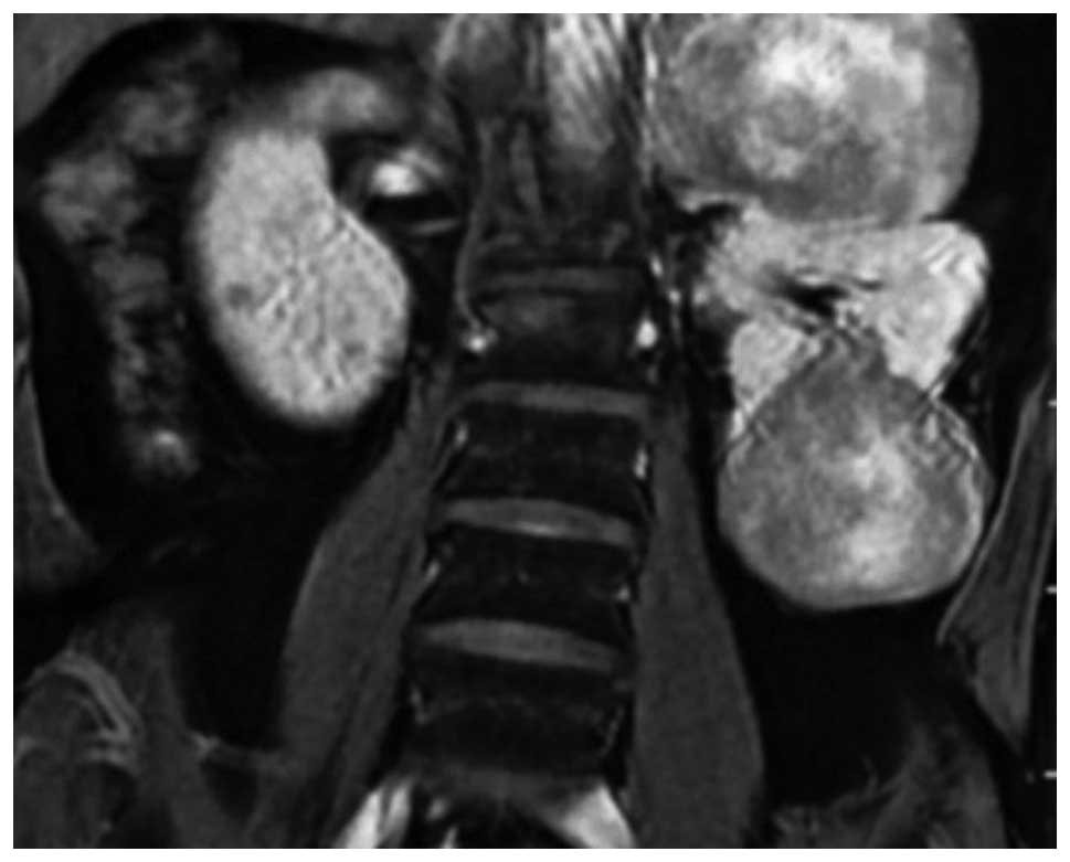
glucose concentration, which was 2.97 mmol/l. A computed tomography scan revealed the presence of two renal masses presenting as soft tissues in the upper and lower pole of the left kidney, measuring 10x8x7 cm and 10x7x7 cm, respectively. The lesions presented with an inhomogeneous density and had no clear boundary, and exhibited slight peripheral enhancement with contrast medium; however, the center enhanced unequally. Magnetic resonance imaging also revealed two renal masses in the upper and lower pole of the left kidney, which presented as elliptical and inhomogeneous densities (Fig. 1).

Figure 1

Magnetic resonance imaging revealing two renal masses in the upper and lower pole of the left kidney, which presented as elliptical and inhomogeneous densities.

An initial diagnosis of space-occupying lesions of the left kidney was given (with suspicions of renal cell carcinoma) and subsequently, the patient underwent left radical nephrectomy. Surgery revealed two masses in the upper and lower pole, with the lower mass showing no clear boundary with the kidney. The cut surface of the tumors was grey to white and crisp in texture with local hemorrhage and necrosis.

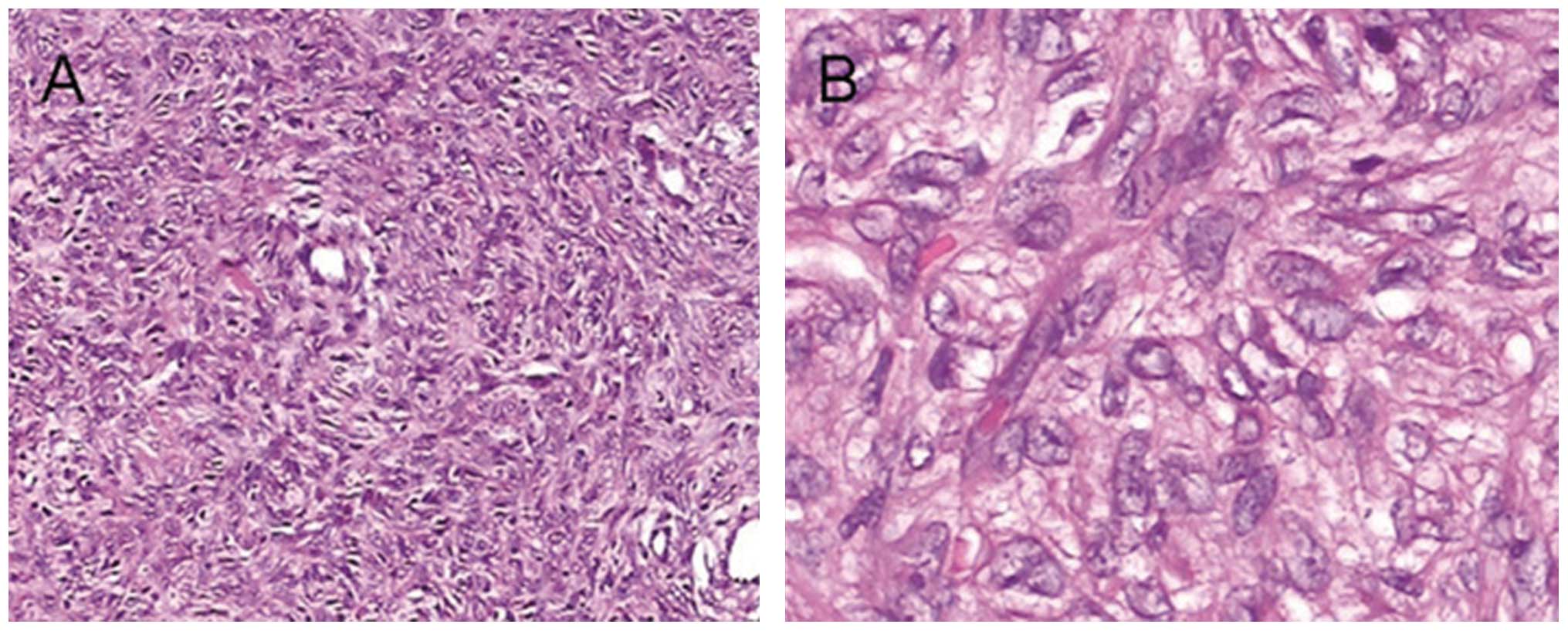
Microscopically, both tumors had a similar histological appearance, consisting of ovoid or spindle cells arranged as bundles, swirls or irregularities. Partial focal areas containing vessels were present, which had a hemangiopericytoma-like pattern. Certain areas in the tumors (particularly the lower one) demonstrated increased cells and cellular atypia and mitotic figures were also focally observed (more than 4 per 10 high-power fields). The edge of the mass demonstrated infiltrating growth, and certain areas revealed hemorrhage and/or necrosis (Fig. 
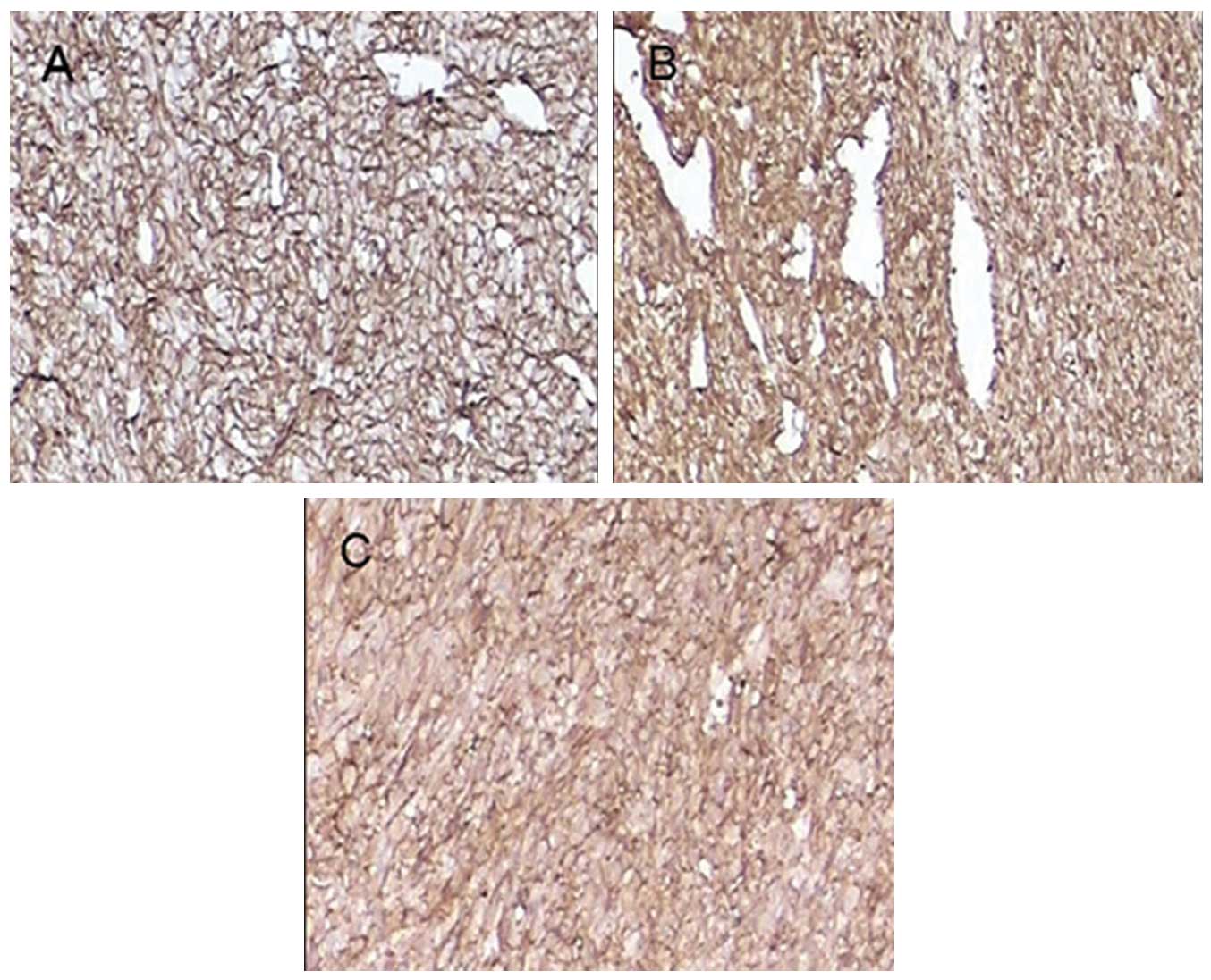
2). Immunohistochemically, the tumor cells were positive for CD34, vimentin and CD99 (Fig. 3). Staining for cytokeratin, α-smooth muscle actin (α-SMA) and S-100 was negative.

Figure 2

Microscopic features of the malignant solitary fibrous tumor of the kidney. Hematoxylin-eosin stained specimen from the left kidney, showing the tumor consisted of spindle or ovoid cells and a hemangiopericytoma-like pattern. Cellular atypia can be seen in some areas. (A) original magnification ×200; (B) original magnification ×400.

Figure 3

Immunohistochemistry of the malignant solitary fibrous tumor of the kidney. The tumor cells were diffusely positive for (A) CD34, (B) vimentin and (C) CD99.

Accordingly, a pathological diagnosis of malignant solitary fibrous tumor of the left kidney (low-grade malignancy) was established based on the microscopic features and the immunohistochemical findings.

A postoperative follow-up of 10 months revealed no signs of tumor recurrence or metastasis and the hypoglycemia was resolved with the removal of the tumors.

Discussion

Solitary fibrous tumors arising in the kidney were first described in 1996 by Gelb et al (2). The majority of renal SFTs are observed in adults in a wide age range (18–85); however, one pediatric case has been reported in a 4-year-old boy (5). In general, SFTs are slow-growing tumors with a favorable prognosis, although there have been some malignant cases (6).

Symptoms of SFTs usually do not differ from those reported by patients with renal cell carcinoma, which include flank or abdominal pain and/or gross hematuria. In certain patients, the tumor is found incidentally and in others the mass is palpable (2,6). Non islet-cell hypoglycemia (Doege Potter syndrome) arises due to the production of high molecular weight insulin-like growth factors and is a frequently associated paraneoplastic syndrome, which may have malignant behavior or poor survival from diagnosis (7). In our case, the patient had symptoms of hypoglycemia; however, it was resolved with the removal of the neoplasm suggesting it was the result of a paraneoplastic syndrome.

Macroscopically, renal SFTs are usually single; however, this was not observed in the present case. Occasionally, myxoid and pseudocapsules are also present. The size of the tumors is usually between 2.0 and 25.0 cm. The majority of cases are without cystic, hemorrhagic or necrotic changes which may be malignant (6). Microscopically, SFTs of the kidney are pathologically characterized by spindle cell proliferation showing a patternless architecture with a combination of alternating hypocellular and hypercellular areas. Furthermore, SFTs are tumors with variable cellularity and are composed of a mixture of haphazard, storiform, short fascicular arrangements of bland spindle cells and collagenous bands. They are highly vascular tumors, with hemangiopericytoma-like patterns observed in certain areas. Myxoid changes and fibrosis may also be observed (6,8,9).

Immunohistochemical study is key to diagnosis. Although it is not specific to SFTs, CD34 immunoreactivity has been reported to be diffusely expressed in many cases of SFTs, thus strong CD34 reactivity is currently regarded as the most prominent characteristic and indispensable finding to aid diagnosis (2–4,8–10). In addition, a positive expression of bcl-2, vimentin and CD99 is also present in the majority of SFTs; however, cytokeratin, α-smooth muscle actin (α-SMA) and S-100 are usually absent (2–4,8–10).

The majority of SFTs are benign; however, a few have revealed histologically malignant features. The criteria for malignant SFTs, first proposed by England et al (10) in 1989, include increased cellularity with crowded/overlapping nuclei, cellular pleomorphism and a mitotic count of more than 4 per 10 high-power fields. However, in spite of cytologic malignancy in SFTs, the clinical outcome is not always poor (10). In the present case, the majority of areas in the tumors (particularly the lower one) demonstrated increase cell density and cellular atypia, and mitotic figures were more than 4 per 10 high-power fields. The edge of the masses showed infiltrating growth and certain areas revealed hemorrhage or necrosis. Accordingly, the diagnosis was confirmed as malignant.

Solitary fibrous tumors should be distinguished from benign and malignant spindle cell tumors of the kidney. Inflammatory myofibroblastic tumors are characterized by spindle cells in an edematous background with blood vessels and inflammation consisting of lymphocytes and plasma cells. Furthermore, a mixture of spindle cells and collagenization with CD34 reactivity differentiates SFTs from inflammatory myofibroblastic tumors. Fibromas are small tumors that do not react with CD34. Leiomyoma can be excluded by the lack of expression of muscle markers. The absence of smooth muscle and adipose tissue components, with the aid of HMB-45 immunonegativity, distinguishes SFT from angiomyolipoma. It is occasionally difficult to differentiate hemangiopericytoma from SFT; however, the presence of spindle cell cytological features, various growth patterns with alternating hypocellular and hypercellular areas and abundant collagenization favors a diagnosis of SFTs. Solitary fibrous tumors may also be differentiated from leiomyosarcoma, malignant nerve sheath tumors, sarcomatoid renal and transitional cell carcinoma by the absence of dense cellularity, severe atypia, high mitotic rates and with the help of immunohistochemical staining of these tumors for α-smooth muscle actin, S-100 and cytokeratin (8).

Surgery is considered the first choice of treatment if possible. The use of chemotherapy is reserved for metastatic or symptomatic non-resectable SFTs; however, there are still no standard chemotherapeutic indications or regimens. The relatively effective drugs appear to be those which are commonly used in soft tissue sarcomas even though SFTs are considered chemoresistant (11,12). Radiation therapy is of some benefit, when applicable, given in combination with chemotherapy (13). The novel targeted drug imatinib mesilate, which exerts some activity on SFTs expressing the wild-type PDGFR-β, has recently been reported (14,15).

The prognosis is generally good for SFTs and the majority are non-recurring and non-metastazing tumors. It is estimated that 10 to 15% of intrathoracic SFTs and 10% of extrathoracic SFTs will recur and/or metastasize (16,17), therefore SFT is regarded as an ‘intermediate malignant, rarely metastasizing neoplasm’ (18). Poor prognosis of SFTs may occur when there is incomplete resection, a large size (>10 cm), presence of malignant cellularity or they are located outside the thoracic cavity. An additional factor conferring a worse prognosis in SFTs is dedifferentiation or sarcomatous overgrowth, which represents an abrupt transition to a morphologically anaplastic component (19). In the present case, a postoperative follow-up of 10 months revealed no recurrence or metastasis. Nevertheless, long-term follow-up is indispensable since metastases may still occur after several years.

To summarize, we presented a case of two malignant SFTs in one kidney with characteristic clinicopathological features. Although malignant SFTs in extrapleural sites remain extremely uncommon, it is indispensable to consider this possibility when renal spindle cell tumors are encountered. In view of characteristic findings which are often not observed in imaging studies including CT and MRI, the histologic features and immunohistochemical staining for CD34, CD99, bcl-2 and vimentin may be helpful in confirming the diagnosis of malignant SFTs of the kidney.

References

1. 

L RobinsonSolitary fibrous tumor of pleuraCancer Control13264269200617075563

2. 

A GelbM SimmonsN WeidnerSolitary fibrous tumor involving the renal capsuleAm J Surg Pathol2012881295199610.1097/00000478-199610000-000168827037

3. 

M MarziP PiraM D’AlpaosA PaiuscoS CanessaMS MinerviniP Di ZittiThe solitary fibrous malignant tumor of the kidney: clinical and pathological considerations on a case revisiting the literatureMinerva Urol Nefrol63109113201121336250

4. 

H TalvitieK AströmO LarssonSolitary fibrous tumor of the prostate: A report of two casesPathol Int61536538201110.1111/j.1440-1827.2011.02696.x21884303

5. 

N FerrariL NieldFinal diagnosis: solitary fibrous tumor of the kidneyClin Pediatr (Phila)45871873200617080561

6. 

SW FineDM McCarthyTY ChanMalignant solitary fibrous tumor of the kidney: report of a case and comprehensive review of the literatureArch Pathol Lab Med130857861200616740040

7. 

B HerrmannB SallerW KiessK MorgenrothK DrochnerT SchröderPrimary malignant fibrous histiocytoma of the lung: IGF-II producing tumor induces fasting hypoglycaemiaExp Clin Endocr Diab108515518200010.1055/s-2000-1100711149628

8. 

G MagroV CavallaroA TorrisiM LopesM Dell’AlbaniS LanzafameIntrarenal solitary fibrous tumor of the kidney report of a case with emphasis on the differential diagnosis in the wide spectrum of monomorphous spindle cell tumors of the kidneyPathol Res Pract1983743200210.1016/S0344-0338(04)70182-X11866209

9. 

T YazakiS SatohT IizumiT UmedaY YamaguchiSolitary fibrous tumor of renal pelvisInt J Urol8504508200110.1046/j.1442-2042.2001.00360.x11683972

10. 

D EnglandL HochholzerM McCarthyLocalized benign and malignant fibrous tumors of the pleura: a clinicopathologic review of 223 casesAm J Surg Pathol13640658198910.1097/00000478-198908000-000032665534

11. 

SR GrobmyerRG MakiGD DemetriM MazumdarE RiedelMF BrennanNeo-adjuvant chemotherapy for primary high-grade extremity soft tissue sarcomaAnn Oncol1516671672200410.1093/annonc/mdh43115520069

12. 

GD DemetriS AntoniaRS BenjaminNCCN Clinical Practice Guidelines in Oncology: Soft Tissue Sarcoma-V. 22009

13. 

J De BoerPL JagerT WiggersP NieboerWAN MachteldE PrasThe therapeutic challenge of a nonresectable solitary fibrous tumor in a hypoglycemic patientInt J Clin Oncol11478481200617180519

14. 

M PrunottoM BoscoL DanieleL Macri’L BonelloL SchirosiImatinib inhibits in vitro proliferation of cells derived from a pleural solitary fibrous tumor expressing platelet-derived growth factor receptor-betaLung Cancer64244246200910.1016/j.lungcan.2008.10.013

15. 

T De PasF ToffalorioP ColomboG TrifiròG PelosiPD VignaBrief report: activity of imatinib in a patient with platelet-derived-growth-factor receptor positive malignant solitary fibrous tumor of the pleuraJ Thorac Oncol3938941200818670317

16. 

SW WeissJR GoldblumSoft tissue tumors of intermediate malignancy of uncertain typeSoft Tissue Tumor5th editionMosby ElsevierPhiladephia109311602008

17. 

AV Vallat-DecouvelaereSM DryCD FletcherAtypical and malignant solitary fibrous tumors in extrathoracic locations: evidence of their comparability to intra-thoracic tumorsAm J Surg Pathol2215011511199810.1097/00000478-199812000-00007

18. 

LFJ GuillouCDM FletcherN MandahiExtrapleural solitary fibrous tumour and hemangiopericytomaWorld Health Organization Classification of Tumours: Pathology and Genetics of Tumours of Soft Tissue and Bone. Fletcher. CDMKK UnniF MertensIARC PressLyon86902002

19. 

JM MosqueraCD FletcherExpanding the spectrum of malignant progression in solitary fibrous tumors: a study of 8 cases with a discrete anaplastic component - is this dedifferentiated SFT?Am J Surg Pathol3313141321200910.1097/PAS.0b013e3181a6cd3319718788

Related Articles

  • Abstract
  • View
  • Download
  • Twitter
Copy and paste a formatted citation
Spandidos Publications style
Zhao G, Li G and Han R: Two malignant solitary fibrous tumors in one kidney: Case report and review of the literature. Oncol Lett 4: 993-995, 2012.
APA
Zhao, G., Li, G., & Han, R. (2012). Two malignant solitary fibrous tumors in one kidney: Case report and review of the literature. Oncology Letters, 4, 993-995. https://doi.org/10.3892/ol.2012.858
MLA
Zhao, G., Li, G., Han, R."Two malignant solitary fibrous tumors in one kidney: Case report and review of the literature". Oncology Letters 4.5 (2012): 993-995.
Chicago
Zhao, G., Li, G., Han, R."Two malignant solitary fibrous tumors in one kidney: Case report and review of the literature". Oncology Letters 4, no. 5 (2012): 993-995. https://doi.org/10.3892/ol.2012.858
Copy and paste a formatted citation
x
Spandidos Publications style
Zhao G, Li G and Han R: Two malignant solitary fibrous tumors in one kidney: Case report and review of the literature. Oncol Lett 4: 993-995, 2012.
APA
Zhao, G., Li, G., & Han, R. (2012). Two malignant solitary fibrous tumors in one kidney: Case report and review of the literature. Oncology Letters, 4, 993-995. https://doi.org/10.3892/ol.2012.858
MLA
Zhao, G., Li, G., Han, R."Two malignant solitary fibrous tumors in one kidney: Case report and review of the literature". Oncology Letters 4.5 (2012): 993-995.
Chicago
Zhao, G., Li, G., Han, R."Two malignant solitary fibrous tumors in one kidney: Case report and review of the literature". Oncology Letters 4, no. 5 (2012): 993-995. https://doi.org/10.3892/ol.2012.858
Follow us
  • Twitter
  • LinkedIn
  • Facebook
About
  • Spandidos Publications
  • Careers
  • Cookie Policy
  • Privacy Policy
How can we help?
  • Help
  • Live Chat
  • Contact
  • Email to our Support Team