Spandidos Publications Logo
  • About
    • About Spandidos
    • Aims and Scopes
    • Abstracting and Indexing
    • Editorial Policies
    • Reprints and Permissions
    • Job Opportunities
    • Terms and Conditions
    • Contact
  • Journals
    • All Journals
    • Oncology Letters
      • Oncology Letters
      • Information for Authors
      • Editorial Policies
      • Editorial Board
      • Aims and Scope
      • Abstracting and Indexing
      • Bibliographic Information
      • Archive
    • International Journal of Oncology
      • International Journal of Oncology
      • Information for Authors
      • Editorial Policies
      • Editorial Board
      • Aims and Scope
      • Abstracting and Indexing
      • Bibliographic Information
      • Archive
    • Molecular and Clinical Oncology
      • Molecular and Clinical Oncology
      • Information for Authors
      • Editorial Policies
      • Editorial Board
      • Aims and Scope
      • Abstracting and Indexing
      • Bibliographic Information
      • Archive
    • Experimental and Therapeutic Medicine
      • Experimental and Therapeutic Medicine
      • Information for Authors
      • Editorial Policies
      • Editorial Board
      • Aims and Scope
      • Abstracting and Indexing
      • Bibliographic Information
      • Archive
    • International Journal of Molecular Medicine
      • International Journal of Molecular Medicine
      • Information for Authors
      • Editorial Policies
      • Editorial Board
      • Aims and Scope
      • Abstracting and Indexing
      • Bibliographic Information
      • Archive
    • Biomedical Reports
      • Biomedical Reports
      • Information for Authors
      • Editorial Policies
      • Editorial Board
      • Aims and Scope
      • Abstracting and Indexing
      • Bibliographic Information
      • Archive
    • Oncology Reports
      • Oncology Reports
      • Information for Authors
      • Editorial Policies
      • Editorial Board
      • Aims and Scope
      • Abstracting and Indexing
      • Bibliographic Information
      • Archive
    • Molecular Medicine Reports
      • Molecular Medicine Reports
      • Information for Authors
      • Editorial Policies
      • Editorial Board
      • Aims and Scope
      • Abstracting and Indexing
      • Bibliographic Information
      • Archive
    • World Academy of Sciences Journal
      • World Academy of Sciences Journal
      • Information for Authors
      • Editorial Policies
      • Editorial Board
      • Aims and Scope
      • Abstracting and Indexing
      • Bibliographic Information
      • Archive
    • International Journal of Functional Nutrition
      • International Journal of Functional Nutrition
      • Information for Authors
      • Editorial Policies
      • Editorial Board
      • Aims and Scope
      • Abstracting and Indexing
      • Bibliographic Information
      • Archive
    • International Journal of Epigenetics
      • International Journal of Epigenetics
      • Information for Authors
      • Editorial Policies
      • Editorial Board
      • Aims and Scope
      • Abstracting and Indexing
      • Bibliographic Information
      • Archive
    • Medicine International
      • Medicine International
      • Information for Authors
      • Editorial Policies
      • Editorial Board
      • Aims and Scope
      • Abstracting and Indexing
      • Bibliographic Information
      • Archive
  • Articles
  • Information
    • Information for Authors
    • Information for Reviewers
    • Information for Librarians
    • Information for Advertisers
    • Conferences
  • Language Editing
Spandidos Publications Logo
  • About
    • About Spandidos
    • Aims and Scopes
    • Abstracting and Indexing
    • Editorial Policies
    • Reprints and Permissions
    • Job Opportunities
    • Terms and Conditions
    • Contact
  • Journals
    • All Journals
    • Biomedical Reports
      • Information for Authors
      • Editorial Policies
      • Editorial Board
      • Aims and Scope
      • Abstracting and Indexing
      • Bibliographic Information
      • Archive
    • Experimental and Therapeutic Medicine
      • Information for Authors
      • Editorial Policies
      • Editorial Board
      • Aims and Scope
      • Abstracting and Indexing
      • Bibliographic Information
      • Archive
    • International Journal of Epigenetics
      • Information for Authors
      • Editorial Policies
      • Editorial Board
      • Aims and Scope
      • Abstracting and Indexing
      • Bibliographic Information
      • Archive
    • International Journal of Functional Nutrition
      • Information for Authors
      • Editorial Policies
      • Editorial Board
      • Aims and Scope
      • Abstracting and Indexing
      • Bibliographic Information
      • Archive
    • International Journal of Molecular Medicine
      • Information for Authors
      • Editorial Policies
      • Editorial Board
      • Aims and Scope
      • Abstracting and Indexing
      • Bibliographic Information
      • Archive
    • International Journal of Oncology
      • Information for Authors
      • Editorial Policies
      • Editorial Board
      • Aims and Scope
      • Abstracting and Indexing
      • Bibliographic Information
      • Archive
    • Medicine International
      • Information for Authors
      • Editorial Policies
      • Editorial Board
      • Aims and Scope
      • Abstracting and Indexing
      • Bibliographic Information
      • Archive
    • Molecular and Clinical Oncology
      • Information for Authors
      • Editorial Policies
      • Editorial Board
      • Aims and Scope
      • Abstracting and Indexing
      • Bibliographic Information
      • Archive
    • Molecular Medicine Reports
      • Information for Authors
      • Editorial Policies
      • Editorial Board
      • Aims and Scope
      • Abstracting and Indexing
      • Bibliographic Information
      • Archive
    • Oncology Letters
      • Information for Authors
      • Editorial Policies
      • Editorial Board
      • Aims and Scope
      • Abstracting and Indexing
      • Bibliographic Information
      • Archive
    • Oncology Reports
      • Information for Authors
      • Editorial Policies
      • Editorial Board
      • Aims and Scope
      • Abstracting and Indexing
      • Bibliographic Information
      • Archive
    • World Academy of Sciences Journal
      • Information for Authors
      • Editorial Policies
      • Editorial Board
      • Aims and Scope
      • Abstracting and Indexing
      • Bibliographic Information
      • Archive
  • Articles
  • Information
    • For Authors
    • For Reviewers
    • For Librarians
    • For Advertisers
    • Conferences
  • Language Editing
Login Register Submit
  • This site uses cookies
  • You can change your cookie settings at any time by following the instructions in our Cookie Policy. To find out more, you may read our Privacy Policy.

    I agree
Search articles by DOI, keyword, author or affiliation
Search
Advanced Search
presentation
Oncology Letters
Join Editorial Board Propose a Special Issue
Print ISSN: 1792-1074 Online ISSN: 1792-1082
Journal Cover
December 2012 Volume 4 Issue 6

Full Size Image

Sign up for eToc alerts
Recommend to Library

Journals

International Journal of Molecular Medicine

International Journal of Molecular Medicine

International Journal of Molecular Medicine is an international journal devoted to molecular mechanisms of human disease.

International Journal of Oncology

International Journal of Oncology

International Journal of Oncology is an international journal devoted to oncology research and cancer treatment.

Molecular Medicine Reports

Molecular Medicine Reports

Covers molecular medicine topics such as pharmacology, pathology, genetics, neuroscience, infectious diseases, molecular cardiology, and molecular surgery.

Oncology Reports

Oncology Reports

Oncology Reports is an international journal devoted to fundamental and applied research in Oncology.

Experimental and Therapeutic Medicine

Experimental and Therapeutic Medicine

Experimental and Therapeutic Medicine is an international journal devoted to laboratory and clinical medicine.

Oncology Letters

Oncology Letters

Oncology Letters is an international journal devoted to Experimental and Clinical Oncology.

Biomedical Reports

Biomedical Reports

Explores a wide range of biological and medical fields, including pharmacology, genetics, microbiology, neuroscience, and molecular cardiology.

Molecular and Clinical Oncology

Molecular and Clinical Oncology

International journal addressing all aspects of oncology research, from tumorigenesis and oncogenes to chemotherapy and metastasis.

World Academy of Sciences Journal

World Academy of Sciences Journal

Multidisciplinary open-access journal spanning biochemistry, genetics, neuroscience, environmental health, and synthetic biology.

International Journal of Functional Nutrition

International Journal of Functional Nutrition

Open-access journal combining biochemistry, pharmacology, immunology, and genetics to advance health through functional nutrition.

International Journal of Epigenetics

International Journal of Epigenetics

Publishes open-access research on using epigenetics to advance understanding and treatment of human disease.

Medicine International

Medicine International

An International Open Access Journal Devoted to General Medicine.

Journal Cover
December 2012 Volume 4 Issue 6

Full Size Image

Sign up for eToc alerts
Recommend to Library

  • Article
  • Citations
    • Cite This Article
    • Download Citation
    • Create Citation Alert
    • Remove Citation Alert
    • Cited By
  • Similar Articles
    • Related Articles (in Spandidos Publications)
    • Similar Articles (Google Scholar)
    • Similar Articles (PubMed)
  • Download PDF
  • Download XML
  • View XML
Case Report

Primary squamous cell carcinoma of the liver: A case report and review of the literature

  • Authors:
    • Rongrong Zhao
    • Kunli Zhu
    • Renben Wang
    • Jie Gao
    • Kai  Cui
    • Fachang Yu
    • Bo Zhang
    • Sheng Li
  • View Affiliations / Copyright

    Affiliations: Department of Hepatobiliary Surgery, Shandong Cancer Hospital, Jinan 276000, P.R. China, Key Laboratory for Rare and Uncommon Diseases of Shandong Province, Jinan 250062, P.R. China, Shandong Academy of Medical Sciences, Jinan 250014, P.R. China
  • Pages: 1163-1166
    |
    Published online on: September 5, 2012
       https://doi.org/10.3892/ol.2012.894
  • Expand metrics +
Metrics: Total Views: 0 (Spandidos Publications: | PMC Statistics: )
Metrics: Total PDF Downloads: 0 (Spandidos Publications: | PMC Statistics: )
Cited By (CrossRef): 0 citations Loading Articles...

This article is mentioned in:


Abstract

Primary squamous cell carcinoma of the liver is rare. It has a high malignancy rate and poor prognosis, and survival is typically no longer than one year. The pathogenesis of the disease is unclear, but is generally considered to be correlated with the long-term inflammation or metaplasia of biliary epithelial cells and congenital cyst of the liver. The current viewpoint is that surgical resection of the tumor, preoperative or postoperative subsidiary chemoembolization, radiotherapy and generalized chemotherapy may extend a patient's survival. In the present case study, a patient suffered from biliary calculosis for over 20 years. We theorized that the mechanism of carcinomatous change was correlated with the persistent stimulation of chronic biliary infection induced by calculus of the intrahepatic duct. However, the transformation mechanism from cholangiocellular carcinoma to squamous carcinoma is undetermined and further studies are needed.

Introduction

Primary squamous cell carcinoma of the liver is rare. More than 20 cases have been reported since the first case was described in 1934 by Imai (1). The pathogenesis of the disease is undetermined. Previous studies have reported that potentially related diseases, including congenital cyst of the liver (2–8), calculus of the intrahepatic duct (9), hepatic cirrhosis (10,11), Caroli’s disease (12) and hepatic teratoma (1), are rare. However, hepatic primary squamous carcinoma without latent hepatic injury has also been reported (13). This disease has a high malignancy rate and poor prognosis, and survival is typically no longer than one year (12). In the present case study, a patient suffered from biliary calculosis for over 20 years. The patient underwent chest, abdominal and pelvic CT scans and gynecological examination to preclude other primary foci. The study was approved by the Ethics Committee of Shandong Cancer Hospital, Jinan, China. Written informed consent was obtained from the patient. The postoperative pathology and immunohistochemistry confirmed the disease as liver primary squamous cell carcinoma. Combined with the patient’s history of calculosis of the biliary tree for 20 years, we determined that the mechanism of carcinogenesis was correlated with persistent stimulation induced by chronic biliary infection caused by calculus of the intrahepatic duct. However, the transformation mechanism from cholangiocellular carcinoma to squamous carcinoma is undetermined and requires further study. In the present study, the patient underwent liver tumor radiofrequency ablation, but the abdominal pain remained evident following the surgery. As the patient’s physical condition was too weak to tolerate generalized chemotherapy, local radiotherapy and supportive therapy is being conducted, and the patient’s pain has already been relieved.

Case report

A 60-year-old female patient underwent cholecystectomy due to cholecystolithiasis with chronic cholecystitis in 1988. In May 2010, the patient experienced repeated pain without obvious cause in the right upper quadrant, accompanied by fever, nausea without vomiting, hypodynamia, anorexia, abdominal distension, diarrhea, shivers, jaundice and lumbar-dorsal radiating pain. A CT examination revealed a calculus of the intrahepatic duct and chronic cholangitis. A partial resection of the hepatic left lobe was carried out on July 29th, 2010. The postoperative pathology indicated left hepatic cholangeitis and calculus of the bile duct. A magnetic resonance imaging (MRI) examination performed in December 2010 indicated that the primary focus was in the left liver at a size of 4.0×3.0×3.0 cm, with reinforcement in the arterial phase. The examination of tumor biomarkers revealed 7.6 μg/l AFP, 1.1 μg/l CEA and 275.9 U/ml CA19-9. The tumor resection of the left hepatic external lobe, exploration of the biliary tract and T-tube drainage were performed on December 31st, 2010. A calculus with a diameter of 0.2 cm and a grey tumor 2.0×1.5 cm in the cross section without peplos in the surroundings of the tumor were observed and the 
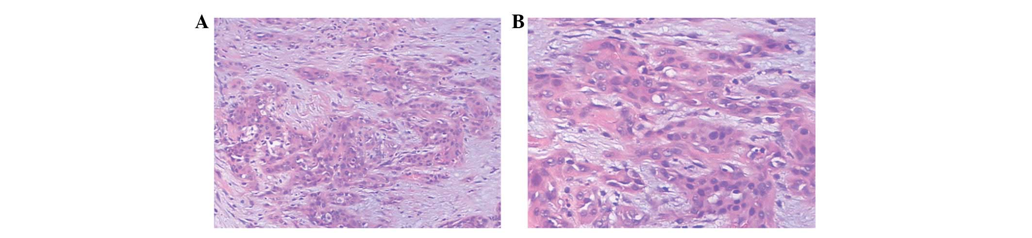
texture was hard. Microscopically (Fig. 2), the tumor tissue was accumulated in the glandular tube, the lumens contained mucilage, the cancer cells were cubic or columnar, the nuclei were round or orbicular-ovate, there was karyomegaly and anachromasis, evident atypia and the surroundings indicated infiltrative growth affecting the nerves. Tumor growth was not observed in cancerous tissue, and there was no pseudo-lobule structure in the remaining liver. Masson (+), AB (+) and reticulum staining (−) were performed. The immunohistochemistry results were 
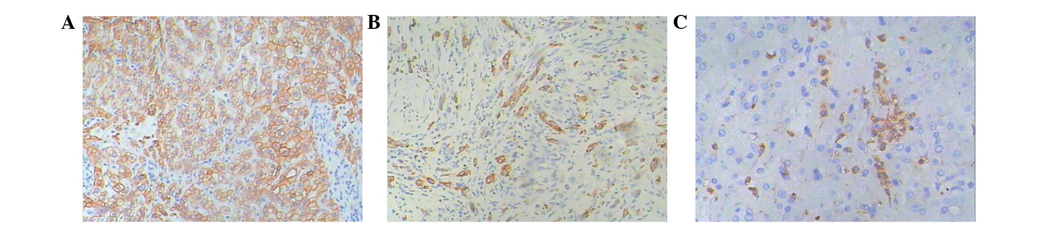
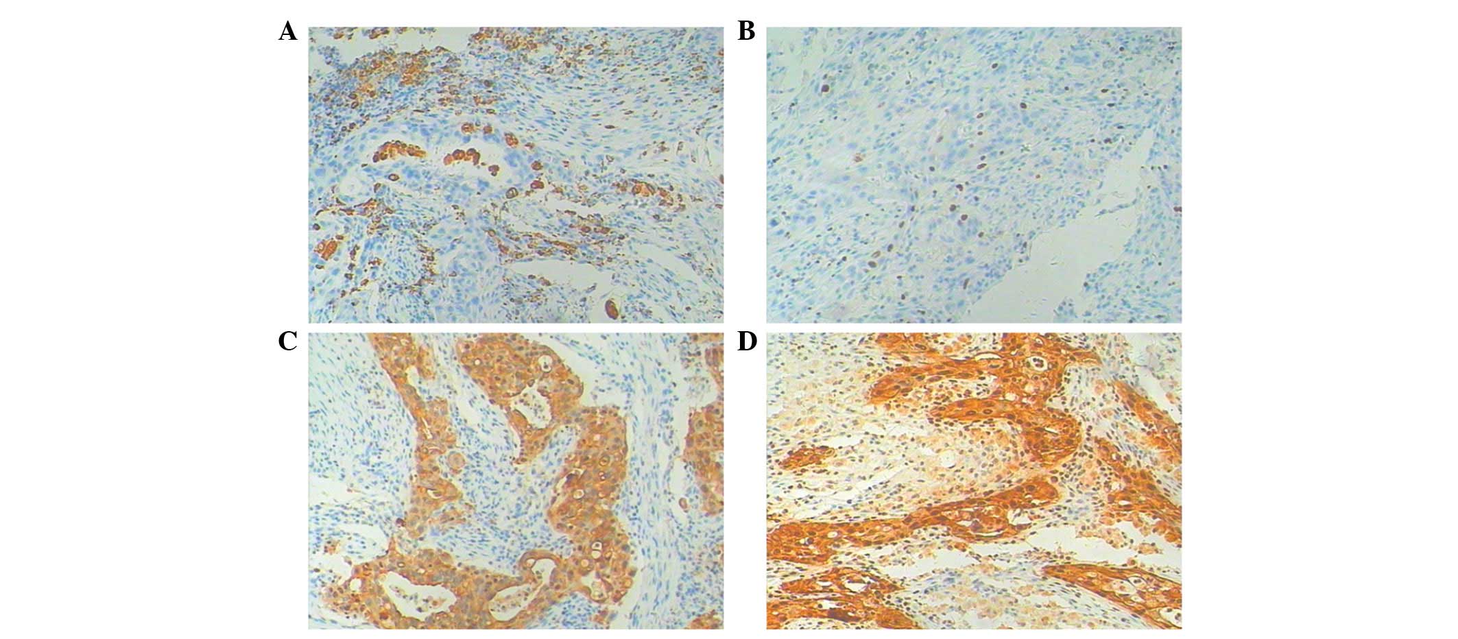
as follows (Fig. 3): Hep-1(+++), HBsAg(−), CK18(+), CK19(++), CD34(−), HbeAg(−), pCEA(+++), β-catenin(−), MUC-1(−), myoglobin(−), Gly-3(−) and MAT1(−). The pathological diagnosis was intrahepatic cholangiocarcinoma of the hepatic left lobe with mild differentiation and calculus of the bile duct. The patient’s symptoms were relieved following the surgery. In June 2011, the patient experienced pain in the right upper quadrant without any evident cause, and the pain started to radiate to the back, accompanied by nausea, vomiting, hypodynamia 
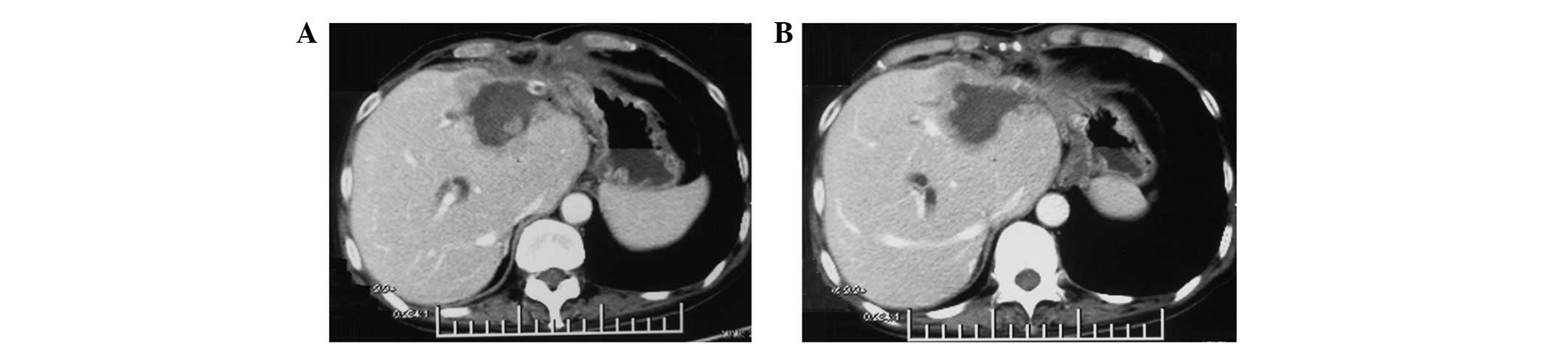
and anorexia. An abdominal CT examination (Fig. 1) was performed and revealed an abnormal density shadow in the medial segment of the left lobe of the liver following the tumor resection of the hepatic left lobe, which was considered to be the postoperative recurrence of cholangiocellular carcinoma of the hepatic left lobe. Examination of the tumor markers indicated 4.63 μg/l AFP, 191.5 U/ml CA125 and 1557.8 U/ml CA19-9. The patient underwent hepatic tumor biopsy and hepatic tumor radiofrequency ablation on July 4th, 2011. The tumor was located in the junctional zone of the hepatic left internal lobe and right anterior lobe with a hard texture at a size of 1.5×1.2×0.5 cm, which was closely adhered to partial gastric serous tunic and peripheral abdominal wall. Considering the extra hepatic encroachment of the tumor, the location and surroundings of the tumor made surgical separation and radical resection difficult; therefore, radiofrequency ablation of the left hepatic tumor was carried out. Postoperative pathology revealed a mass of grayish-white and grayish-yellow tissue with a hard texture and a 
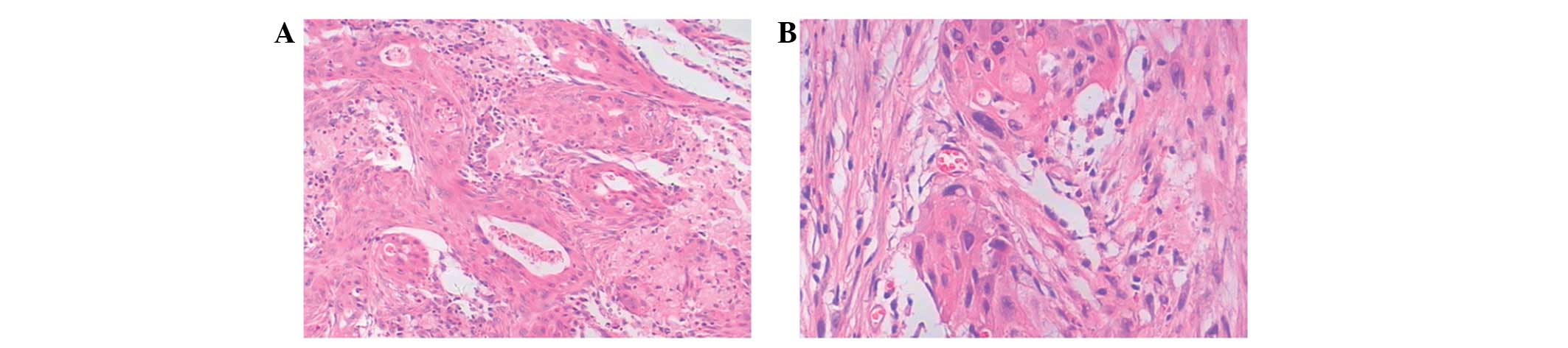
size of 1.2×1.2×0.4 cm. Microscopically, infiltration of poorly differentiated carcinoma tissue in the proliferative fibrous tissue and interaction between foam cells and multinucleated giant cells in the mesenchyme were observed (Fig. 4). The immunohistochemical staining of the tumor cells revealed CD68(−), Ki-67(+, <25%), CK(+), CK590(+) (Fig. 5), which is consistent with liver squamous cell carcinoma.

Figure 2.

Microscopically, the tumor tissue accumulated in the glandular tube. The cancer cells were cubic or columnar, the nuclei were round or orbicular-ovate, and there was karyomegaly, anachromasis and evident atypia. (A) H&E staining; magnification, x100. (B) H&E staining; magnification, x200. H&E hematoxylin and eosin.

Figure 3.

Immunohistochemistry results for (A) CK18(+); (B) CK19(++); (C) pCEA(+++). Magnification, x100.

Figure 1.

Abdominal CT examination. An abnormal density shadow in the medial segment of the left lobe of the liver following the tumor resection of the hepatic left lobe, which was considered to be the postoperative recurrence of cholangiocellular carcinoma of the hepatic left lobe.

Figure 4.

Microscopically, infiltration of poorly differentiated carcinoma tissue in the proliferative fibrous tissue and interaction between foam cells and multinucleated giant cells in the mesenchyme were observed. H&E staining. (A) Magnification, x100; (B) magnification, x200. H&E hematoxylin and eosin.

Figure 5.

Immunohistochemistry results for (A) CD68(−); (B) Ki-67(+, < 25%); (C) CK(+); (D) CK5(+). Magnification, x100.

Currently, the patient continues to feel pain in the right upper quadrant of the abdomen, accompanied by fever, nausea, vomiting, hypodynamia, anorexia without jaundice, abdominal distension and diarrhea, with poor diet and sleeping. The patient’s weight has reduced by ∼15 kg in 1 year. Nutritional therapy was administered, and radiotherapy of the hepatic tumor focus and the periphery of the invaded structure were performed at a total dose of 36 Gy 20 times. The patient’s abdominal pain was markedly relieved and the abdominal mass was reduced to a size of 4.0×5.0 cm, and we are currently following up the patient.

Discussion

Incidence of primary squamous cell carcinoma of the liver is rare and the pathogenesis of the disease is undetermined. Previous studies have reported that potentially related diseases, including congenital cyst of the liver (7,8), calculus of the intrahepatic duct (9), hepatic cirrhosis (11), Caroli’s disease (12) and hepatic teratoma (1), are also rare.

In the present study, the patient underwent chest, abdominal and pelvic CT scans and gynecological examination to preclude other primary foci. The postoperative pathology and immunohistochemistry confirmed the disease as liver primary squamous cell carcinoma. The patient’s primary operative pathology revealed hepatic cholangiocellular carcinoma. The tumor recurrence occurred in half a year, and tumor pathological biopsy and immunohistochemistry revealed the tumor to be hepatic primary squamous carcinoma. The transformation from hepatic cholangiocellular carcinoma to liver squamous carcinoma is extremely rare, and there are no similar studies. Combined with the patient’s history of calculosis of the biliary tree for 20 years, we determined that the mechanism of carcinogenesis was correlated with persistent stimulation induced by chronic biliary infection caused by calculus of the intrahepatic duct. However, the transformation mechanism from cholangiocellular carcinoma to squamous carcinoma is undetermined and requires further study.

The morbidity of hepatic primary squamous has no evident gender differentiation and mainly occurs in 50–70-year-old patients. The clinical symptoms are commonly reported as stomach ache or jaundice, accompanied by anorexia, hypodynamia, nausea, vomiting and physical athrepsia. Swelling of the liver may be palpable in the physical examination. Currently, the definitive diagnosis mainly relies on pathology and immunohistochemistry.

In cases of liver primary squamous carcinoma, due to its high malignancy, poor prognosis and late definitive diagnosis, survival is generally no longer than 1 year (11). As the incidence of liver primary squamous carcinoma is extremely low, its therapeutic regime has not been agreed. Reported therapeutic methods include surgical resection, generalized chemotherapy, radiotherapy, hepatic arterial chemoembolization (HACE) and combinations of these therapies. In the present study, the patient underwent liver tumor radiofrequency ablation, but the patient’s abdominal pain continued after the surgery. As the patient’s physical condition was too weak to tolerate generalized chemotherapy, local radiotherapy and supportive therapy is being carried out, and the patient’s pain has already been relieved. The current viewpoint is that HACE combined with radiotherapy or generalized chemotherapy may extend survival in patients with primary squamous carcinoma, whereas the timely total surgical resection of tumor is the key to preventing invasive tumor growth and further surgical resection, systematic generalized chemotherapy or HACE may be used for patients with postoperative recurrence of the disease. We determined that local radiotherapy may be used to relieve symptoms in patients who are unable to undergo resection of the tumor or tolerate generalized chemotherapy.

Acknowledgements

This study was financially supported by grant no. 2011CB504302 from the 973 Program of China and grant nos. ZR2010HL018 and Y2008C30 from the Natural Science Foundation of Shandong. We appreciate the valuable comments from other members of our laboratories.

References

1. 

T ImaiEin Fall von zystischem Teratom der Leber, in welchem Plattenepithelkrebs entstandTrans Soc Pathol Jap245785801934(In German).

2. 

MJ LynchMK McLeodL WeatherbeeJR GilsdorfKS GuiceFE EckhauserSquamous cell cancer of the liver arising from a solitary benign nonparasitic hepatic cystAm J Gastroenterol8342643119883279761

3. 

J BanburyKC ConlonR GhosseinMF BrennanPrimary squamous cell carcinoma within a solitary nonparasitic hepatic cystJ Surgl Oncol57210212199410.1002/jso.29305703167967614

4. 

M MonteagudoG VidalM MorenoSquamous cell carcinoma and infection in a solitary hepatic cystEur J Gastroenterol Hepatol1010511053199810.1097/00042737-199812000-000129895053

5. 

DJ VickZD GoodmanKG IshakSquamous cell carcinoma arising in a ciliated hepatic foregut cystArch Pathol Lab Med12311151117199910539920

6. 

A FurlanettoAP Dei TosSquamous cell carcinoma arising in a ciliated hepatic foregut cystVirchows Archiv441296298200210.1007/s00428-002-0668-z12242527

7. 

H YagiM UedaS KawachiSquamous cell carcinoma of the liver originating from non-parasitic cysts after a 15 year follow-upEur J Gastroenterol Hepatol1610511056200415371931

8. 

X ZhangZ WangY DongSquamous cell carcinoma arising in a ciliated hepatic foregut cyst: case report and literature reviewPathol Res Pract205498501200910.1016/j.prp.2008.12.00319410383

9. 

E SongMC KewT GrieveC IsaacsonJA MyburghPrimary squamous cell carcinoma of the liver occurring in association with hepatolithiasisCancer53542546198410.1002/1097-0142(19840201)53:3%3C542::AID-CNCR2820530328%3E3.0.CO;2-36692259

10. 

Y AraseY EndoM HaraH KumadaK IkedaA YoshibaHepatic squamous cell carcinoma with hypercalcemia in liver cirrhosisActa Pathol Jpn3864365019882463731

11. 

N YukiY HijikataM KatoK KawaharaK WakasaSquamous cell carcinoma as a rare entity of primary liver tumor with grave prognosisHepatol Res36322327200610.1016/j.hepres.2006.08.00416978916

12. 

M SpaggiariF Di BenedettoR BallarinPrimary squamous cell carcinoma of the liver associated with Caroli’s disease: a case reportOnkologie34193195201121447978

13. 

R KajiN SasakiI TateishiA case report of primary hepatic squamous cell carcinoma that remarkably responded to low dose arterial injection of anti-cancer drugsKurume Med J507175200310.2739/kurumemedj.50.7112971268

Related Articles

  • Abstract
  • View
  • Download
  • Twitter
Copy and paste a formatted citation
Spandidos Publications style
Zhao R, Zhu K, Wang R, Gao J, Cui K, Yu F, Zhang B and Li S: Primary squamous cell carcinoma of the liver: A case report and review of the literature. Oncol Lett 4: 1163-1166, 2012.
APA
Zhao, R., Zhu, K., Wang, R., Gao, J., Cui, K., Yu, F. ... Li, S. (2012). Primary squamous cell carcinoma of the liver: A case report and review of the literature. Oncology Letters, 4, 1163-1166. https://doi.org/10.3892/ol.2012.894
MLA
Zhao, R., Zhu, K., Wang, R., Gao, J., Cui, K., Yu, F., Zhang, B., Li, S."Primary squamous cell carcinoma of the liver: A case report and review of the literature". Oncology Letters 4.6 (2012): 1163-1166.
Chicago
Zhao, R., Zhu, K., Wang, R., Gao, J., Cui, K., Yu, F., Zhang, B., Li, S."Primary squamous cell carcinoma of the liver: A case report and review of the literature". Oncology Letters 4, no. 6 (2012): 1163-1166. https://doi.org/10.3892/ol.2012.894
Copy and paste a formatted citation
x
Spandidos Publications style
Zhao R, Zhu K, Wang R, Gao J, Cui K, Yu F, Zhang B and Li S: Primary squamous cell carcinoma of the liver: A case report and review of the literature. Oncol Lett 4: 1163-1166, 2012.
APA
Zhao, R., Zhu, K., Wang, R., Gao, J., Cui, K., Yu, F. ... Li, S. (2012). Primary squamous cell carcinoma of the liver: A case report and review of the literature. Oncology Letters, 4, 1163-1166. https://doi.org/10.3892/ol.2012.894
MLA
Zhao, R., Zhu, K., Wang, R., Gao, J., Cui, K., Yu, F., Zhang, B., Li, S."Primary squamous cell carcinoma of the liver: A case report and review of the literature". Oncology Letters 4.6 (2012): 1163-1166.
Chicago
Zhao, R., Zhu, K., Wang, R., Gao, J., Cui, K., Yu, F., Zhang, B., Li, S."Primary squamous cell carcinoma of the liver: A case report and review of the literature". Oncology Letters 4, no. 6 (2012): 1163-1166. https://doi.org/10.3892/ol.2012.894
Follow us
  • Twitter
  • LinkedIn
  • Facebook
About
  • Spandidos Publications
  • Careers
  • Cookie Policy
  • Privacy Policy
How can we help?
  • Help
  • Live Chat
  • Contact
  • Email to our Support Team