Introduction
Thyroid cancer is the most common endocrine
neoplasm. Histopathologically, thyroid cancers can be classified
into papillary, follicular, medullary and anaplastic carcinomas.
Papillary and follicular carcinomas are derived from thyroid
follicular epithelial cells and account for the vast majority of
thyroid cancer. These two types of thyroid cancers are
differentiated tumors with low grades. The prognosis of thyroid
cancer is associated with the histological type of the cancer and
the stage at diagnosis. For papillary thyroid cancer (PTC), the
overall prognosis is excellent following proper treatment,
including thyroidectomy, lobectomy or radioactive iodine (RAI)
therapy (1).
RAI ablation, which removes all remnant or residual
normal thyroid tissues, is an important element of therapy
following initial surgery in patients with papillary and follicular
thyroid carcinomas (1). The
underlying mechanism of RAI treatment of thyroid cancer is based on
the ability of thyroid follicular cells to concentrate iodine,
which is dependent on the functional Na+/I−
symporter (NIS) (2). NIS is a
transmembrane glycoprotein with a molecular weight of 87 kDa, which
transports two Na+ for each I− into thyroid
follicular cells. The loss of NIS expression in thyroid follicular
cells may cause goiters or hypothyroidism. During thyroid cancer
development, NIS expression has been reported to be reduced or
lost. Thus, the detection of NIS expression was able to predict the
outcome of RAI treatment in patients with thyroid cancer. Previous
studies have demonstrated that the NIS protein was differentially
expressed in differentiated thyroid carcinomas compared with normal
thyroid tissues (2–4). By contrast, certain previous studies
have found that PTC patients with normal NIS expression respond to
RAI therapy (3,5,6).
However, to date, few studies reporting NIS expression in PTC and
their surrounding tissues have been published. Therefore, in the
present study, we evaluated NIS expression in adjacent normal
thyroid tissues in comparison to nodular goiter in PTC patients.
The detection of NIS expression in surrounding normal thyroid
tissues was able to predict the iodine uptake activity during RAI
therapy of differentiated thyroid carcinomas.
Materials and methods
Study population
In this study, we first identified and reviewed the
medical records from 600 patients who were diagnosed with PTC and
underwent total or near-total thyroidectomy at the Affiliated
Hospital of Qingdao Medical College between January 1, 2008 and
January 1, 2011. Histology sections from these 600 patients were
carefully re-examined by two pathologists for confirmation of the
original diagnosis of PTC. Specimens of PTC and the surrounding
normal tissues or the surrounding nodular goiter tissues were used
in the current study, which generated two groups of cases: a group
of 60 patients (52.63%; 9 males and 51 females with a mean age of
49.55±11.29 years) whose PTC had surrounding nodular goiter tissues
(abbreviated as GNG) and a second group of 54 patients
(47.37%; 8 males and 46 females with a mean age of 45.78±12.11
years) whose PTC had surrounding normal tissues (abbreviated as
Gnormal) (Table I). 1.
The study was approved by the Ethics Committee of The Affiliated
Hospital of Qingdao Medical College, Qingdao, China. Written
informed patient consent was obtained from the patient.
 | Table IExpression of NIS protein and the
clinicopathological characteristics of 114 PTC patients. |
Table I
Expression of NIS protein and the
clinicopathological characteristics of 114 PTC patients.
| IHS points in
GNG cases
| IHS points in
Gnormal cases
|
|---|
| Characteristics | Total n=60 | Cancer tissue | Surrounding
tissue | Total n=54 | Cancer tissue | Surrounding
tissue |
|---|
| Total | | 348 | 276 | | 366 | 351 |
| Age (years) | | | | | | |
| ≥45 | 42 | 219 | 198 | 30 | 198 | 119 |
| <45 | 18 | 129 | 78 | 24 | 168 | 132 |
| Gender | | | | | | |
| Male | 9 | 48 | 33 | 8 | 30 | 33 |
| Female | 51 | 300 | 243 | 46 | 336 | 318 |
| Tumor size (cm) | | | | | | |
| ≥1 | 33 | 234 | 53 | 48 | 330 | 330 |
| <1 | 27 | 114 | 223 | 6 | 36 | 21 |
| N | | | | | | |
| + | 12 | 63 | 45 | 36 | 114 | 144 |
| - | 48 | 285 | 231 | 18 | 252 | 107 |
| DM | | | | | | |
| + | 0 | 0 | 0 | 3 | 24 | 36 |
| - | 60 | 348 | 276 | 51 | 342 | 315 |
| pTNM stage | | | | | | |
| I | 48 | 279 | 219 | 42 | 300 | 267 |
| II | 0 | 0 | 0 | 0 | 0 | 0 |
| III | 12 | 69 | 57 | 9 | 42 | 48 |
| IV | 0 | 0 | 0 | 3 | 24 | 36 |
Immunohistochemistry
Formalin-fixed and paraffin-embedded tissue blocks
from PTCs with GNG and Gnormal were obtained
from the Department of Pathology at the Affiliated Hospital of
Qingdao Medical College, and prepared in 5-μm thick sections for
immunohistochemistry. Briefly, all tissue sections were
deparaffinized in xylene, rehydrated in graded alcohol (100-50%)
and endogenous peroxidase activity was blocked in 3%
H2O2 solution in methanol for 5 min and the
sections were washed with PBS three times for 2 min each. Next, the
sections were incubated with 20% normal serum for 30 min and then
with a rabbit polyclonal anti-NIS antibody (#696557; American Basic
Gene Associate Bioscience, Inc., Chicago, IL, USA) at a dilution of
1:400 in PBS at 4°C overnight. The next day, the sections were
washed three times with PBS for 2 min each and incubated with a
secondary antibody from a PV-6000 kit (Zhongshan Golden Bridge
Biotechnology, Beijing, China) for 15 min at room temperature.
Next, the sections were incubated with 3,3′-diaminobenzidine
tetrahydrochloride solution (DAB; Zhongshan Golden Bridge
Biotechnology) after washing three times with PBS. The color
reaction was stopped after a suitable color had developed or after
a maximum of 10 min. The sections were briefly counterstained with
hematoxylin. Finally, all sections were washed with distilled
water, dehydrated through ascending alcohol and xylene washes and
mounted with cover slips with a drop of mounting medium. Both
positive and negative controls were used for each sample of tumor
tissues and surrounding tissues.
Review and score of the immunostained
tissue sections
All the immunostained tissue sections were reviewed
and scored under a microscope for expression and localization of
NIS protein by two pathologists independently and blindly. The
scores of each section were compared and if there was a
discrepancy, the two pathologists reviewed them again and reached a
consensus. Briefly, five high-powered fields under the microscope
were randomly chosen and one hundred cells in each field were
counted. The staining scores (IHS) were calculated by combining an
estimate of the percentage of immunoreactive cells (quantity score)
with an estimate of the staining intensity (staining intensity
score) (7). For the percentage of
staining, 0 indicated no staining; 1, 1–10% of cells stained; 2,
11–50%; 3, 51–80%; 4, 81–100%. Staining intensity scores were as
follows: 0, negative; 1, weak; 2, moderate; and 3, strong. The raw
data were converted to the IHS by multiplying the quantity and
staining intensity scores. The scores of IHS ranged from 0 to 12,
with 0 indicating negative, 1 to 4 indicating weak, 5 to 8
indicating moderate and 9 to 12 indicating strong immunoreactivity.
For multi-focal immunoreactivity or significant differences in
staining intensities between foci, the average of the least and
most intense staining was recorded.
Statistical analyses
All statistical analyses were performed with SPSS
version 17.0 (SPSS, Inc., Chicago, IL, USA). Statistical
independent sample t-tests or t’-tests were used to determine the
correlation of NIS expression between tumor and normal tissues.
P<0.05 was considered to indicate a statistically significant
result.
Results
Clinicopathological characteristics
In this study, we collected a total of 114 cases of
PTC and divided them into two groups: 60 patients with PTC whose
resected tissues contained adjacent normal thyroid epithelium
(Gnormal) and 54 patients with PTC whose surgical
specimens contained nodular goiter tissues (GNG). We
found that tumor size (P=0.0004) and lymph node metastases
(P=0.0000) were significantly different between these two groups.
Additionally, Gnormal patients had larger tumor sizes
and more lymph node metastases than GNG patients
(Tables I and II).
| Table IIComparison of NIS expression with
clinicopathological data between PTC, GNG and
Gnormal tissues. |
Table II
Comparison of NIS expression with
clinicopathological data between PTC, GNG and
Gnormal tissues.
| Characteristic | P1 | P2 | P3 | P4 | P5 |
|---|
| Total | | 0.008 | 0.675 | 0.075 | 0.002 |
| Age (years) | | | | | |
| ≥45 | | 0.394 | 0.483 | 0.800 | 0.003 |
| <45 | 0.110 | 0.000 | 0.065 | 0.781 | 0.107 |
| Gender | | | | | |
| Male | | 0.389 | 0.361 | 0.000 | 0.000 |
| Female | 0.84 | 0.003 | 1.000 | 0.326 | 0.000 |
| Tumor size (cm) | | | | | |
| ≥1 | | 0.000 | 1.000 | 0.693 | 0.002 |
| <1 | 0.0004 | 0.879 | 0.037 | 0.310 | 0.358 |
| N | | | | | |
| + | | 0.026 | 0.175 | 0.284 | 0.000 |
| - | 0.000 | 0.037 | 0.107 | 0.111 | 0.162 |
| DM | | | | | |
| + | | - | - | - | - |
| - | 0.065 | 0.008 | 0.432 | 1.108 | 0.008 |
| pTNM stage | | | | | |
| I | 0.172 | 0.017 | 0.270 | 0.031 | 0.006 |
| II | | - | - | - | - |
| III | | 0.278 | 0.725 | 0.417 | 0.696 |
| IV | | - | - | - | - |
Expression of NIS
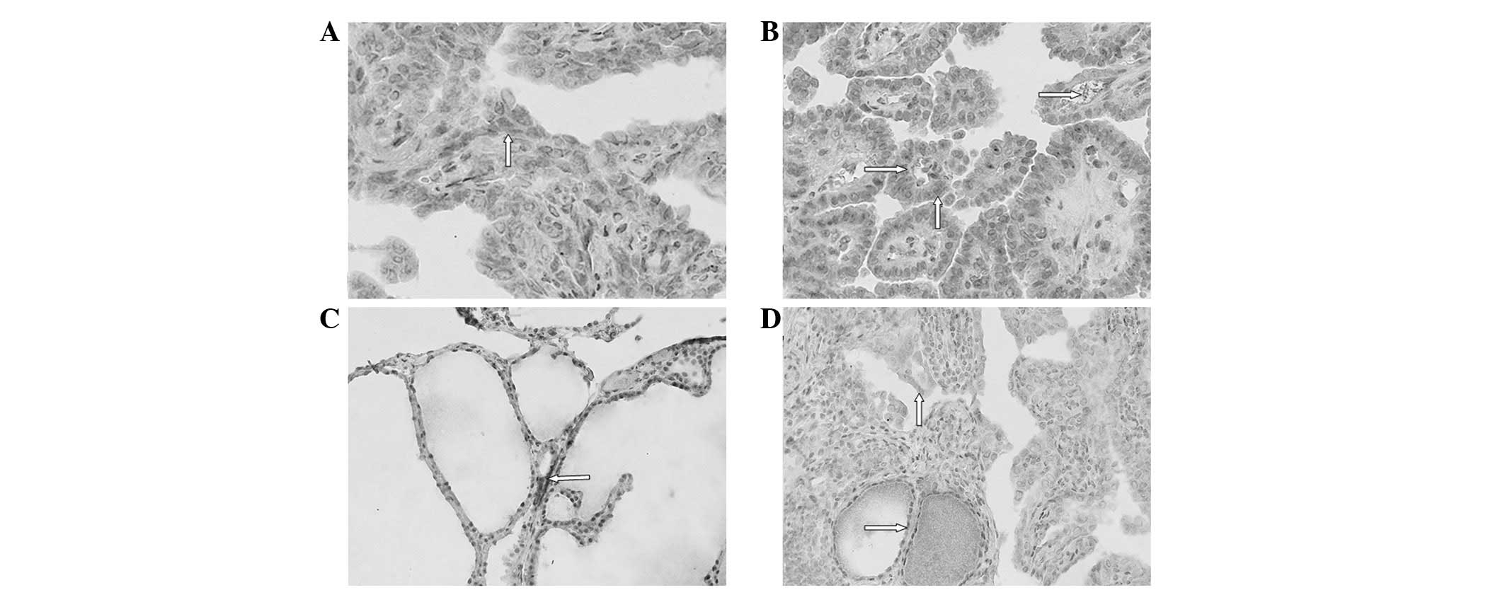
We then assessed NIS expression in these tissues and
found that NIS protein was expressed in the baso-lateral membrane
of the normal epithelium, while nodular goiter cells expressed NIS
in the cytoplasm and occasionally in the basolateral membrane
(Fig. 1). By contrast, NIS protein
was mostly expressed in the cytoplasm and rarely in the basolateral
membrane of PTC cells (Fig. 1).
Based on the percentage of staining and the staining intensity of
the NIS antibody, we summarized NIS expression levels as ‘points’
in each section of PTC, Gnormal and GNG
tissues. The 114 cases of thyroid cancer exhibited NIS staining
with a score ranging between 0 and 12 points. The total NIS protein
scores of the PTC and GNG tissues were 348 and 276
points, respectively (P=0.008). In addition, we associated NIS
expression with clinicopathological data from the PTC patients with
surrounding GNG tissue. We found that age <45 years
(P=0.000), female gender (P=0.003), tumor size ≥1 cm (P= 0.000),
TNM stage I (P= 0.017) and lymph node and distant metastases
(P=0.026 and P=0.008, respectively) were associated with NIS
expression.
IHS scores
Furthermore, as shown in Table II, the total scores of NIS
expression in PTC and Gnormal tissues were 366 and 351
points, respectively, which was not statistically significant (P=
0.675). By contrast, the expression levels of NIS protein in
Gnormal tissue of tumors <1 cm in size were lower
(21/351 points) compared with cancerous tissue (36/366; P= 0.037).
Moreover, NIS expression in cancerous tissue of Gnormal
and GNG did not reach statistical significance (366 and
348 points, respectively, P=0.075), indicating that the
cancer-surrounding tissues may play an important role in mediating
the sensitivity of PTC patients to RAI treatment.
Gnormal tissue from male patients had lower NIS
expression (30/366 points) compared with GNG tissue
(48/348 points; P=0.000). Indeed, the total score of NIS expression
of GNG tissues were lower than that of
Gnormal tissues (276 vs. 351 points; P= 0.002) (Table II) and were not dependent on gender
(P=0.000), tumor size (P<0.05) or lymph node and distant
metastasis (P<0.05).
Discussion
In the present study, we detected expression levels
of NIS protein in cases of PTC with adjacent normal or adjacent
nodular goiter tissues. We observed the differential expression of
NIS protein among these three types of tissues, suggesting that
adjacent thyroid tissue may play an important role in the uptake of
iodide during RAI therapy and may aid the prediction of treatment
outcomes. NIS is an integral plasma membrane glycoprotein that is
mostly expressed in the thyroid gland and mediates the active
transport of I− into the thyroid follicular cells, which
is the crucial first step for thyroid hormone biosynthesis
(8). Differentiated thyroid
carcinoma (DTC) retains this iodide-concentrating ability due to
the functional integrity of the NIS (2). However, certain DTCs are incapable of
concentrating iodide and are therefore resistant to RAI therapy.
The reason for this may be due to tumor or adjacent tissues that do
not express functional NIS protein. Normally, NIS protein is
localized in the cell membrane, otherwise, it loses the ability to
uptake iodide (4,9).
The results of our current study demonstrated that
the NIS protein was mostly localized to the cytoplasm of thyroid
cancer, indicating non- or low-functional NIS protein, whereas
adjacent normal glands or even goiters expressed NIS in the plasma
membrane, suggesting a functional NIS protein. Localization of NIS
protein at the basolateral plasma membrane is important for NIS
function to transport iodide in the thyroid gland and radioiodide
in thyroid cancer therapy. Dohán et al(9) demonstrated that improvements in
131I radioablation therapy for thyroid carcinoma
patients resulted from the introduction of NIS targeting the plasma
membrane. Another study showed that iodine supply also influenced
expression and localization of human NIS (hNIS) protein (10). For example, thyroid nodules from the
iodine-sufficient area had absent or only weak NIS protein
expression, whereas almost all the nodules from the
iodine-deficient area expressed NIS protein. Our current data agree
with these previous data (9,10) and
show that few cases of PTC cells expressed functional NIS protein
in the bilateral membrane, indicating that the tumor cells had lost
the ability to intake iodine. From this point of view, these
tissues play an important role in predicting the sensitivity of RAI
treatment.
Furthermore, we observed differential NIS expression
in different surrounding tissues from PTC patients. This is the
first study to report that expression levels of NIS protein were
lower in surrounding nodular goiter tissues than in surrounding
normal thyroid tissues. A previous study revealed that
immunodetection of NIS protein predicted radioiodine uptake in
thyroid cancer tissues (3),
recurrent lesions (5), metastatic
and recurrent disease (6). Thus, we
suggest that the impaired NIS protein localization or various
expression levels in the surrounding tissues may also affect the
sensitivity of PTC to RAI therapy. Moreover, the current data
further indicated that plasma membrane localization of NIS protein
or induction of NIS cell membrane expression may improve the
sensitivity of PTC to RAI therapy. Thus, detection of NIS protein
expression and the localization of NIS in surrounding thyroid
tissues may be useful to predict RAI therapy outcomes in PTC
patients. In addition, based on NIS protein expression and
localization, a physician administering nuclear medicine may be
able to modify the 131I dose when treating PTC.
In conclusion, NIS protein was differentially
expressed in surrounding normal thyroid tissues and nodular goiter
tissues from PTC patients and may regulate the sensitivity of PTC
patients to RAI therapy. Future studies should evaluate the
association between NIS protein expression, sensitivity of RAI
treatment and serum levels of iodine in PTC patients.
Acknowledgements
The authors would like to thank
Professor Xiaoming Xing of the Department of Pathology, The
Affiliated Hospital of Qingdao Medical College (Qingdao, China),
for her excellent assistance with pathology.
References
|
1
|
American Thyroid Association (ATA)
Guidelines Taskforce on Thyroid Nodules and Differentiated Thyroid
Cancer; Cooper DS, Doherty GM, Haugen BR, Kloos RT, Lee SL, Mandel
SJ, Mazzaferri EL, McIver B, Pacini F, Schlumberger M, Sherman SI,
Steward DL and Tuttle RM: Revised American Thyroid Association
management guidelines for patients with thyroid nodules and
differentiated thyroid cancer. Thyroid. 19:1167–1214. 2009.
View Article : Google Scholar : PubMed/NCBI
|
|
2
|
Arturi F, Russo D, Schlumberger M, du
Villard JA, Caillou B, Vigneri P, Wicker R, Chiefari E, Suarez HG
and Filetti S: Iodide symporter gene expression in human thyroid
tumors. J Clin Endocrinol Metab. 83:2493–2496. 1998.PubMed/NCBI
|
|
3
|
Caillou B, Troalen F, Baudin E, Talbot M,
Filetti S, Schlumberger M and Bidart JM: Na+/I- symporter
distribution in human thyroid tissues: an immunohistochemical
study. J Clin Endocrinol Metab. 83:4102–4106. 1998.
|
|
4
|
Saito T, Endo T, Kawaguchi A, Ikeda M,
Katoh R, Kawaoi A, Muramatsu A and Onaya T: Increased expression of
the sodium/iodide symporter in papillary thyroid carcinomas. J Clin
Invest. 101:1296–1300. 1998. View
Article : Google Scholar : PubMed/NCBI
|
|
5
|
Min JJ, Chung JK, Lee YJ, Jeong JM, Lee
DS, Jang JJ, Lee MC and Cho BY: Relationship between expression of
the sodium/iodide symporter and I-131 uptake in recurrent lesions
of differentiated thyroid carcinoma. Eur J Nucl Med. 28:639–645.
2001. View Article : Google Scholar
|
|
6
|
Castro MR, Bergert ER, Goellner JR, Hay ID
and Morris JC: Immunohistochemical analysis of sodium iodide
symporter expression in metastatic differentiated thyroid cancer:
correlation with radioiodine uptake. J Clin Endocrinol Metab.
86:5627–5632. 2001. View Article : Google Scholar
|
|
7
|
Soslow RA, Dannenberg AJ, Rush D, Woerner
BM, Khan KN, Masferrer J and Koki AT: COX-2 is expressed in human
pulmonary, colonic, and mammary tumors. Cancer. 89:2637–2645. 2000.
View Article : Google Scholar : PubMed/NCBI
|
|
8
|
Carrasco N: Iodide transport in the
thyroid gland. Biochim Biophys Acta. 1154:65–82. 1993. View Article : Google Scholar : PubMed/NCBI
|
|
9
|
Dohán O, Baloch Z, Bánrévi Z, Livolsi V
and Carrasco N: Rapid communication: predominant intracellular
overexpression of the Na(+)/I(-) symporter (NIS) in a large
sampling of thyroid cancer cases. J Clin Endocrinol Metab.
86:2697–2700. 2001.PubMed/NCBI
|
|
10
|
Scipioni A, Ferretti E, Soda G, Tosi E,
Bruno R, Costante G, Meringolo D, Arturi F, Durante C, Amorosi A,
Foschini MP, Nardi F, Russo D and Filetti S: hNIS protein in
thyroid: the iodine supply influences its expression and
localization. Thyroid. 17:613–618. 2007. View Article : Google Scholar : PubMed/NCBI
|