Introduction
Carcinoma of the tongue is characterized by a
malignancy with rapid and invasive growth and is the most common
type of oral and maxillofacial malignant tumor. Metastasis to the
lymph nodes on the neck occurs frequently (1,2). In
addition to surgery, chemotherapy is another important treatment
method (3,4). These malignant tumors are typically
treated by multi-drug combinations (5,6).
Pingyangmycin (PYM) is produced by Streptomyces
pingyangensisn (S.P.) and has antitumor roles in the cell cycle
without specificity. The mechanism of PYM is understood to
introduce breaks into DNA which makes it a broad-spectrum
antibiotic with anticancer potential. By preferentially acting on
the dividing phase of cells with fast metabolisms, it is able to
destroy tumor cells preferentially. Disturbing the metabolism of
tumor cells results in apoptosis, degeneration and necrosis. PYM
has a clear chemotherapeutic effect for carcinoma of the tongue
(7,8), but recurrence and metastasis present
challenges in certain cases. Furthermore, severe and irreversible
side-effects, such as pulmonary fibrosis, have been shown to occur
with large dosages (9).
Hydroxycamptothecin (HCPT), an alkaloid, is
distilled from tree roots, bark or fruit. This type of tree is
unique to China and belongs to the Nyssaceae family. HCPT is a cell
cycle-specific botanical drug. It is able to inhibit DNA
topoisomerase II (TOPO II) ligating the broken ends of DNA, causing
DNA breaks and preventing the synthesis of RNA (10,11).
HCPT acts directly during the S phase and prevents progression in
the G2/M phase (12).
HCPT exhibits a marked effect on tumor cells with low toxicity and
a wide spectra. It has been reported to potently inhibit several
tumor cell lines and transplanted tumor cells and has mainly been
applied to glandular epithelium cancers in the head and neck,
particularly adenoid cystic carcinoma.
It has been shown that PYM has a reasonable potency
in oral squamous carcinomas with low toxicity. There have been no
reports of HCPT being used for the treatment of oral squamous
carcinoma or its combined usage with PYM. In the present study,
HCPT and PYM were used in combination to strengthen the
chemotherapeutic effect and decrease the toxicity of the treatment
of squamous cell carcinoma of the tongue as modeled in cell lines
in a mouse model. Although preliminary, these data suggest there is
promise for future clinical studies of this combined therapy in
humans.
Materials and methods
Materials
HCPT was provided by HSFY Pharmaceutical Industries
(Hubei, China). PYM was provided by Taihe Pharmaceutical Industries
(Tianjin, China). The MTT agent was purchased from Sigma Chemical
Company (St. Louis, MO, USA). DMSO was purchased from Chemical
Reagents Company (Beijing, China). The Telomerase PCR-ELISA kit was
obtained from Boehringer (Mannheim, Germany). A TUNEL apoptosis
detection kit was obtained from Boster Company (Wuhan, China). The
Tca8113 cell line was obtained from the Ninth People’s Hospital,
Shanghai Second Medical University (China). BALB/C nude mice (6
weeks old, 20 g average weight) were supplied by the laboratory
animal research center of the General Hospital of PLA (Beijing,
China). This study was approved by the Ethics Committee of the
General Hospital of PLA.
Cell culture
Tca8113 cells cultured in RPMI-1640 supplemented
with 100 U/ml streptomycin, 100 U/ml penicillin and 10% FCS were
incubated at 37°C in a 5% CO2 humidified atmosphere. The
cells were digested by 0.25% parenzyme, washed with cold PBS and
counted in parallel, then collected and resuspended in PBS, to
prepare for inoculation.
Nude mice
A total of 20 BALB/C nude mice were randomly
assigned into four groups, each with five mice.
Inhibition of cell growth
To test the effect of the drugs on cell growth,
Tca8113 cells were expanded and harvested in the exponential growth
phase. The cells were digested by 0.25% parenzyme to make a
unicellular suspension. The cell density was adjusted and cells
were inoculated in three 96-well plates. Each well was inoculated
with 2×10⁵ cells/0.1ml and incubated at 37°C in 5%
CO2, with saturated humidity. According to the
experimental objectives, four treatment groups were prepared the
next day: i) HCPT group: each well was dosed with 50 μl HCPT
solution of varying drug concentrations (final concentrations, 10,
32, 100 and 1,000 ng/ml), then an additional 50 μl fresh
culture was added; ii) PYM group: each well was dosed with 50
μl PYM solution of varying drug concentrations (final
concentrations tested, 32, 100, 320 and 10,000 ng/ml), then an
additional 50 μl fresh culture media was added; iii)
combined medicine group: a total of 50 μl HCPT and PYM of
various concentrations were added to each well in combination; and
iv) blank group: cells were cultivated without the addition of
drugs under the same culture conditions described previously. After
92 h, 20μl MTT (0.5%) was added for 4 h to each well in each
of the four groups. After removing the supernatant, 150 μl
DMSO was added to resolve the crystals and determine the
OD490nm with a spectrophotometer according to the
manufacturer’s instructions. Using the blank group to establish a
100% survival signal, the relative ODs were calculated for each
group. Microsoft Excel software was used to draw a curve of the
dosage effect. From this curve, the 30% cell growth inhibition
concentration (IC30) was calculated for HCPT, PYM and
the combination of the two by graphical means. This was used to
select the drug concentration with the best IC30s, which
had a limited impact on cell survival. The IC30s of HCPT
and PYM were used for additional experiments.
Cell cloning efficiency
The double-deck agar culture method was used in
24-well plates. Each group had four parallel wells, with 600
Tca8113 cells either treated as described previously or not treated
and all were suspended in 1 ml 0.3% agar in culture media.
Physiological saline (1 ml) was added into the surrounding eight
wells. These cells were cultivated under normal conditions for two
weeks. After two weeks, an inverted microscope was used to count
the number of colonies and from this the cloning efficiency was
calculated according to the following formula: cloning efficiency =
number of clones / number of inoculated cells × 100%.
Cell ultrastructure
Cell specimens were prepared for four days with
either the blank treatment or IC30 concentrations of the
drugs. These were prepared for use with a JEM-2000EX transmission
electron microscope to observe the cell ultrastructure.
Cell cycle of Tca8113
Cells were treated as described for four days,
1×10⁶ cells were collected for each group and 70%
precooled alcohol was used to create single cell suspensions. These
were stored at 4°C prior to FACS analysis. Before analysis, the
cells were pelleted and the supernatant was discarded. Propidium
iodide (PI) was used to dye the cells for 30 min, cell lumps were
filtered out through a 300 screen mesh and the DNA density of the
cells was read via flow cytometry.
Telomerase activity assay
After 92 h of compound treatment, Tca8113 cells were
prepared with telomerase extracting buffer. Based on the
TRAP-PCR-ELISA method improved by Kim and Wu (13) to measure the telomerase activity,
the assay was performed according to the kit instructions. After
processing the lysates, the A450 was measured. The
negative control group was protein extracted from Tca8113 cells
after a 65°C-heat shock. The positive control group was provided by
the kit. The experiment was repeated three times to produce an
average value. If the change in A450 between an
experimental group and the negative control group was >0.2, this
indicated that the sample was telomerase-positive.
Nude mouse Tca8113 cell transplantation
tumors
All mice were injected subcutaneously in the right
sciatic nerve with 0.2 ml 1×10⁷ Tca8113 cells/ml per
nude mouse. The animals underwent chemotherapy 10 days after
implantation. Four mice were randomly assigned to each treatment
group and were then treated with physiological saline, PYM, HCPT or
PYM/HCPT. Mice were injected with 0.2 ml physiological solution for
each of the four treatment conditions, 0.05 mm from the base of the
tumor. The physiological solution was administered twice each week
and the drugs were administered continuously for five weeks. The
dosages were as follows: PYM 4.16 mg/kg, HCPT 1.09 mg/kg, PYM/HCPT
1.00/0.35 mg/kg, respectively. The gross tumor volume was measured
twice each week and was used to calculate the tumor control rate
(a, long diameter; b, short diameter). Gross tumor volume =
0.5×b2; tumor control rate = 1 −
(T1−T2) / (C1−C2) ×100,
where T1 and T2 represent the average tumor
volume at the beginning and end for the treatment group and
C1 and C2 represent the average tumor volume
at the beginning and end for the control group, respectively. The
observed survival time of the athymic mice with tumors in each
group was recorded.
Apoptotic features of nude mouse Tca8113
cell transplantation tumors
When the nude mice died, a specimen was obtained for
each athymic mouse tumor. The biopsies were dried,
paraffin-embedded and serial sections were prepared on glass slides
coated with poly-lysine. From these sections, one was randomly
selected from the sections of each specimen, so that each treatment
group was represented by five tumor samples which were dyed for a
TUNEL assay. The sections were observed under a 20× objective lens.
Five visual fields were randomly selected from each section and in
each the total cell number and number of apoptotic cells was
counted, in order to calculate the proportion of apoptotic cells.
Those cells showing amethyst granules in the cell nucleus were
positive for apoptosis.
Statistical analysis
The differences between each group were compared
using the simplex factor analysis of variance. P<0.01 was
considered to indicate significant differences. Analysis was
performed using the SPSS 11.0 statistical package.
Results
HCPT and/or PYM inhibit Tca8113
cells
To assess the potential anticancer effects of HCPT
and PYM, the two drugs were first tested in a cell culture system
with Tca8113 cells. In vitro HCPT and PYM exhibited
significant growth inhibitory effects on Tca8113 cells. A greater
inhibition occurred with the combination of the two compounds
(Table I). After 96 h of treatment
with PYM, HCPT and PYM+HCPT, the IC30s were determined
to be 416 ng/ml, 109 ng/ml and 100+35 ng/ml, respectively.
 | Table IEffect on survival rate of HCPT and
PYM in Tca8113 cells, as indicated by MTT assays after 96 h of
treatment (mean, %). |
Table I
Effect on survival rate of HCPT and
PYM in Tca8113 cells, as indicated by MTT assays after 96 h of
treatment (mean, %).
| HCPT (ng/ml) | PYM (ng/ml)
|
|---|
| 0 | 32 | 100 | 320 | 10,000 |
|---|
| 0 | 100.00 | 91.32 | 82.76 | 73.54 | 40.07 |
| 10 | 89.97 | 88.75 | 90.07 | 65.54 | 38.32 |
| 32 | 75.43 | 80.65 | 72.78 | 61.02 | 35.47 |
| 100 | 72.75 | 71.98 | 54.32 | 53.31 | 32.43 |
| 1,000 | 42.67 | 38.78 | 33.79 | 30.43 | 19.69 |
HCPT and PYM decrease Tca8113 cell
cloning efficiency
Since the two compounds appear to be toxic to
Tca8113 cells, we hypothesized that this would result in a
decreased cloning efficiency in these cells. To test this, Tca8113
cells were allowed to grow on soft agar for two weeks in the
presence or absence of HCPT and PYM. The cloning efficiency of the
untreated group on Tca8113 cell was 31.57%, while it was 15.92,
11.46 and 4.18% for PYM, HCPT and PYM/HCPT groups, respectively.
Significant differences were observed among the groups
(P<0.01).
PYM and HCPT alter the ultrastructure of
Tca8113 cells
To understand the cytotoxic effects of these
compounds on the Tca8113 cells, the changes to the cells as a
result of treatment were observed. The effects on the
ultrastructure of Tca8113 cells are shown in Fig. 1. Compared with the control group,
cells treated with PYM exhibited the following changes: an elevated
nucleus to cell size ratio, multinucleation, shrinking of the
nucleolus, reduction in the number of intracytoplasmic organelles
and ribosomes, increased number of lysosomes and glycogen granules
and different sizes of cytoplasmic vesicles. The HCPT treatment
group also exhibited significant changes compared with the control,
including increased cell sizes, apocytes and swollen nuclei. There
were no clear changes to the nucleolus sizes or cytoplasmic
vesicles, while the cytoplasmic organelles decreased sharply in
number. The number of ribosomes was also observed to be reduced.
The endoplasmic reticulum expanded and the number of lysosomes and
glycogen granules increased significantly. Cytoplasmic vesicles of
different sizes formed, even forming cellular bridges in one place.
For the PYM/HCPT group, the increase in lysosome volume was more
significant, with more formation of cytoplasmic vesicles. The
majority of other characteristics were similar to the other
treatment two groups.
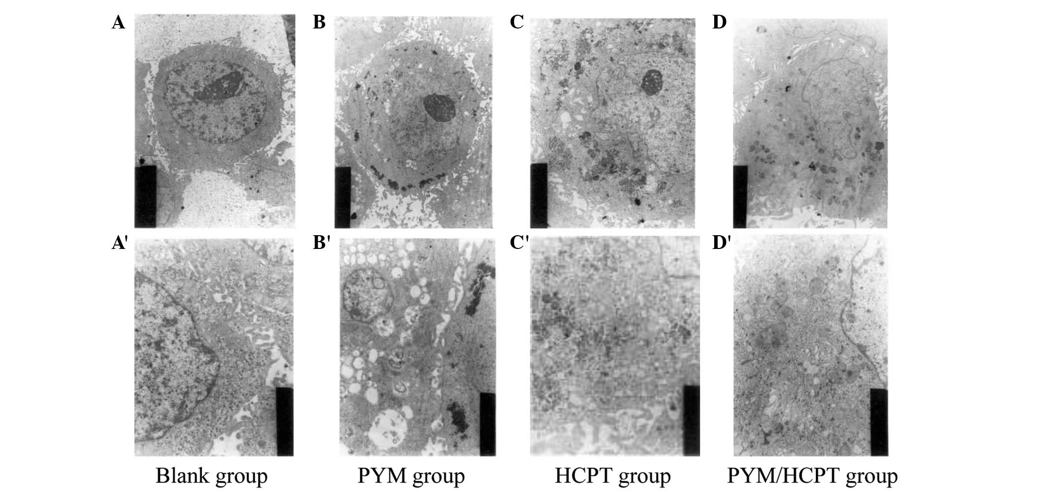 | Figure 1Ultrastructure of Tca8113 cells:
(A,A′) blank group; (B,B′) PYM group; (C,C′) HCPT group; (D,D′)
PYM/HCPT group. (A,B,C,D) Magnification, 3,000×1.45; (A′,B′,C′,D′)
Magnification, 10,000×1.45. HCPT, hydroxycamptothecin; PYM,
pingyangmycin. |
HCPT and PYM alter distribution of the
cell cycle
What is known about the mechanism of the two
compounds suggests that they cause DNA damage, so it was expected
that treatment with these compounds would disrupt the cell cycle.
To test this, unsynchronized Tca8113 cells were treated as
previously with HCPT and/or PYM and then assessed by FACS analysis.
The two compounds significantly altered the distribution in the
cell cycle. Table II shows the
effect on the cell cycle of Tca8133 cells.
| Table IIEffect on growth cycle of Tca8113
cells treated with HCPT and/or PYM. |
Table II
Effect on growth cycle of Tca8113
cells treated with HCPT and/or PYM.
| Proportion of Tca8113
cell cycle phase (%)
|
|---|
| Group | G1
phase | S phase | G2
phase |
|---|
| Blank | 69.2 | 21.8 | 9.1 |
| PYM | 79.9 | 14.0 | 6.2 |
| HCPT | 21.7 | 34.8 | 43.5 |
| PYM/HCPT | 39.0 | 39.4 | 21.5 |
HCPT and PYM inhibit telomerase activity
in Tca8113 cells
The telomerase activity of Tca8113 cells following
treatment with HCPT and/or PYM was tested as described previously
and the results are shown in Table
III. The two drugs inhibit this activity compared with the
positive control.
| Table IIIEffect on telomerase activity of
Tca8113 cells following treatment with HCPT and/or PYM. |
Table III
Effect on telomerase activity of
Tca8113 cells following treatment with HCPT and/or PYM.
| Groups | A450 (mean
±SD) |
|---|
| Positive | 1.73±0.04 |
| Negative | 0.12±0.02 |
| Blank | 1.89±0.03 |
| PYM | 0.77±0.02 |
| HCPT | 0.82±0.02 |
| PYM/HCPT | 0.53±0.03 |
Effect on the growth of Tca8113 cell
transplantation tumors in nude mice
To test the effect of the compounds on a growing
tumor, Tca8113 cells were transplanted into nude mice. The
transplanted tumors were treated with the compounds as described
previously and the tumor volume was measured. The changes in
transplanted tumor volume of the blank and experimental groups is
shown in Table IV. The nude mice of
the blank group all died by day 48, at which point the survival
rate of the PYM and HCPT groups was 60%, while it was 80% for the
PYM/HCPT group.
| Table IVEffect of treatment with PYM, HCPT or
PYM/HCPT on transplanted tumor growth in nude mice. |
Table IV
Effect of treatment with PYM, HCPT or
PYM/HCPT on transplanted tumor growth in nude mice.
| Gross tumor volume
(mm3, mean ± SD)
|
|---|
| Groups | Pre-therapy | Post-treatment | Rate of depression
tumor growth (%) |
|---|
| Blank | 121±34 | 2452±456 | - |
| PYM | 108±23 | 1356±342 | 45±18.2 |
| HCPT | 119±29 | 1456±406 | 41±16.1 |
| PYM/HCPT | 123±22 | 634±145 | 74±14.7 |
The effect on apoptosis in athymic mice
with transplanted Tca8113 cells
After the transplanted mice died, the tumors were
sampled for further analysis. We hypothesized that the compounds
may inhibit tumor growth by increasing apoptosis and a TUNEL assay
was used on the tumor samples to test this. Counts of the apoptotic
cells in each treatment group showed that the proportion of
apoptotic cells in the combined treatment group was significantly
higher than treatment with either compound alone (P<0.01),
suggesting synergy. A significant difference was also observed
between treatment with each compound alone and the control
(P<0.01). No significant differences were observed between the
PYM and HCPT groups (P>0.05; Table
V).
| Table VPercentage of apoptotic cells in all
groups (n=5). |
Table V
Percentage of apoptotic cells in all
groups (n=5).
| Group | Percentage (mean
±SD) |
|---|
| Blank | 5.7±2.1 |
| PYM | 18.3±5.4 |
| HCPT | 21.6±3.2 |
| PYM/HCPT | 57.4±5.3 |
Discussion
Given the challenges of treating tongue cancer, the
present study evaluated the effects of HCPT and PYM on cancer cell
growth. In the present study, HCPT showed a marked inhibitory
effect on cultured Tca8113 tongue cancer cells, particularly when
used in combination with PYM. This combined effect was expected
given that the two compounds are known to have separate targets at
different phases of the cell cycle. The combined application of PYM
and HCPT enhanced the cytotoxic effects on the tumor cells,
simultaneously enabling a decrease in the dosage of the two drugs
which reduced the side-effects. These effects were tested and
confirmed in several assays.
First, the effects of these compounds cultured
Tca8113 tongue cancer cells in vitro were evaluated. HCPT
exhibited an inhibitory effect on the cell line and this inhibition
was improved when combined with PYM. In addition to this cytotoxic
effect, it was observed that these compounds reduced cell cloning
efficiency and increased the proportion of cells in S to
G2 phase and multinucleation. Clear changes in the cell
shape, decreased numbers of cell organelles, increased lysosomal
volumes, cell degeneration and reduced telomerase activity were
also observed, as well as the inhibition of the growth of
transplantation tumors in athymic mice which resulted in an
increased survival rate and increased apotosis.
A clonal colony formation experiment is an efficient
way to detect the proliferation ability of single cell and its
adaptability to the environment in vitro. A cell with higher
cloning efficiency has greater ability to survive alone (14). Studies have shown that colony
formation efficiency reflects the tumor cells’ ability to
transplant in surgery (15). The
present study revealed that the combination of PYM and HCPT reduced
the clonal formation efficiency of Tca8113 cells. The possible
clinical results of treatment with PYM and/or HCPT, are inhibition
of the transformation of tumor cells at the tumor site and
circulating tumor cells.
From the observed ultrastructural changes in the
tumor cells, it appeared that the cellular activity was decreasing
and there were signs of autophagy and programmed cell death.
In the present study, PYM, HCPT and combined
treatment at IC30 were able to reduce the telomerase
activation of cells, with the best results from the combination.
This showed that inhibition decreases the telomerase activity and
replication of cells. The growth inhibiting and cytotoxic effects
of PYM and HCPT on tumor cells may be associated with inhibition of
telomerase activity. The presumed mechanism of inhibition of
telomerase activity may result from the inhibiting DNA synthesis or
destroying the integrity of the DNA (16–19).
The two chemicals combined increased the inhibition of the cells,
promoting the inhibition of telomerase activity. Further study is
necessary to better understand the mechanism of this
inhibition.
Zhu et al reported that telomerase activity
is correlated with the cell cycle phase (20). Typically, telomerase activity
increases gradually after the cells enter into the G1/S
period and reaches its highest at the S phase. During the
G2/M phase there is no telomerase activity. However,
there has been dispute about whether telomerase activity is limited
to specific phases of the cell cycle. In the present study, there
was no significant difference in the S to G2/M
proportion of cells between the PYM group and the control cells.
The proportion of the S to G2/M cells decreased
significantly in the HCPT group compared with the PYM group without
a significant difference being observed in the telomerase activity
between these two groups. This is indicative of a connection
between telomerase activity and the cell cycle. Analyzing the
experiments associated with telomerase activity and cell phase, we
propose that the compounds reduced not only the proportion of S to
G2/M cells but also the telomerase activity. In this
experiment, these specific and non-specific cell cycle inhibitors
were administered simultaneously, which highlighted the synergy of
these compounds. Further research should address the connection
between telomerase activity and the cell cycle.
PYM or HCPT were used separately and in combination
to treat transplanted Tca8113 tumors in athymic mice. All three
treatment conditions inhibited the tumor growth and the combined
treatment showed the greatest increase in survival. From this, we
concluded that PYM and HCPT have inhibitory effects on tongue
cancer and that this effect may be improved by using the drugs in
combination. In addition, using the two compounds at a lower dose
decreased the toxicity. One possible reason for this synergy is
that PYM and HCPT have separate targets, as demonstrated by the
impact on the cell cycle phase. PYM affected tumor cells in all
phases, while HCPT sensitized cells in the S phase. The combined
effect of the compounds allowed the overall treatment to overcome
the shortcomings of each. Due to the low clinical efficiency of
chemotherapy drugs, a large number of normal cells are usually
killed while attempting to target tumor cells. This toxicity leads
to serious side-effects as the dose of compounds is increased to
target the tumor. The various mechanisms of chemotherapies result
in harmful side-effects which harms different target organs. The
combined application of PYM and HCPT decreased the dosages of each,
and achieved or even exceeded the expected effects based on the
separate application of each while avoiding the general
toxicity.
The advantage of the TUNEL method is that it is able
to detect the early period of apoptosis at a cellular level by
marking the apoptotic cells (21,22).
The present study used a TUNEL kit to demonstrate that the rate of
apoptosis in the combined treatment group was significantly higher
than that of either of the separate treatment groups (P<0.01).
In addition, there was a marked difference between the two separate
treatment groups and control group (P<0.01). There was no
significant difference between the two separate treatment groups
(P>0.05). This suggests that the two medicines accelerate cell
apoptosis by separate mechanisms, resulting in a more potent effect
when used in combination. Inducing the apoptosis of tumor cells is
the standard mechanism of chemotherapy. The combined treatment with
PYM and HCPT enhanced the apoptosis of tumor cells, resulting in
improved inhibition of tumor growth. The combination of PYM and
HCPT triggered apoptosis signals at different cell cycle phases and
targeted the signals by different means to kill tumor cells.
Specific mechanisms of action are not yet known for either
compound. Further research is required to understand how these
compounds act to inhibit tongue cancer.
In conclusion, combined chemotherapy with PYM and
HCPT exhibited a marked effect on oral squamous carcinoma. Further
research may reveal more effective dosing schemes for the clinical
application of this combination chemotherapy.
References
|
1
|
Miyahara M, Tanuma J, Sugihara K and Semba
I: Tumor lymphangiogenesis correlates with lymph node metastasis
and clinicopathologic parameters in oral squamous cell carcinoma.
Cancer. 110:1287–1294. 2007. View Article : Google Scholar : PubMed/NCBI
|
|
2
|
Huang X, Wei Y, Li L, et al: Serum
proteomics study of the squamous cell carcinoma antigen 1 in tongue
cancer. Oral Oncology. 42:26–30. 2006. View Article : Google Scholar : PubMed/NCBI
|
|
3
|
Bagan J, Sarrion G and Jimenez Y: Oral
cancer: clinical features. Oral Oncol. 46:414–417. 2010. View Article : Google Scholar
|
|
4
|
Pederson AW, Haraf DJ, Witt ME, et al:
Chemoradiotherapy for locoregionally advanced squamous cell
carcinoma of the base of tongue. Head Neck. 32:1519–1527. 2010.
View Article : Google Scholar : PubMed/NCBI
|
|
5
|
Kurita H, Yamamoto E, Nozaki S, et al:
Multicenter phase I trial of induction chemotherapy with docetaxel
and nedaplatin for oral squamous cell carcinoma. Oral Oncol.
40:1000–1006. 2004. View Article : Google Scholar : PubMed/NCBI
|
|
6
|
Cranmer LD, Engelhardt C and Morgan SS:
Treatment of unresectable and metastatic cutaneous squamous cell
carcinoma. Oncologist. 15:1320–1328. 2010. View Article : Google Scholar : PubMed/NCBI
|
|
7
|
Sun ML, Wang CM and Wen YM: Anticancer
effects of Pingyang mycin-activated carbon nanoparticles against
human oral squamous carcinoma Tca8113 and BcaCD885 cell lines in
vitro. Hua Xi Kou Qiang Yi Xue Za Zhi. 28:257–260. 2010.(In
Chinese).
|
|
8
|
Li WZ, Wang XY, Li ZG, et al: Celecoxib
enhances the lethal effects of bleomycin in human tongue squamous
carcinoma cell line Tca8113. Zhonghua Kou Qiang Yi Xue Za Zhi.
44:140–143. 2009.(In Chinese).
|
|
9
|
Xu Y, Luchsinger L, Lucey EC and Smith BD:
The effect of class II transactivator mutations on
bleomycin-induced lung inflammation and fibrosis. Am J Respir Cell
Mol Biol. 44:898–905. 2011. View Article : Google Scholar : PubMed/NCBI
|
|
10
|
Gupta M, Fujimori A and Pommier Y:
Eukaryotic DNA topoisomerases I. Biochim Biophys Acta. 1262:1–14.
1995. View Article : Google Scholar
|
|
11
|
Saleem A, Ibrahim N, Patel M, et al:
Mechanisms of resistance in a human cell line exposed to sequential
topoisomerase poisoning. Cancer Res. 57:5100–5106. 1997.PubMed/NCBI
|
|
12
|
Zhang XW, Jiang JF and Xu B:
Differentiation-inducing action of 10-hydroxycamptothecin on human
hepatoma HepG2 cells. Acta Pharmacol Sin. 21:364–368.
2000.PubMed/NCBI
|
|
13
|
Kim NW and Wu F: Advances in
quantification and characterization of telomerase activity by the
telomeric repeat amplification protocol (TRAP). Nucleic Acid Res.
25:2595–2697. 1997. View Article : Google Scholar : PubMed/NCBI
|
|
14
|
Zhang P, Zhang Y, Mao L, et al: Side
population in oral squamous cell carcinoma possesses tumor stem
cell phenotypes. China Journal of Oral and Maxillofacial Surgery.
8:149–153. 2010.PubMed/NCBI
|
|
15
|
Wang LJ, Yan M, Ye DX, et al: Preliminary
study on the sphere-forming stem-like cell populations of oral
squamous cell carcinoma. Zhongguo Kouqiang Hemian Waike Zazhi.
8:149–153. 2010.
|
|
16
|
Nishikawa T, Nakajima T, Katagishi T, et
al: Oxidative stress may enhance the malignant potential of human
hepatocellular carcinoma by telomerase activation. Liver Int.
29:846–856. 2009. View Article : Google Scholar : PubMed/NCBI
|
|
17
|
Iliopoulos D, Satra M, Drakaki A, et al:
Epigenetic regulation of hTERT promoter in hepatocellular
carcinomas. Int J Oncol. 34:391–399. 2009.PubMed/NCBI
|
|
18
|
Saini N, Srinivasan R, Chawla Y, et al:
Telomerase activity, telomere length and human telomerase reverse
transcriptase expression in hepatocellular carcinoma is independent
of hepatitis virus status. Liver Int. 29:1162–1170. 2009.
View Article : Google Scholar
|
|
19
|
Jiang W, Wang XW, Unger T, et al:
Cooperation of tumor-derived HBx mutants and p53-249(ser) mutant in
regulating cell proliferation, anchorage-independent growth and
aneuploidy in atelomerase-immortalized normal human
hepatocyte-derived cell line. Int J Cancer. 127:1011–1020. 2010.
View Article : Google Scholar
|
|
20
|
Zhu X, Kumar R, Mandal M, et al: Cell
cycle-dependent modulation of telomerase activity in tumor cells.
Proc Natl Acad Sci USA. 93:6091–6095. 1996. View Article : Google Scholar : PubMed/NCBI
|
|
21
|
Niculescu T, Weerth S, Soane L, et al:
Effects of membrane attack complex of complement on apoptosis in
experimental autoimmune encephalomyelitis. Ann NY Acad Sci.
1010:530–533. 2003. View Article : Google Scholar : PubMed/NCBI
|
|
22
|
Pulera MR, Adams LM, Liu H, et al:
Apoptosis in a neonatal rat model of cerebral hypoxia-ischemia.
Stroke. 29:2622–2630. 1998. View Article : Google Scholar : PubMed/NCBI
|


