Introduction
Gastric cancer is the fourth most common type of
cancer and the second most common cause of cancer-related mortality
worldwide (1,2). At present, the management of gastric
cancer includes surgery, radiotherapy, conventional chemotherapy,
molecular targeted therapy and biological therapy. Despite
therapeutic advances, the 5-year survival rate of gastric cancer is
generally <20% (3). Thus, it is
necessary to identify more effective therapeutic agents for gastric
cancer to improve the survival rate.
A number of studies have drawn attention to natural
products extracted from Chinese medicinal herbs as anti-cancer
agents in gastric cancer therapy (4–6).
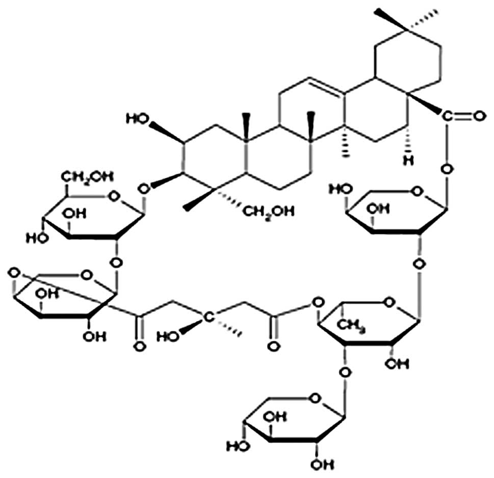
Tubeimoside-1 (TBMS1; Fig. 1) is a
natural compound isolated from the Chinese medicinal herb
Bolbostemma paniculatum (Maxim.) Franquet
(Cucurbitaceae). Previous studies have shown that TBMS1
exhibits a variety of biological activities, including potent
anticancer effects in several cancer cell lines, and it has been
reported that TBMS1 exerts anticancer effects through the
inhibition of cancer cell proliferation and the induction of G2/M
phase arrest and apoptosis (7–11).
Previously, we reported that TBMS1 inhibits proliferation and
induces apoptosis by increasing the Bax to Bcl-2 ratio and
decreasing COX-2 expression levels in lung cancer A549 cells
(12). However, the effects of
TBMS1 on human gastric cancer cells remain unclear.
In the present study, the effects of TBMS1 on the
growth of BGC823 gastric cancer cells and the cellular mechanism
involved in TBMS1-induced apoptosis were investigated. The findings
suggest that TBMS1 may be developed as an anti-cancer agent for
gastric cancer therapy.
Materials and methods
Reagents and chemicals
TBMS1 was purchased from the National Institute for
the Control of Pharmaceutical and Biological Products (Beijing,
China) and a 1 mmol/l stock solution of TBMS1 was dissolved in PBS
and stored at −20°C. Fetal bovine serum (FBS) was purchased from
Solarbio Science and Technology Co., Ltd. (Beijing, China).
3-(4,5-Dimethylthiazol-2-yl)-2,5-diphenyltetrazolium bromide (MTT),
Hoechst 33342 and dimethyl sulfoxide (DMSO) were purchased from
Sigma-Aldrich (St. Louis, MO, USA). The Annexin V-FITC and PI
double staining kit were purchased from KeyGene (Nanjing, China).
Antibodies were purchased from Santa Cruz Biotechnology Inc. (Santa
Cruz, CA, USA). All other reagents were procured locally.
Cell culture
The human gastric cancer cell line BGC823 was
obtained from the China Center for Type Culture Collection (Wuhan,
China) and maintained in RPMI-1640 supplemented with 10% FBS, 100
U/ml penicillin and 100 μg/ml streptomycin at 37°C in a
humidified atmosphere of 5% CO2.
MTT assay
The effect of TBMS1 on the proliferation of BGC823
cells was measured by MTT assay. Briefly, BGC823 cells were plated
at a density of 1×10⁴ cells per well in 96-well plates
overnight and then treated with various concentrations of TBMS1 (0,
5, 10, 15 and 20 μmol/l) for 24 and 48 h. MTT solution (20
μl, 2 mg/ml in PBS) was added to each well and the cells
were cultured for a further 4 h at 37°C. The medium was then
removed and 150 μl DMSO was added to solubilize the MTT
formazan crystals. Finally, the plates were agitated and the
optical density was determined at 570 nm (OD570) using an ELISA
plate reader (Model 550, Bio-Rad, Hercules, CA, USA). At least
three independent experiments were performed.
Fluorescence microscopy
BGC823 cells (1×10⁶) were seeded in
6-well plates overnight and then treated with different
concentrations of TBMS1 (0 and 10 μmol/l) for 24 h. The
cells were washed twice with cold PBS, fixed with cold methanol and
acetic acid (3/1, v/v) for 30 min and then stained with Hoechst
33342 (1 mg/ml) for 30 min in the dark. The stained cells were
observed with a fluorescence microscope (×400 magnification, Nikon,
Tokyo, Japan).
Flow cytometric analysis
The apoptotic rates of the BGC823 cells were
determined by flow cytometric analysis using an Annexin V-FITC
Apoptosis kit. Briefly, BGC823 cells (1×10⁶) were seeded
in 6-well plates overnight and then treated with various
concentrations of TBMS1 (0, 5, 10 and 15 μmol/l) for 24 h.
Cells (1×10⁶) were then harvested by centrifuging (1,000
rpm) and washed twice with cold PBS. The staining was performed
according to the instructions of the manufacturer (KeyGene) and
then the cells were analyzed using a FACScan flow cytometer
(Becton-Dickinson, San Jose, CA, USA). At least three independent
experiments were performed.
Western blot analysis
The expression of apoptosis-related proteins was
evaluated by western blot analysis. Briefly, BGC823 cells
(1×10⁶) were seeded in 6-well plates overnight, then
treated with various concentrations of TBMS1 (0, 5, 10 and 15
μmol/l). After treatment for 24 h, the total proteins were
solubilized and extracted with lysis buffer (20 mM HEPES, pH 7.9,
20% glycerol, 200 mM KCl, 0.5 mM EDTA, 0.5% NP-40, 0.5 mM DTT and
1% protease inhibitor cocktail). The protein concentration was
determined using a bicinchoninic acid (BCA) protein assay. All
samples were separated by SDS-PAGE to determine the expression
levels of Bax, Bcl-2 and β-actin proteins. Blots were developed
using an ECL kit.
Statistical analysis
Statistical analyses were performed using the SPSS
13.0 package (SPSS Inc., Chicago, IL, USA). All experiments were
conducted at least three times. All data are expressed as the mean
± SD. The statistical correlations of the data were tested for
significance using ANOVA and the Student’s t-test. P<0.05 and
P<0.01 were considered to indicate statistically significant
differences.
Results
TBMS1 inhibited BGC823 cell
proliferation
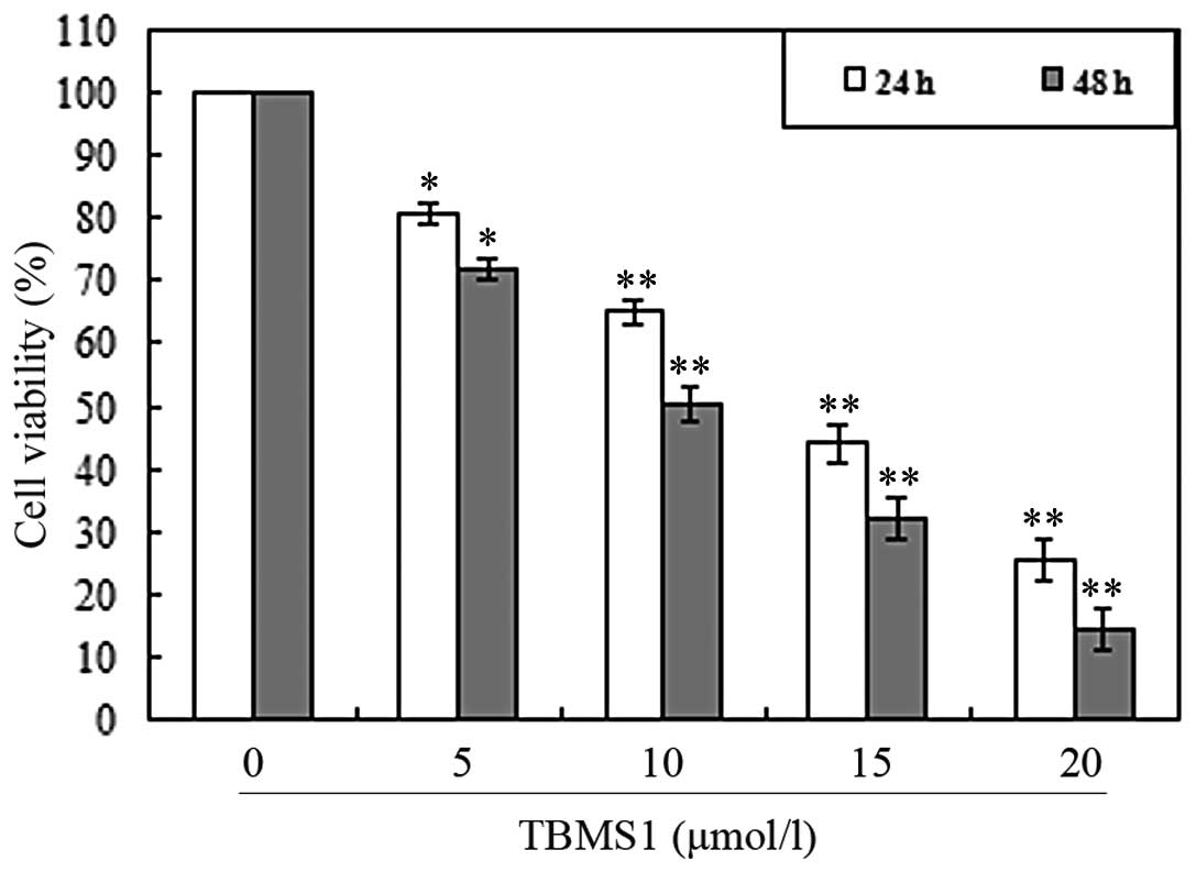
To investigate the growth inhibiting effects of
TBMS1, the BGC823 cells were treated with various concentrations of
TBMS1 for 24 and 48 h and the rate of inhibition was determined by
MTT assays. As shown in Fig. 2, it
was observed that the growth of BGC823 cells was inhibited in a
concentration- and time-dependent manner.
TBMS1 induced BGC823 cell apoptosis
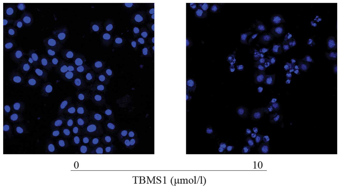
To investigate the apoptosis-inducing effect of
TBMS1, the BGC823 cells were treated with various concentrations of
TBMS1. After treatment with TBMS1 (0 and 10 μmol/l) for 24
h, the cells were examined by fluorescent microscopy using Hoechst
33324 staining. As shown in Fig. 3,
chromatin condensation, nuclear fragmentation and apoptotic bodies
were observed clearly in the treated cells. The results revealed
that when exposed to TBMS1, BGC823 cells underwent the typical
morphological changes of apoptosis.
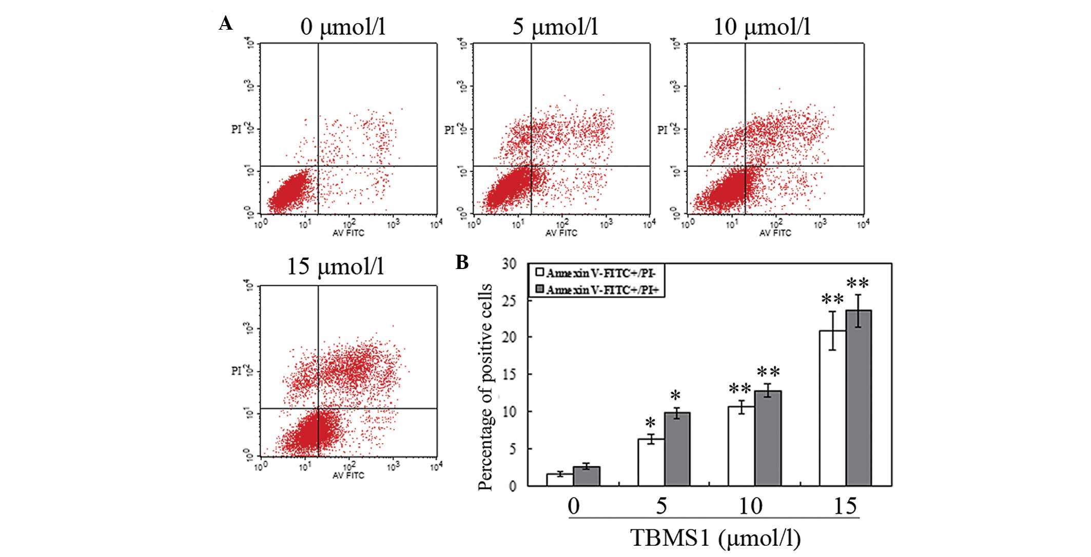
The ratio of apoptotic cells induced by TBMS1 was
measured by flow cytometry. BGC823 cells were treated with various
concentrations of TBMS1 (0, 5, 10 and 15 μmol/l) for 24 h
and analyzed by flow cytometry using Annexin V and PI staining. As
shown in Fig. 4, the ratio of early
and late apoptotic cells was significantly increased in the
TBMS1-treated cells compared with the control group. The results
show that when treated with TBMS1 for 24 h, the ratio of apoptotic
cells significantly increased in a concentration-dependent
manner.
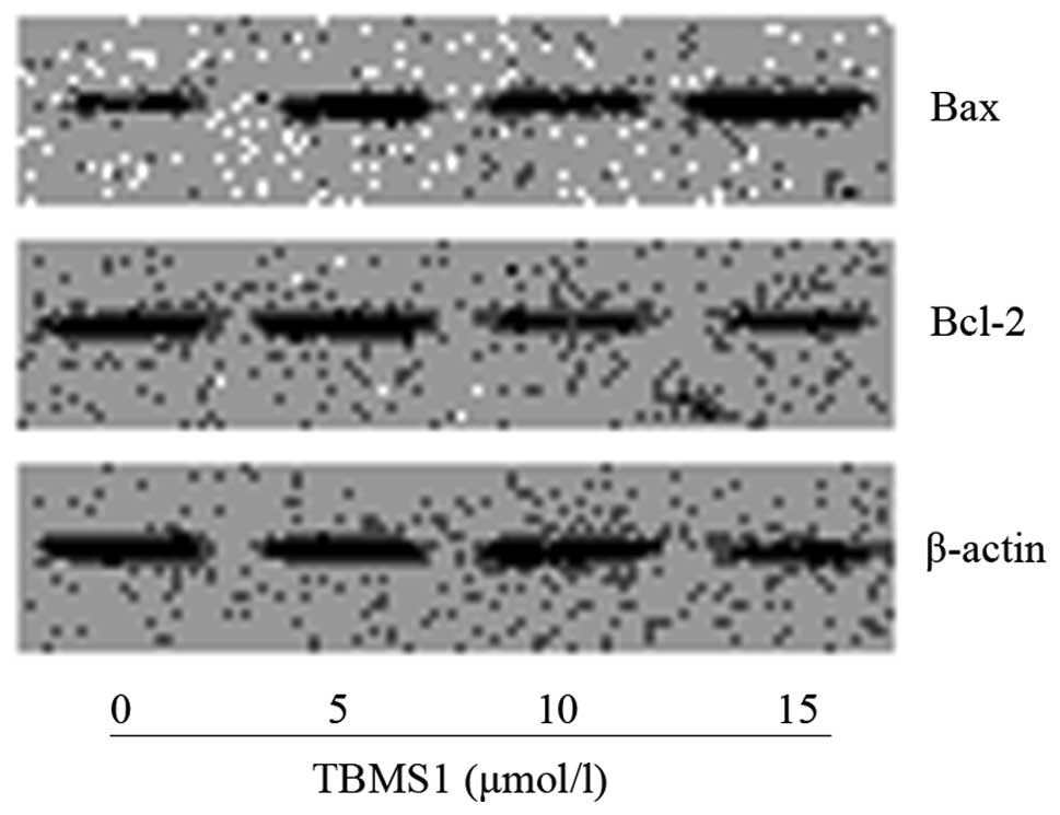
Effect of TBMS1 on expression levels of
the Bcl-2 gene family
The expression of apoptosis-related proteins was
evaluated by western blot analysis. As shown in Fig. 5, TBMS1 treatment led to an increase
in Bax levels and a reduction in Bcl-2 levels compared with those
in the control cells. The ratio of Bax to Bcl-2 increased in a
concentration-dependent manner.
Discussion
An increasing amount of attention has been focused
on the use of natural products isolated from Chinese medicinal
herbs for gastric cancer therapy (4–6). TBMS1
is a natural compound extracted from the Chinese medicinal herb
Bolbostemma paniculatum (Maxim.) Franquet
(Cucurbitaceae), which has been used for a long time in the
treatment of numerous diseases and possesses well-documented
antiviral, anti-inflammatory and immunosuppressive activities
(13–15). The anticancer effects of TBMS1 have
been documented in numerous types of human cancer (7–12).
These studies have revealed that TBMS1 inhibits cell growth and
induces G2/M phase arrest and the apoptosis of cancer cells.
However, the effects of TBMS1 on human gastric cancer cells remain
unclear.
In the present study, TBMS1 inhibited BGC823 gastric
cancer cell proliferation in a concentration- and time-dependent
manner. Chromosome condensation, nuclear fragmentation and
apoptotic bodies were observed by fluorescent microscopy. Flow
cytometric analysis revealed that TBMS1 induced BGC823 cell
apoptosis in a concentration-dependent manner. Moreover, the
results from the western blot analysis showed that the molecular
basis of the TBMS1-induced apoptosis in the BGC823 cells was via
the downregulation of Bcl-2 protein levels and the upregulation of
Bax protein expression.
Apoptosis, or programmed cell death, is critical in
developmental processes, the maintenance of homeostasis and
elimination of damaged cells (16,17).
Resistance to apoptosis is a significant hallmark of cancer cells
and the induction of apoptosis is one of the major goals of
anticancer therapy (18). Several
genes are involved in the regulation of apoptosis, such as the
Bcl-2 gene family. The Bcl-2 gene family, which is significantly
involved in the regulation of cell apoptosis, includes
anti-apoptotic genes such as Bcl-2 and Bcl-xl and pro-apoptotic
genes including, Bax, Bak, Bik, Bid and Bad (19,20).
The ratio of Bax to Bcl-2 is a decisive factor for the induction of
apoptosis and the balance between the expression levels of the
proteins Bax and Bcl-2 is critical for cell survival or death.
Certain anticancer drugs extracted from Chinese medicinal herbs
induce cancer cell apoptosis through the upregulation of the ratio
of Bax to Bcl-2 (21–24). Similarly, the present results showed
that the expression of Bax was upregulated by TBMS1, whereas that
of Bcl-2 was downregulated, leading to an upregulation of the ratio
between Bax and Bcl-2. This indicates the involvement of the Bcl-2
gene family in the regulation of TBMS1-induced cell apoptosis.
In conclusion, the present study demonstrated that
TBMS1 inhibited proliferation and promoted apoptosis in BGC823
gastric cancer cells. This apoptotic response is associated with
the regulation of the expression of the Bcl-2 gene family. The
findings indicate that TBMS1 may be developed as a possible
therapeutic agent for the management of gastric cancer.
References
|
1
|
Parkin DM, Bray F, Ferlay J and Pisani P:
Global cancer statistics, 2002. CA Cancer J Clin. 55:74–108. 2005.
View Article : Google Scholar
|
|
2
|
Compare D, Rocco A and Nardone G: Risk
factors in gastric cancer. Eur Rev Med Pharmacol Sci. 14:302–308.
2010.
|
|
3
|
Jemal A, Siegel R, Ward E, Murray T, Xu J
and Thun MJ: Cancer statistics, 2007. CA Cancer J Clin. 57:43–66.
2007. View Article : Google Scholar
|
|
4
|
Li N, Fan LL, Sun GP, Wan XA, Wang ZG, Wu
Q and Wang H: Paeonol inhibits tumor growth in gastric cancer in
vitro and in vivo. World J Gastroenterol. 16:4483–4490. 2010.
View Article : Google Scholar : PubMed/NCBI
|
|
5
|
Chen J, Shi DY, Liu SL and Zhong L:
Tanshinone IIA induces growth inhibition and apoptosis in gastric
cancer in vitro and in vivo. Oncol Rep. 27:523–528. 2012.PubMed/NCBI
|
|
6
|
Onoda C, Kuribayashi K, Nirasawa S, Tsuji
N, Tanaka M, Kobayashi D and Watanabe N:
(−)-Epigallocatechin-3-gallate induces apoptosis in gastric cancer
cell lines by down-regulating survivin expression. Int J Oncol.
38:1403–1408. 2011.
|
|
7
|
Yin Y, Chen W, Tang C, Ding H, Jang J,
Weng M, Cai Y and Zou G: NF-κB, JNK and p53 pathways are involved
in tubeimoside-1-induced apoptosis in HepG2 cells with oxidative
stress and G2/M cell cycle arrest. Food Chem Toxicol.
49:3046–3054. 2011.
|
|
8
|
Wang Y, Deng L, Zhong H, Jiang X and Chen
J: Natural plant extract tubeimoside I promotes apoptosis-mediated
cell death in cultured human hepatoma (HepG2) cells. Biol Pharm
Bull. 34:831–838. 2011. View Article : Google Scholar : PubMed/NCBI
|
|
9
|
Huang P, Yu C, Liu XQ, Ding YB, Wang YX
and He JL: Cytotoxicity of tubeimoside I in human choriocarcinoma
JEG-3 cells by induction of cytochrome c release and apoptosis via
the mitochondrial-related signaling pathway. Int J Mol Med.
28:579–587. 2011.PubMed/NCBI
|
|
10
|
Xu Y, Chiu JF, He QY and Chen F:
Tubeimoside-1 exerts cytotoxicity in HeLa cells through
mitochondrial dysfunction and endoplasmic reticulum stress
pathways. J Proteome Res. 8:1585–1593. 2009. View Article : Google Scholar : PubMed/NCBI
|
|
11
|
Chen WJ, Yu C, Yang Z, He JL, Yin J, Liu
HZ, Liu HT and Wang YX: Tubeimoside-1 induces G2/M phase arrest and
apoptosis in SKOV-3 cells through increase of intracellular
Ca2+ and caspase-dependent signaling pathways. Int J
Oncol. 40:535–543. 2012.PubMed/NCBI
|
|
12
|
Zhang Y, Xu X and He P: Tubeimoside-1
inhibits proliferation and induces apoptosis by increasing the Bax
to Bcl-2 ratio and decreasing COX-2 expression in lung cancer A549
cells. Mol Med Rep. 4:25–29. 2011.PubMed/NCBI
|
|
13
|
Yu TX, Ma RD and Yu LJ: Structure-activity
relationship of tubeimosides in anti-inflammatory, antitumor, and
antitumor-promoting effects. Acta Pharmacol Sin. 22:463–468.
2001.PubMed/NCBI
|
|
14
|
Zhang XH, Sun NX, Guo RX, Xing JL and Liu
XN: Efficacy research of tubeimoside against the experimental
herpes simplex keratitis. Rec Adv Ophthalmol. 22:373–376. 2002.(In
Chinese).
|
|
15
|
Li XH, Wang P, Fu ZC, et al: Effects of
Bolbastemmosaponins A on immunologic functions of experimental
animals. China Pharm. 9:131998.(In Chinese).
|
|
16
|
Yagi Y, Fushida S, Harada S, Kinoshita J,
Makino I, Oyama K, Tajima H, Fujita H, Takamura H, Ninomiya I,
Fujimura T, Ohta T, Yashiro M and Hirakawa K: Effects of valproic
acid on the cell cycle and apoptosis through acetylation of histone
and tubulin in a scirrhous gastric cancer cell line. J Exp Clin
Cancer Res. 29:1492010. View Article : Google Scholar : PubMed/NCBI
|
|
17
|
Xiao R, Ferry AL and Dupont-Versteegden
EE: Cell death-resistance of differentiated myotubes is associated
with enhanced anti-apoptotic mechanisms compared to myoblasts.
Apoptosis. 16:221–234. 2011. View Article : Google Scholar : PubMed/NCBI
|
|
18
|
Hanahan D and Weinberg RA: Hallmarks of
cancer: the next generation. Cell. 144:646–674. 2011. View Article : Google Scholar : PubMed/NCBI
|
|
19
|
Robertson JD and Orrenius S: Molecular
mechanisms of apoptosis induced by cytotoxic chemicals. Crit Rev
Toxicol. 30:609–627. 2000. View Article : Google Scholar : PubMed/NCBI
|
|
20
|
Tamm I, Schriever F and Dörken B:
Apoptosis: implications of basic research for clinical oncology.
Lancet Oncol. 2:33–42. 2001. View Article : Google Scholar : PubMed/NCBI
|
|
21
|
Yu J, Zhou X, He X, Dai M and Zhang Q:
Curcumin induces apoptosis involving bax/bcl-2 in human hepatoma
SMMC-7721 cells. Asian Pac J Cancer Prev. 12:1925–1929.
2011.PubMed/NCBI
|
|
22
|
Xu YH, Zhao LJ and Li Y: Alisol B acetate
induces apoptosis of SGC7901 cells via mitochondrial and
phosphatidylinositol 3-kinases/Akt signaling pathways. World J
Gastroenterol. 15:2870–2877. 2009. View Article : Google Scholar : PubMed/NCBI
|
|
23
|
Jiang T, Zhou L, Zhang W, Qu D, Xu X, Yang
Y and Li S: Effects of sinomenine on proliferation and apoptosis in
human lung cancer cell line NCI-H460 in vitro. Mol Med Rep.
3:51–56. 2010.PubMed/NCBI
|
|
24
|
Xu X, Zhang Y, Qu D, Jiang T and Li S:
Osthole induces G2/M arrest and apoptosis in lung cancer A549 cells
by modulating PI3K/Akt pathway. J Exp Clin Cancer Res. 30:332011.
View Article : Google Scholar : PubMed/NCBI
|