Introduction
Endometrial cancer is the most frequent
gynecological malignancy and the fourth most common type of cancer
in females in the United States (1). When endometrial cancer is
localized to the uterus, it is often detected at an early stage;
therefore, the overall survival rate is >80% (1). However, the
prognosis of advanced endometrial cancer remains poor (2). Surgery, radiotherapy and multidrug
chemotherapy have all been used to treat advanced endometrial
cancer with little success. Therefore, limited improvements in
overall treatment outcomes have been observed in endometrial cancer
over the past 30 years (1). Therefore, novel strategies, such as
anti-angiogenic therapy and targeted molecular therapy, may be
useful in improving the prognosis of endometrial cancer.
Angiogenesis is a hallmark of malignant tumor
development, and is important in the growth of primary, metastatic
and disseminated lesions of endometrial cancer (3). Therefore, anti-angiogenic therapy may
be effective in treating endometrial cancer. At present,
anti-angiogenic therapy has been approved for several types of
cancer, including colon, lung, breast and kidney cancer.
Furthermore, several drugs that target vascular endothelial growth
factor (VEGF) signals are already in clinical use (4). While the effectiveness of these drugs
is encouraging, resistance to anti-angiogenic therapy has been
demonstrated in several reviews (5–8). To
overcome these problems, novel molecular targets for
anti-angiogenic therapy need to be discovered.
The vasohibin family includes vasohibin-1 (VASH1)
and vasohibin-2 (VASH2). In endothelial cells (ECs), VASH1 is
selectively induced by angiogenesis stimulators, such as VEGF and
basic fibroblast growth factor (bFGF). VASH1 functions as an
intrinsic negative feedback regulator at the termination zone of
angiogenesis (9). VASH2 is a
homolog of VASH1, and the VASH2 gene is highly expressed in bone
marrow-derived mononuclear cells and weakly expressed in ECs. In
contrast to VASH1, VASH2 has been found to promote angiogenesis at
the sprouting front in a mouse model of hypoxia-induced
subcutaneous angiogenesis (10). To
date, there are a limited number of studies available in the
literature concerning the correlation between VASH2 and tumor
angiogenesis (11,12). Recently, we demonstrated that VASH2
contributes to the development of tumor growth and peritoneal
dissemination by promoting angiogenesis in human ovarian serous
adenocarcinoma (12). However, the
specific role of VASH2 in endometrial cancer remains unknown.
In this study, we used a short hairpin RNA (shRNA)
vector to silence VASH2 expression in a VASH2-expressing
endometrial cancer cell line, to further elucidate the relationship
between VASH2 expression and endometrial cancer progression.
Moreover, we investigated the function of VASH2 in endometrial
cancer angiogenesis to develop a VASH2-targeted anti-angiogenic
molecular therapy for endometrial cancer.
Materials and methods
Cell culture
Human endometrial cancer cell lines, HEC1A and
HEC50B, were obtained from the Japanese Collection of Research
Bioresources (JCRB; Osaka, Japan), and were maintained as described
previously (13,14). The Ishikawa cell line (clone 3H12)
was a gift from Dr M. Nishida (Department of Obstetrics and
Gynecology, National Hospital Organization, Kasumigaura Medical
Center, Ibaraki, Japan), and was maintained as described previously
(15). Cells were cultured in
RPMI-1640 medium (Wako Pure Chemical Industries, Ltd., Osaka,
Japan) supplemented with 10% heat-inactivated fetal bovine serum
(FBS; BioWest S.A.S, Nuaillé, France). Human umbilical vein
endothelial cells (HUVECs) were obtained from Kurabo Industries,
Ltd. (Osaka, Japan) and were cultured in type I collagen-coated
dishes (Iwaki, Chiba, Japan) in endothelial basal medium (EBM)-2
(Lonza, Walkersville, MD, USA) supplemented with
EGM-2-MV-SingleQuots (Lonza) containing VEGF, bFGF, insulin-like
growth factor-1, epidermal growth factor and 5% FBS. All cells were
cultured at 37°C in a humidified atmosphere with 5%
CO2.
shRNA stable cell line and control cell
line
The DNA oligo-nucleotide sequences encoding shRNA
targeting VASH2 included forward:
5′-CACGGGGCAGATTATAAGAATTACGTGTGCTGTCCGTAATTCTTGTAGTCTGCTCCTTTTT-3′
and reverse:
5′-CCCCGTCTAATATTCTTAATGCACACGACAGGCATAAGAACATCAGACGAGGAAAAATACG-3′.
The oligonucleotides were synthesized, annealed and inserted into
the BspMI site of the piGENE PURhU6 vector (16), which contained a human U6 promoter
and a puromycin resistance gene. The shRNA expression plasmid
(piGENE PURhU6/shVASH2) and control plasmid (piGENE PURhU6) were
transfected into HEC50B cells by the use of Lipofectamine LTX and
Plus Reagent (Invitrogen Life Technologies, Carlsbad, CA, USA)
according to the manufacturer’s instructions. Following
transfection, the cells were selected in puromycin-containing
medium (Calbiochem, La Jolla, CA, USA). Subsequently,
VASH2-knockdown clones and control clones were established.
Reverse transcription-polymerase chain
reaction (RT-PCR)
Total RNA was extracted from cell cultures using
Isogen (Nippon Gene, Toyama, Japan) according to the manufacturer’s
instructions. The concentration of extracted RNA was determined
using the Nanodrop 2000c spectrophotometer (Thermo Scientific,
Wilmington, DE, USA). First-strand cDNA was generated with ReverTra
Ace (Toyobo Co., Ltd., Osaka, Japan). The RT-PCR procedure was
performed in a DNA thermal cycler (Takara Bio, Inc., Tokyo, Japan).
PCR conditions consisted of an initial denaturation step at 94°C
for 5 min, followed by 35 cycles comprising a 15-sec phase at 94°C
(denaturation), a 30-sec phase at 56°C (annealing) and a 30-sec
phase at 72°C (extension). PCR products were separated on a 2%
agarose gel and visualized under ultraviolet rays by ethidium
bromide staining. The primer pairs used were as follows: forward,
5′-ACCACAGTCCATGCCATCAC-3′ and reverse, 5′-TCCACCACCCTGTTGCTGTA-3′
for human GAPDH gene; and forward, 5′-ACGTCTCAAAGATGCTGAGG-3′ and
reverse, 5′-CTCTCCGACCCAAGTGAGAA-3′ for human VASH2.
Quantitative real-time RT-PCR
cDNA was synthesized as described previously. The
specific primer for human VASH2 was the same as that for RT-PCR.
The CFX96 real-time PCR detection system was used with the reagent
SYBR Premix Ex Taq™ (Takara Bio, Inc.) for real-time PCR. PCR
conditions included an initial denaturation step at 95°C for 3 min,
followed by 50 cycles comprising a 10-sec phase at 95°C, a 10-sec
phase at 56°C and a 30-sec phase at 72°C. Amplification of GAPDH
was used as an endogenous control. Relative gene expression levels
were calculated using the comparative Ct method.
Proliferation of tumor cells
Proliferation of tumor cells was measured by
performing the TetraColor ONE cell proliferation assay (17). Briefly, cells were seeded at a
density of 2×10³ cells/well in a 96-well plate and
incubated at 37°C. After 24, 48, 72 and 96 h, 5 μl of
TetraColor ONE (Seikagaku Co., Tokyo, Japan) was added to each
well. The mixture was subsequently incubated for an additional 2 h
and absorbance at 450 nm was monitored.
Proliferation of ECs
The VASH2-knockdown clones of HEC50B and HEC50B
cells transfected with empty vector were cultured for 24 h. The
conditioned medium (CM) was obtained from each culture.
Subsequently, cellular components were removed from the CM using a
Millex GP filter (0.22 μm; PES, 33MM; Millipore, Billerica,
MA, USA). HUVECs were plated in a 96-well plate at a density of
2×10³ cells/well and cultured in medium containing the
CM obtained previously. After 48 h, the proliferation of HUVECs was
measured using the TetraColor ONE as previously described.
Mouse xenograft model of human
endometrial cancer
Female 6- to 8-week old BALB/c nude mice were
obtained from CLEA Japan, Inc. (Tokyo, Japan). The experimental
protocols were approved and conducted according to the guidelines
for animal experimentation of Jichi Medical University. Tumor cells
were subcutaneously transplanted into the back of mice at a
concentration of 4×10⁶ cells/mouse. The dimensions of
the tumor were measured twice per week and the volume was
calculated using the following formula: Volume=1/2× (long
diameter)x(short diameter)2. For the immunohistochemical
analysis of tumor angiogenesis, the tumors were frozen in optimal
cutting temperature (OCT) compound (Sakura, Tokyo, Japan), cut into
7-μm sections, fixed in methanol for 20 min at −20°C and
blocked with 1% bovine serum albumin in phosphate-buffered saline
(PBS) for 45 min at room temperature. Primary antibody reactions
were performed overnight at 4°C with rat monoclonal antibody
against mouse CD31 (BD Biosciences, San Diego, CA, USA) at a
dilution of 1:500. Secondary antibody reactions were performed for
1 h at room temperature with Alexa Fluor 488-conjugated donkey
anti-rat IgG (Molecular Probes, Eugene, OR, USA) at a dilution of
1:500. After washing 3 times with PBS, the sections were covered
with fluorescent mounting medium (Dako, Carpintera, CA, USA). All
samples were analyzed with a BZ-9000 fluorescence microscope
(Keyence, Osaka, Japan) at room temperature. To evaluate tumor
angiogenesis, the vascular luminal area was calculated using five
different fields of each tumor section with BZ-H1C software
(Keyence).
Statistical analysis
A Student’s t-test was used to test for a
significant difference between the 2 groups. P<0.05 was
considered to indicate a statistically significant difference.
Results
VASH2 expression
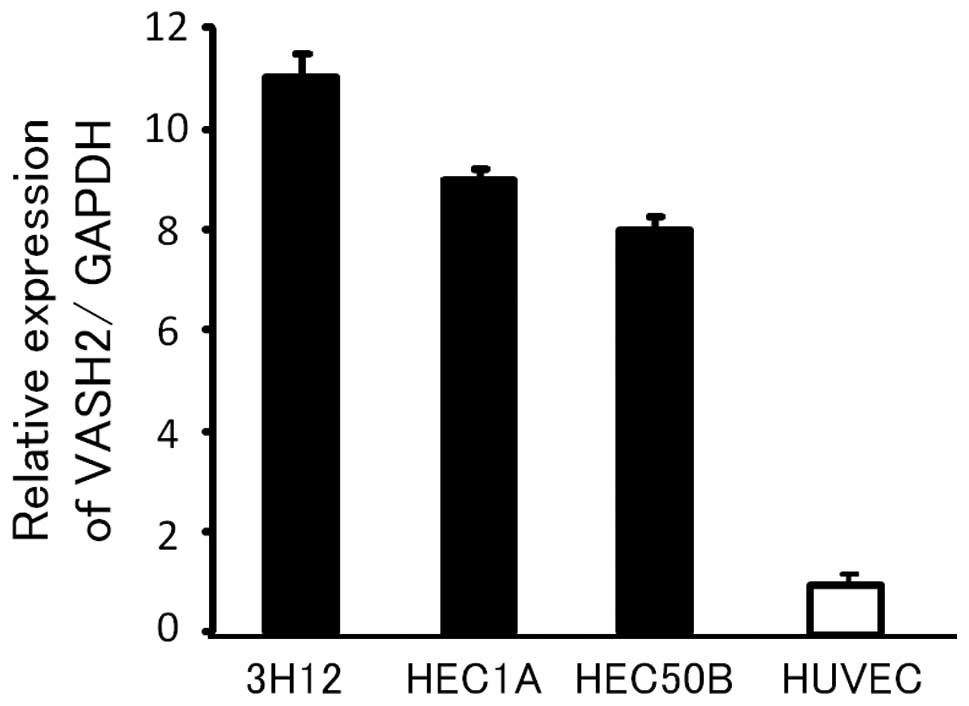
To examine the possible involvement of VASH2 in
endometrial cancer, we analyzed human endometrial cancer cell lines
by quantitative RT-PCR. As demonstrated in Fig. 1, VASH2 mRNA expression was
considerably higher in several human endometrial cancer cell lines
than in the HUVECs.
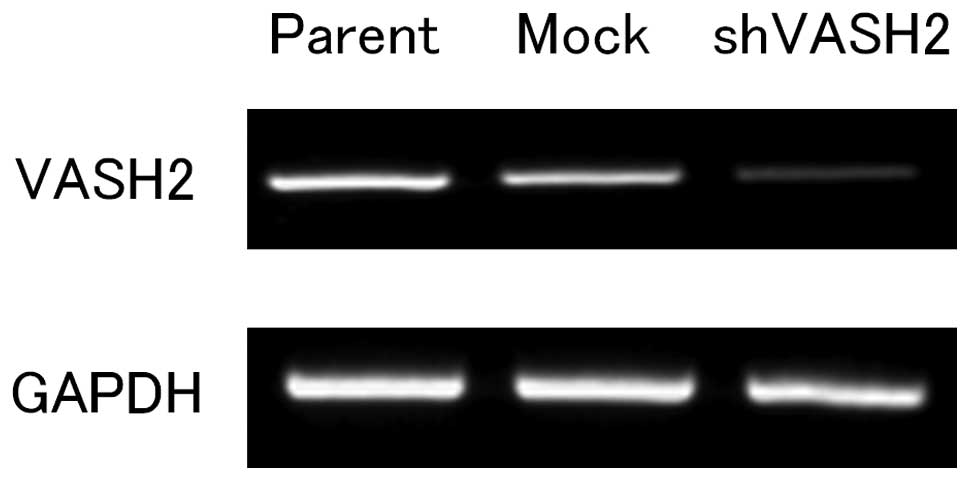
Knockdown of VASH2 and its effects in
vitro and in vivo
To clarify the function of VASH2 in endometrial
cancer, we performed a loss-of-function experiment by knocking down
VASH2 expression. We used HEC50B, an endometrial cancer cell line
with high VASH2 expression, for the following experiments. By the
transfection of shRNA, we established the VASH2-knockdown (shVASH2)
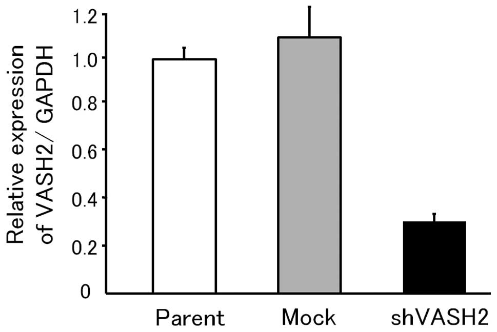
clone from HEC50B (Fig. 2). The
efficacy of knockdown was >70% (Fig.
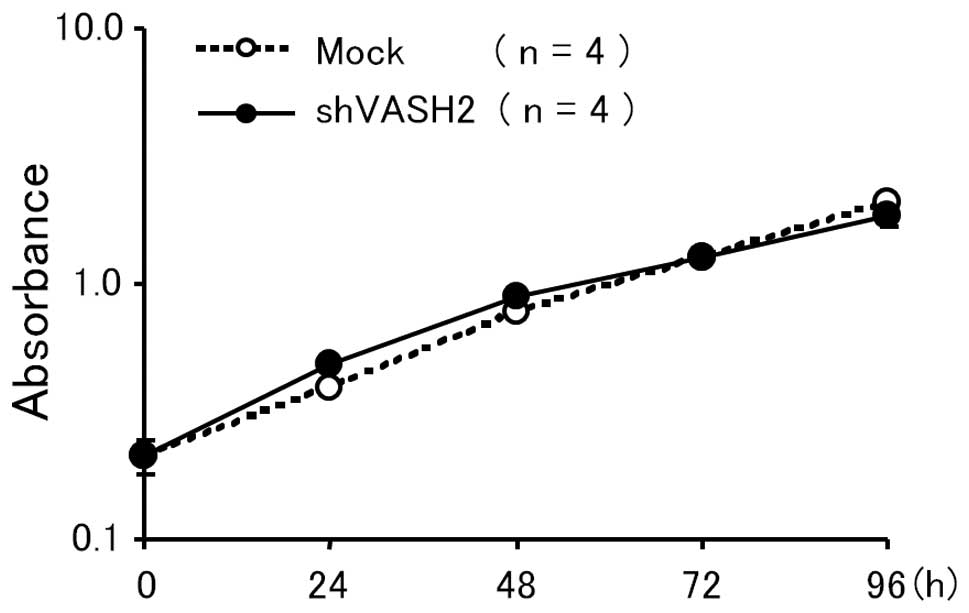
3). Knockdown of VASH2 did not affect the in vitro
proliferation of HEC50B cells (Fig.
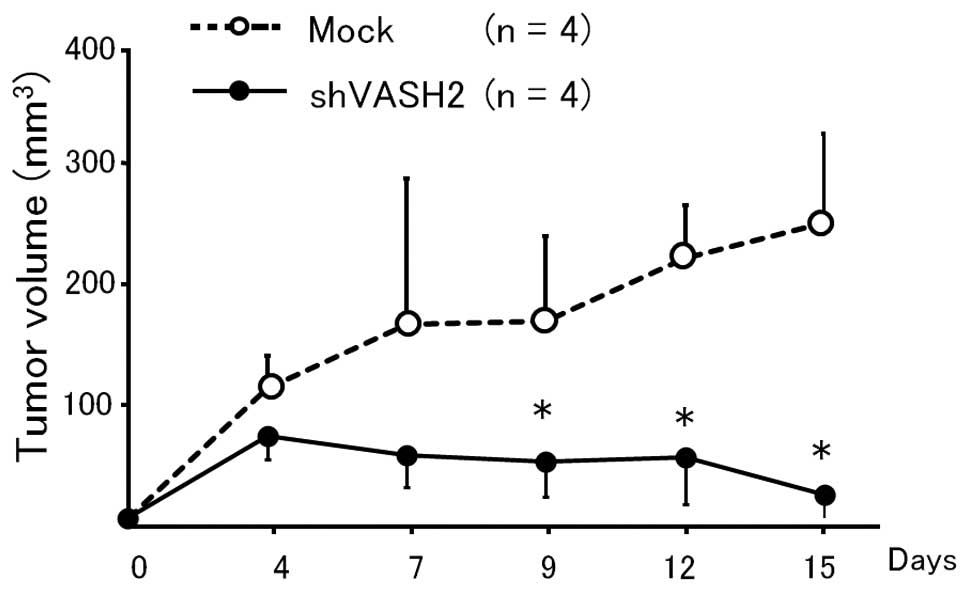
4). We then inoculated the shVASH2 clone subcutaneously into
nude mice, and observed a significant inhibition of tumor growth in
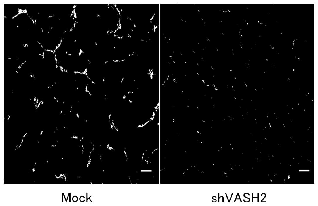
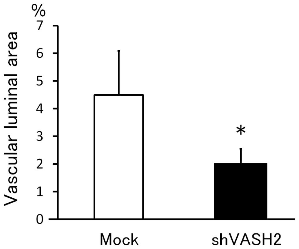
the shVASH2 group compared with the control mock group (Fig. 5). Furthermore, we analyzed
angiogenesis in the tumors of the mouse xenograft model. As
expected, tumor angiogenesis was significantly inhibited in the
shVASH2 tumors, as assessed by immunofluorescent staining of CD31
(Figs. 6 and 7).
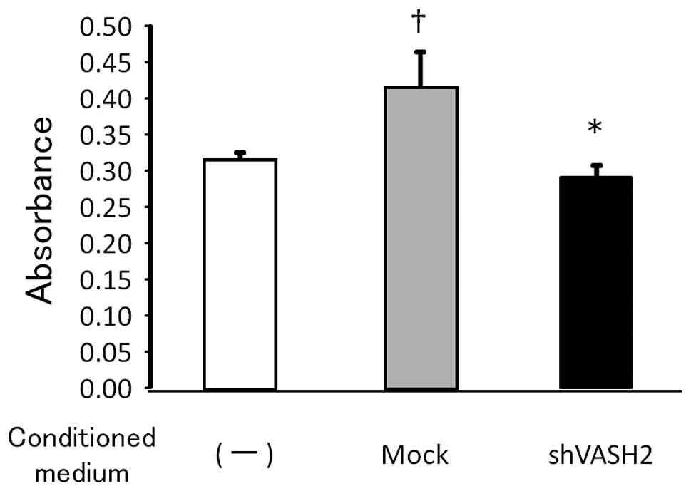
Effect of secreted VASH2 on EC
proliferation
Members of the vasohibin family are secretory
proteins that bind to the intracellular small vasohibin-binding
protein (SVBP) (18). To
investigate the effect of VASH2 on the ECs, we evaluated the
proliferation of HUVECs by using the CM from shVASH2 clones or from
mock transfectants of HEC50B. As demonstrated in Fig. 8, secreted VASH2 significantly
promoted the proliferation of HUVECs, whereas knockdown of VASH2
significantly attenuated the proliferative effect.
Discussion
In the present study, we examined the correlation
between VASH2 and endometrial cancer cell for the first time. VASH2
was expressed in human endometrial cancer cell lines, and the
specific knockdown of VASH2 from the endometrial cancer cell line,
HEC50B, significantly inhibited tumor growth by decreasing tumor
angiogenesis. In addition, the experiment using the CM revealed
that secreted VASH2 significantly promoted the proliferation of
HUVECs. These results suggest that VASH2 secreted from the cancer
cells acts on neighboring ECs to stimulate angiogenesis in a
paracrine manner, and thus contributes to the development of
endometrial cancer.
Angiogenesis is recognized as a principal hallmark
of various types of cancer (19).
There are a number of angiogenic stimulators, one of the most
important of which is VEGF, which stimulates EC migration and
proliferation, as well as EC tube formation. VEGF is the prototype
of the VEGF family, and its pro-angiogenic signals are mainly
transmitted via its type 2 receptor (VEGFR2) on ECs (20). In endometrial cancer tissues, VEGF
expression is associated with elevated tumor vascularization as
measured by microvessel density (3), and is a predictive marker for
decreased 5-year survival in patients with advanced endometrial
carcinoma (21–23). Thus, anti-angiogenic therapy is
considered to be a promising option for treating endometrial
cancer. Current VEGF-targeted therapeutic drugs, including
bevacizumab (a monoclonal antibody against VEGF-A), have yielded
promising results in animal models and clinical trials of
endometrial cancer (3,24). However, resistance to such
therapeutics may occur, owing to the development of compensatory
mechanisms for producing angiogenic factors other than VEGF, or the
recruitment of bone marrow-derived angiogenic cells. Therefore,
alternative targets for anti-angiogenic therapy, a number of which
are being investigated, ought to be sought (25). Taking its stimulatory effect on
angiogenesis into account, VASH2 may be a novel molecular target
for the treatment of endometrial cancer.
While the putative VASH2 receptor and its downstream
signaling are currently under investigation, a number of studies
have examined the novel function of VASH2. In one study, an
autocrine and paracrine mode of action for VASH2 was found to
enhance the expression of FGF-2 and VEGF by nuclear factor-κB
upregulation in hepatocellular carcinoma (HCC) cells (11). Furthermore, VASH2 has been found not
only to accelerate angiogenesis but also to promote HCC cell
proliferation (11). In ovarian
serous adenocarcinoma cells, the expression of VASH2 was inversely
correlated with that of miR-200b, which represses the expression of
ZEB1 and ZEB2, the products of which are key to
epithelial-to-mesenchymal transition (26). Therefore, VASH2 may possess other
tumor-promoting functions, such as invasion and migration. These
features of VASH2 require further investigation.
The local balance between angiogenesis stimulators
and inhibitors, both of which are activated simultaneously during
angiogenesis, determines the occurrence and progression of
angiogenesis. In contrast to VEGF and VASH2, VASH1 has been shown
to both inhibit EC angiogenesis and protect EC from apoptosis
(11). As a VEGF-independent and
EC-extrinsic angiogenesis regulator, VASH2 is considered to be a
novel target for anti-angiogenic therapy that enables the toxic
side effects of anti-VEGF therapy, such as hypertension and
proteinuria, to be avoided. Moreover, the twin combination of VASH2
inhibition and VASH1 upregulation would be a powerful anti-cancer
strategy.
In summary, VASH2 contributes to the development of
endometrial cancer by regulating angiogenesis through paracrine
effects. As such, it constitutes a promising molecular target for
endometrial cancer therapy.
Acknowledgements
This study was supported by The
Research Award to JMU Graduate Students (T.K. and Y.T.).
References
|
1
|
Jemal A, Siegel R, Xu J and Ward E: Cancer
Statistics, 2010. CA Cancer J Clin. 60:277–300. 2010. View Article : Google Scholar
|
|
2
|
Wolfson AH, Sightler SE and Markoe AM: The
prognostic significance of surgical staging for carcinoma of the
endometrium. Gynecol Oncol. 45:142–146. 1992. View Article : Google Scholar : PubMed/NCBI
|
|
3
|
Kamat AA, Merritt WM, Coffey D, et al:
Clinical and biological significance of vascular endothelial growth
factor in endometrial cancer. Clin Cancer Res. 13:7487–7495. 2007.
View Article : Google Scholar : PubMed/NCBI
|
|
4
|
Kowanetz M and Ferrara N: Vascular
endothelial growth factor signaling pathways: therapeutic
perspective. Clin Cancer Res. 12:5018–5022. 2006. View Article : Google Scholar : PubMed/NCBI
|
|
5
|
Ortega J, Vigil CE and Chodkiewicz C:
Current progress in targeted therapy for colorectal cancer. Cancer
Control. 17:7–15. 2010.PubMed/NCBI
|
|
6
|
Koutras AK, Fountzilas G, Makatsoris T,
Peroukides S and Kalofonos HP: Bevacizumab in the treatment of
breast cancer. Cancer Treat Rev. 36:75–82. 2010. View Article : Google Scholar : PubMed/NCBI
|
|
7
|
Tamaskar I and Pili R: Update on novel
agents in renal cell carcinoma. Expert Rev Anticancer Ther.
9:1817–1827. 2009. View Article : Google Scholar : PubMed/NCBI
|
|
8
|
Norden AD, Drappatz J and Wen PY:
Antiangiogenic therapies for high-grade glioma. Nat Rev Neurol.
5:610–620. 2009. View Article : Google Scholar : PubMed/NCBI
|
|
9
|
Watanabe K, Hasegawa Y, Yamashita H, et
al: Vasohibin as an endothelium-derived negative feedback regulator
of angiogenesis. J Clin Invest. 114:898–907. 2004. View Article : Google Scholar : PubMed/NCBI
|
|
10
|
Kimura H, Miyashita H, Suzuki Y, et al:
Distinctive localization and opposed roles of vasohibin-1 and
vasohibin-2 in the regulation of angiogenesis. Blood.
113:4810–4818. 2009. View Article : Google Scholar : PubMed/NCBI
|
|
11
|
Xue X, Gao W, Sun B, et al: Vasohibin 2 is
transcriptionally activated and promotes angiogenesis in
hepatocellular carcinoma. Oncogene. May 21–2012.(Epub ahead of
print).
|
|
12
|
Takahashi Y, Koyanagi T, Suzuki Y, et al:
Vasohibin-2 expressed in human serous ovarian adenocarcinoma
accelerates tumor growth by promoting angiogenesis. Mol Cancer Res.
Jul 23–2012.(Epub ahead of print).
|
|
13
|
Kuramoto H, Tamura S and Notake Y:
Establishment of a cell line of human endometrial adenocarcinoma in
vitro. Am J Obstet Gynecol. 114:1012–1019. 1972.PubMed/NCBI
|
|
14
|
Suzuki M, Kuramoto H, Hamano M, Shirane H
and Watanabe K: Effects of oestradiol and progesterone on the
alkaline phosphatase activity of a human endometrial cancer
cell-line. Acta Endocrinol (Copenh). 93:108–113. 1980.PubMed/NCBI
|
|
15
|
Nishida M: The Ishikawa cells from birth
to the present. Hum Cell. 15:104–117. 2002. View Article : Google Scholar : PubMed/NCBI
|
|
16
|
Miyagishi M and Taira K: Strategies for
generation of an siRNA expression library directed against the
human genome. Oligonucleotides. 13:325–333. 2003. View Article : Google Scholar : PubMed/NCBI
|
|
17
|
Yamamoto O, Hamada T, Tokui N and Sasaguri
Y: Comparison of three in vitro assay systems used for assessing
cytotoxic effect of heavy metals on cultured human keratinocytes. J
UOEH. 23:35–44. 2001.PubMed/NCBI
|
|
18
|
Suzuki Y, Kobayashi M, Miyashita H, Ohta
H, Sonoda H and Sato Y: Isolation of a small vasohibin-binding
protein (SVBP) and its role in vasohibin secretion. J Cell Sci.
123:3094–3101. 2010. View Article : Google Scholar : PubMed/NCBI
|
|
19
|
Hanahan D and Weinberg RA: The hallmarks
of cancer. Cell. 100:57–70. 2000. View Article : Google Scholar
|
|
20
|
Ferrara N: Vascular endothelial growth
factor. Arterioscler Thromb Vasc Biol. 29:789–791. 2009. View Article : Google Scholar
|
|
21
|
McMeekin DS, Sill MW, Benbrook D, et al
Gynecologic Oncology Group: A phase II trial of thalidomide in
patients with refractory endometrial cancer and correlation with
angiogenesis biomarkers: a Gynecologic Oncology Group study.
Gynecol Oncol. 105:508–516. 2007. View Article : Google Scholar : PubMed/NCBI
|
|
22
|
Sanseverino F, Santopietro R, Torricelli
M, et al: pRb2/p130 and VEGF expression in endometrial carcinoma in
relation to angiogenesis and histopathologic tumor grade. Cancer
Biol Ther. 5:84–88. 2006. View Article : Google Scholar : PubMed/NCBI
|
|
23
|
Hirai M, Nakagawara A, Oosaki T, Hayashi
Y, Hirono M and Yoshihara T: Expression of vascular endothelial
growth factors (VEGF-A/VEGF-1 and VEGF-C/VEGF-2) in postmenopausal
uterine endometrial carcinoma. Gynecol Oncol. 80:181–188. 2001.
View Article : Google Scholar : PubMed/NCBI
|
|
24
|
Cerezo L, Cardenes H and Michael H:
Molecular alterations in the pathogenesis of endometrial
adenocarcinoma. Therapeutic implications. Clin Transl Oncol.
8:231–241. 2006. View Article : Google Scholar : PubMed/NCBI
|
|
25
|
Saranadasa M and Wang ES: Vascular
endothelial growth factor inhibition: conflicting roles in tumor
growth. Cytokine. 53:115–129. 2011. View Article : Google Scholar : PubMed/NCBI
|
|
26
|
Korpal M and Kang Y: The emerging role of
miR-200 family of microRNAs in epithelial-mesenchymal transition
and cancer metastasis. RNA Biol. 5:115–119. 2008. View Article : Google Scholar : PubMed/NCBI
|