Introduction
Colorectal cancer (CRC) is a common lethal
malignancy around the world. The initiation and progression of CRC
is a complex process that results from the loss of the normal
regulatory pathways between cell proliferation, differentiation and
apoptosis. Previous studies have identified a small subset of
cancer-initiating cells within tumors that drive tumor growth and
recurrence, termed cancer stem cells (CSCs) (1–3). CSCs
possess self-renewal capabilities and the ability to generate tumor
bulk. Several signaling pathways appear to be key to the
self-renewal behavior of CSCs, including the Wnt/β-catenin, Notch
and Hedgehog pathways (4–6). The ultimate failure of numerous
current cancer treatments, including chemo- and radiation therapy,
is due to a failure to eliminate CSCs (7–11). The
surviving CSCs regenerate recurrent tumors. Therefore, new drugs
and novel therapies are required for the treatment of cancer
patients. Several natural products have been demonstrated to be
effective against CSCs, including curcumin, sulforaphane and
epigallocatechin-3-gallate (12–14).
Huaier aqueous extract (obtained from Trametes
robiniophila) has been used for the treatment of diseases such
as viral hepatitis in China for many years (15). It is isolated from the extract of
the officinal fungi and the effective ingredient has been
identified as proteoglycan (containing 8.72% water, 12.93% amino
acids and 41.53% polysaccharides) (16,17).
It has been reported that Huaier extract has anticancer activity
against various cancer types through the inhibition of tumor
growth, induction of apoptosis and anti-angiogenic effects
(16,18,19).
There are, however, no studies dealing with the effect of Huaier
extract on colorectal CSCs at present.
The Wnt/β-catenin pathway is one of the critical
pathways demonstrated to mediate the self-renewal of CSCs. The
activation of Wnt target genes depends on mediation by β-catenin,
which enters the nucleus to transactivate the TCF/LEF transcription
factor (20,21). The level of intracellular β-catenin
is regulated by the axin-adenomatous polyposis coli-glycogen
synthase kinase-3β complex and β-catenin is degraded via the
ubiquitin-proteasome pathway (22,23).
Activation of the Wnt/β-catenin pathway in CSCs has been shown to
mediate the resistance to chemo- and radiation therapy (24,25).
This indicates that the dysregulation of β-catenin is crucial in
CSCs. If β-catenin transcriptional activity is markedly
down-regulated, tumor growth is likely to be suppressed. Therefore,
it is of great importance to find agents that are able to directly
target this pathway and its downstream targets.
The present study examined the effects of Huaier
aqueous extract on colorectal CSCs. The results showed that Huaier
eliminated CSCs, partially by downregulating β-catenin and
consequently inhibiting the Wnt pathway. The present study, for the
first time, identified Huaier as an effective agent for eradicating
CSCs and implicated the Wnt pathway as a potential target of Huaier
in CRC.
Materials and methods
Materials
Huaier aqueous extract was obtained from Gaitianli
Pharmacy Co. (Qidong, China). Dulbecco’s modified Eagle’s medium,
nutrient mixture F-12 (DMEM/F12), was purchased from Invitrogen
(Carlsbad, CA, USA). Fetal bovine serum (FBS) was supplied by
Sijiqing Biological Engineering Materials Co., Ltd. (Hangzhou,
China). The anti-β-catenin (1:3,000), anti-cyclin D1 (1:3,000) and
anti-β-actin (1:1,000) antibodies were purchased from Cell
Signaling Technology (Beverly, MA, USA). The collagenase and
hyaluronidase were obtained from Sigma Chemical (Balcatta, WA,
Australia).
Tumor cell preparation
Primary CRC cells (T1 and T2 cells) were established
from patients’ cancer tissues following surgery as described
previously (26). In brief,
resected CRC tissues were obtained in accordance with the Research
Ethics Board on Human Experimentation at the Second Affiliated
Hospital, Zhejiang University School of Medicine (Hangzhou, China)
from two patients with informed consent. The histological diagnosis
was based on microscopic features of the carcinoma cells. The
cancer tissues were intensively washed four times in PBS solution
containing antibiotics. Enzymatic digestion was performed using
collagenase (1.5 mg/ml) and hyaluronidase (20 mg/ml) in PBS for 1
h. The cancer cells were then used for culturing in DMEM/F12
supplemented with 10% FBS and 1X antibiotic-antimycotic. The cells
were finally incubated at 37°C in a 5% CO2 humidified
incubator. Cultures contaminated with fibroblasts were removed and
cancer cells were identified in NOD/SCID mice.
Viability assays
The proliferation rates and sensitivity to Huaier
extract were assessed by MTS assays using the CellTiter 96 Aqueous
MTS kit (Promega, Fitchburg, WI, USA). The colorectal primary
cancer cells were seeded in 100 μl medium at a density of
2-5×10⁴ cells per well in 96-well plates (Corning, New
York, NY, USA). Following exposure to the Huaier extract for 48 h,
the MTS assay was performed according to the manufacturer’s
instructions.
Detection of Huaier-induced changes in
cell morphology
CRC cells were plated in 24-chamber culture plates
at 10,000 cells per well, allowed to adhere and incubated with the
Huaier aqueous extract at 8 mg/ml for 48 h. The morphology of the
cells was visualized and photomicrographs were obtained with an
Olympus light microscope (×10).
Spheroid formation assay
Cells were cultured in DMEM/F12 basal serum-free
medium supplemented with 20 ng/ml EGF (Invitrogen), 10 ng/ml bFGF,
B27, 100 U/ml penicillin and 100 μg/ml streptomycin. Single
cells were prepared by enzymatic dissociation and seeded at low
densities (100–200 cells/well) in 96-well low-adhesion plates
(Corning). Various concentrations of the Huaier extract were added.
After 14 days of culturing, the number and size of spheroids were
assessed.
Aldehyde dehydrogenase (ALDH) assay
Colorectal CSCs are highly enriched with cells
having a high ALDH enzyme activity (27). The Aldefluor assay was performed
using an Aldefluor kit (Stemcell Technologies, Durham, NC, USA).
Single cells obtained from cell cultures were incubated in an
Aldefluor assay buffer containing an ALDH substrate for 40 min at
37°C according to the manufacturer’s instructions. A fraction of
the cells was incubated with the ALDH inhibitor
diethylaminobenzaldehyde under the same conditions as a negative
control. The cells were analyzed with a FACSCalibur instrument
(Becton Dickinson, Franklin Lakes, NJ, USA).
Protein isolation and western blot
analysis
The cells were treated with Huaier aqueous extract
(2 or 4 mg/ml) for 48 h. At the end of the incubation period, the
cells were harvested and lysed using the radioimmunoprecipitation
assay buffer [20 mmol/l Tris-HCl, 150 mmol/l NaCl, 1% NP-40, 5
mmol/l EDTA and 1 mmol/l Na3VO4 (pH 7.5)]
supplemented with a protease inhibitor cocktail. Proteins were
detected by western blot analysis using anti-β-catenin, anti-cyclin
D1 and anti-β-actin. Goat anti-rabbit IgG conjugated to horseradish
peroxidase (HRP) was used as the secondary antibody. Immunoreactive
bands were detected using a chemiluminescent substrate.
TCF/LEF transcription assay
To evaluate the TCF/LEF transcriptional activity
induced by activated β-catenin, TOPflash and the negative control
FOPflash, a pair of luciferase reporter constructs, were used. The
reporter system was a gift from Dr Yongliang Zhu (Zhejiang
University School of Medicine). In brief, the cells were
transiently infected with either TOPflash reporter plasmids (10
μg/100 μl) or FOPflash plasmids (10 μg/100
μl) together with Renilla-tk plasmids (1 μg/100
μl; encoding Renilla luciferase). After 24 h, various
concentrations of the Huaier extract were added and the cells were
continually incubated in the same medium for 24 h. Cells were
washed with PBS and the luciferase activity was measured with the
dual-luciferase reporter assay system (Promega).
Statistical analysis
The statistical analysis was performed with GraphPad
Prism 5.0 software and statistical differences were determined by
one-way ANOVA. The data were expressed as the mean ± standard
deviation and all experiments were performed in triplicate.
P<0.05 was considered to indicate a statistically significant
difference.
Results
Huaier aqueous extract suppresses the
growth of CRC cells
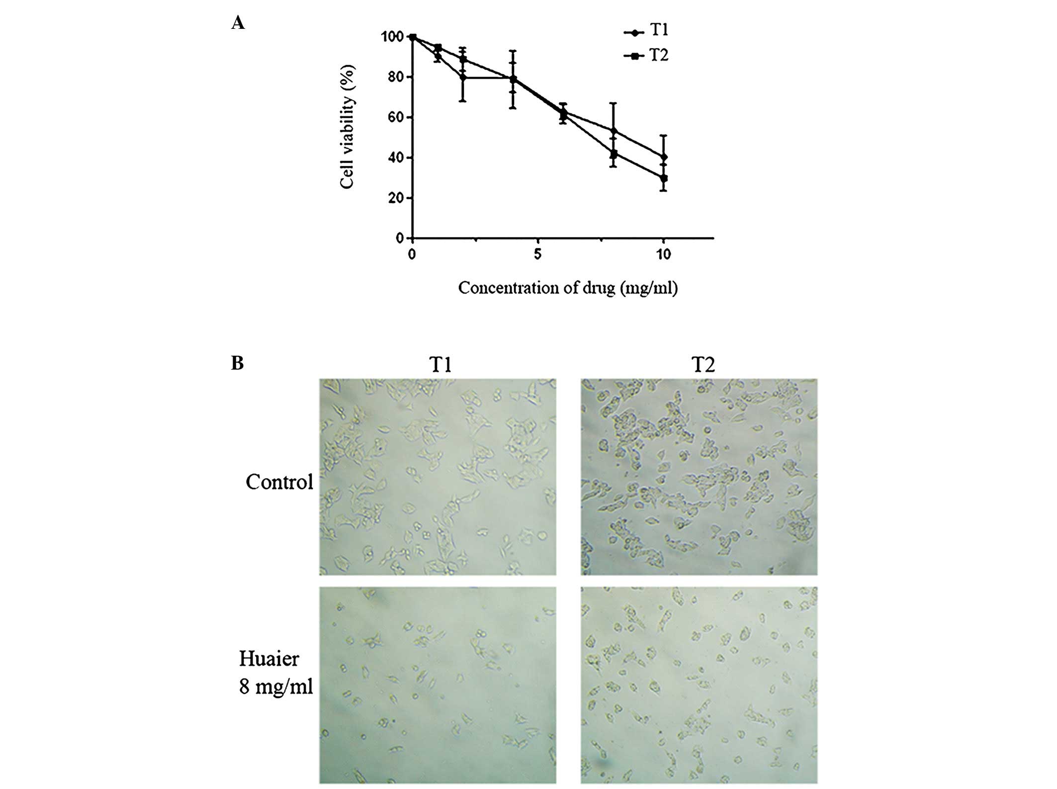
To evaluate the biological activity of Huaier
aqueous extract on cancer cells, cell viability was measured using
the MTS assay following treatment with various concentrations of
the extract. Cell survival decreased with increasing concentrations
of the Huaier extract and the IC50 was determined to be
∼8 mg/ml for T1 and T2 cells (colorectal primary cells; Fig. 1A). The results showed the Huaier
extract to be an antiproliferative agent against CRC cells.
In addition to the antiproliferative effects, the
morphological effects of the Huaier extract on CRC cells were also
investigated. The control cells were epithelial cells with radial
spread and large cell size, indicating typical epithelial
morphology (Fig. 1B). Following the
addition of the Huaier extract, the cell morphology was markedly
altered. In the majority of the Huaier-treated colorectal cells,
the membranes shrank and the cells became irregularly shaped or
round with no visible radial spread. These alterations were
accompanied by a decrease in cell number.
Huaier aqueous extract inhibits the
formation of spheroids
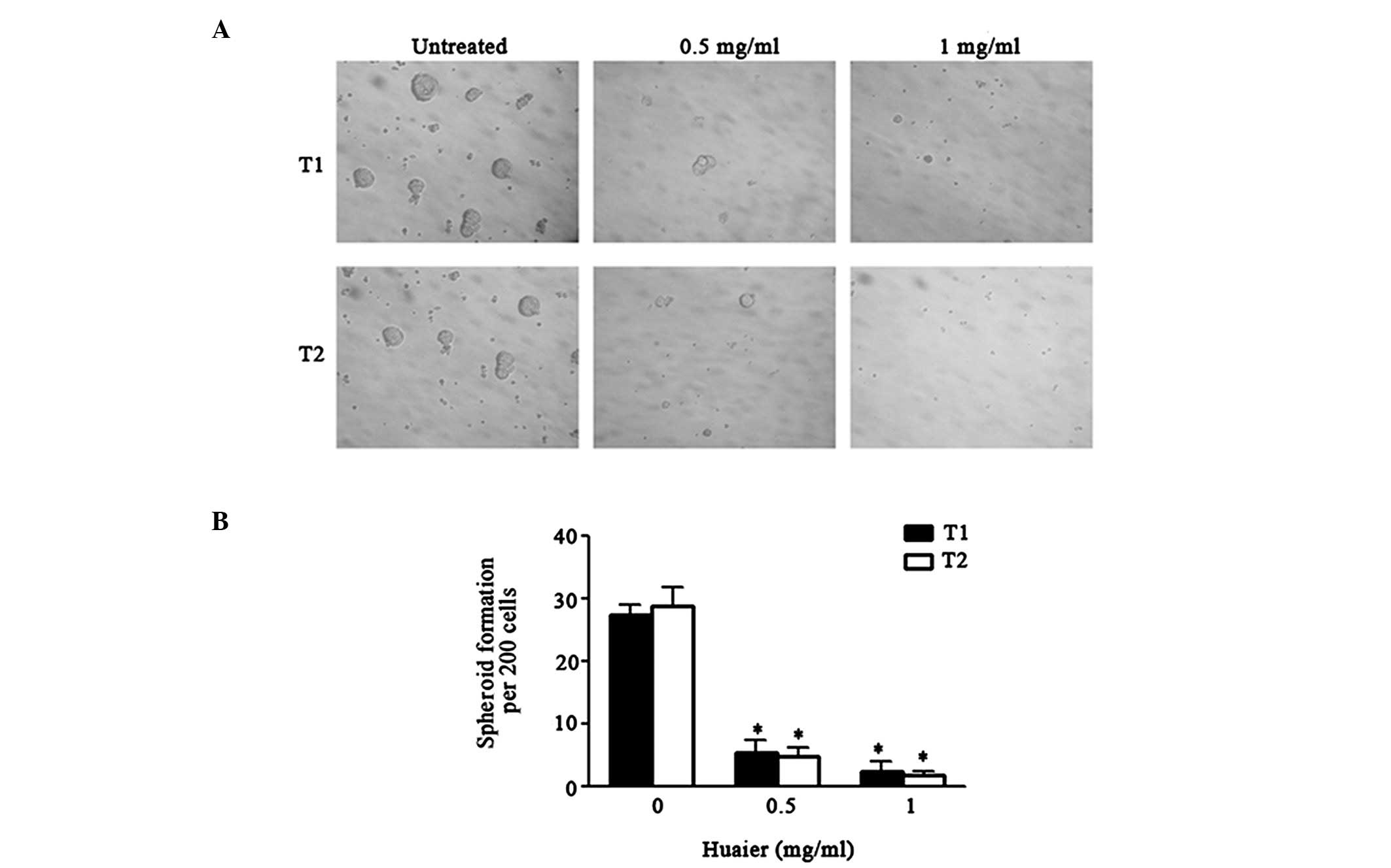
To evaluate its direct effects on the formation of
spheroids, spheroidal growing cells were treated with Huaier
aqueous extract. CRC cells were exposed to varying concentrations
of the Huaier extract and then cultured for 14 days. The Huaier
extract inhibited spheroid formation by the cancer cells (Fig. 2). Not only did the number of
spheroids decline significantly, but also the size of the spheroids
was reduced (P<0.05). It was noteworthy that the concentrations
of Huaier capable of suppressing spheroid formation were ∼15-fold
lower than those exhibiting antiproliferative effects in the MTS
assay (IC50, ∼8 mg/ml for T1 and T2 cells).
Huaier aqueous extract effectively
eliminates ALDH-positive cells in vitro
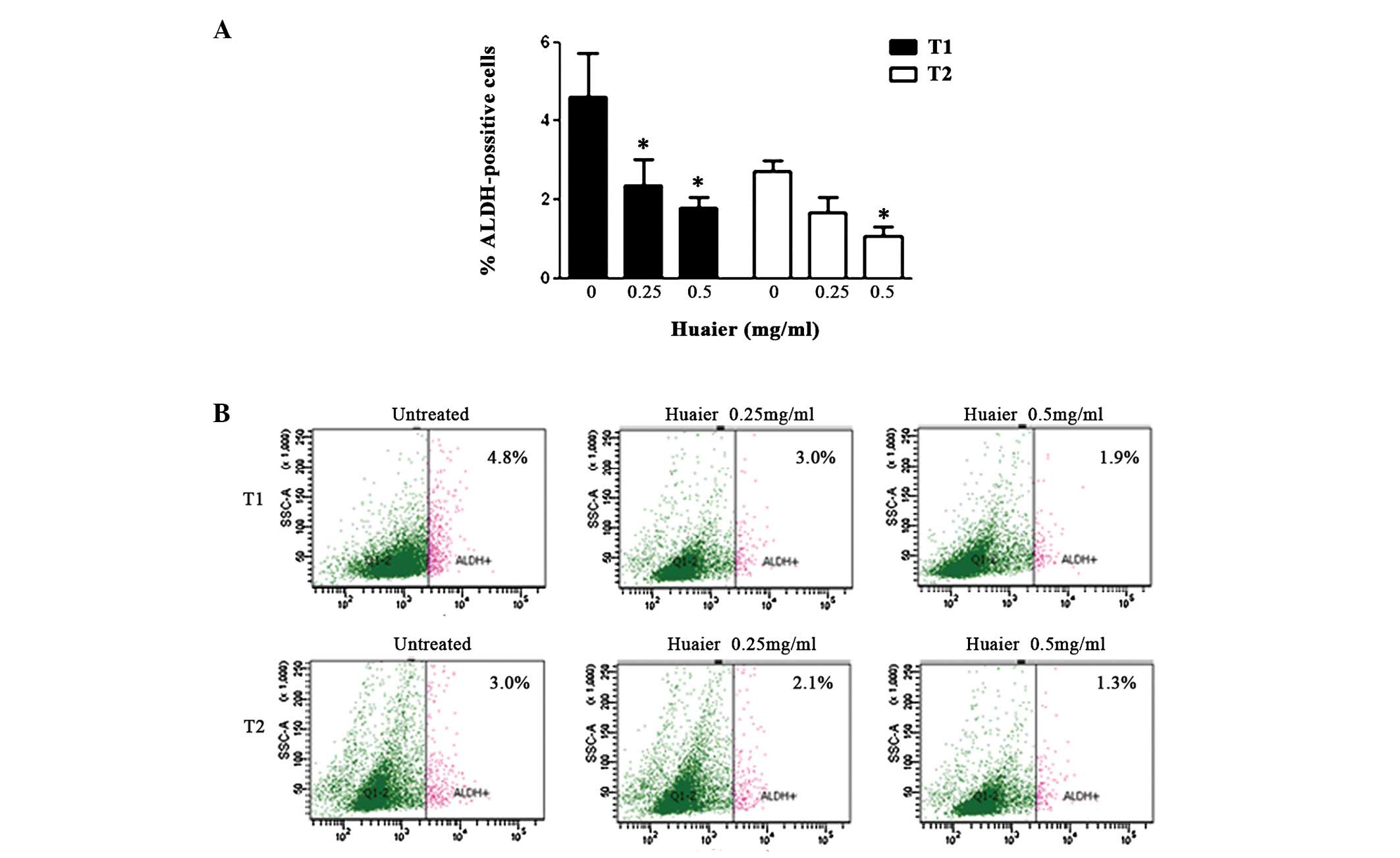
ALDH1 activity, a marker associated with CSCs, was
further tested by the Aldefluor assay. Cell populations with high
ALDH activity have been demonstrated to enrich CSCs. Long-term
treatment with Huaier extract for seven days led to a selective
decrease in the ALDH-positive populations of the CRCs (Fig. 3; P<0.05). This finding
demonstrated that the Huaier extract was able to eliminate CSCs
in vitro. A notable observation was that the Huaier extract
was able to kill CSCs at concentrations (0.25–0.5 mg/ml) that
hardly affected the bulk cancer cells, suggesting that it may
preferentially target CSCs compared with the bulk cancer cells.
Huaier aqueous extract downregulates the
Wnt/β-catenin pathway
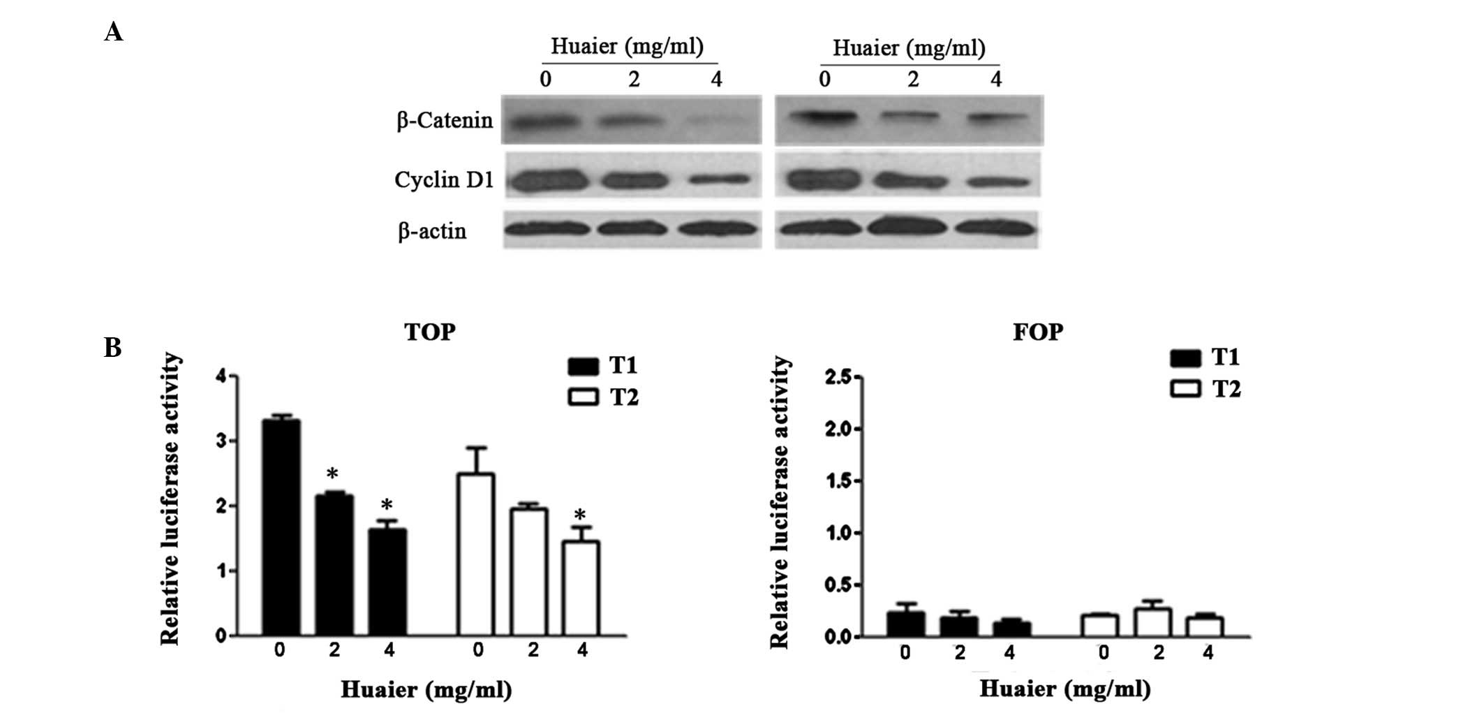
The Wnt/β-catenin pathway is a key pathway in CSC
self-renewal that is activated via β-catenin and results in the
phosphorylation of numerous downstream molecules. Whether the
Huaier extract altered the Wnt/β-catenin pathway in human CRC cells
was studied. The β-catenin protein was constitutively expressed in
cancer cells. Treating the CRC cells with Huaier led to
dose-dependent downregulation of the levels of total β-catenin
protein and also decreased the expression of cyclin D1 (Fig. 4A), one of the Wnt/β-catenin target
genes. To further investigate whether the downregulation of
β-catenin reduced the activity of downstream molecules, a TOP/FOP
flash reporter system was used. Activation of TCF/LEF in the
nucleus mediated by β-catenin was detected and quantified with a
luciferase assay system. A decrease in the TOP levels was observed
in the CRC cells treated with Huaier extract (Fig. 4B; P<0.05). These data indicate
that the downregulation of the Wnt/β-catenin self-renewal pathway
may be a potential target of the Huaier extract.
Discussion
The anticancer effects of Huaier aqueous extract, a
traditional Chinese medicine obtained from the extract of the
officinal fungi, has been evaluated in various types of cancer. For
instance, treatment with Huaier has been shown to inhibit the
proliferation of breast cancer cells by inducing apoptosis
(16). Additionally, it has been
suggested that Huaier has various biological activities related to
metastasis inhibition, immune system activation and drug resistance
reversal (15). These findings
provide a clear rationale for investigating the preventive and
therapeutic properties of Huaier in clinical trials. Huaier has
been used for treating primary liver cancer for many years in China
(28). However, the effects of
Huaier on colorectal CSCs and its mechanism require further
validation.
In the present study, the Huaier extract
demonstrated potent anti-CSC effects in CRC cells. At present,
although there are no exact criteria that directly identify CSCs,
several techniques are used to isolate and characterize colorectal
CSCs. One method is to use a particular stem cell marker such as
ALDH1 (29,30). Unlike the CD molecules, ALDH1 is a
recently identified cancer stem cell marker which has greater
specificity than CD133 and CD44 for colorectal CSCs (31,32).
Previous studies have shown that in normal crypts, ALDH1 positive
cells are sparse and limited to the normal crypt bottom. During the
conversion from normal epithelium to adenoma, the number of
ALDH-positive cells increases and distributes in the crypt and as
few as 25 cells are able to form a tumor in NOD/SCID mice. Thus,
the Aldefluor assay was used to evaluate the effects of the Huaier
extract in eliminating colorectal CSCs. The present results
revealed that the Huaier extract was able to kill the
tumor-initiating ALDH-positive cells. Another method of
characterizing CSCs is spheroid culture under the absence of serum
and without attachment to culture plates. CSCs form spheroids while
differentiated cells fail to survive (33,34).
The Huaier extract was observed to inhibit the spheroid-forming
capacity of CRC cells. These results suggest that Huaier is able to
eliminate the small percentage of cells in a tumor responsible for
chemo-resistance and recurrence.
In the current study, it was demonstrated that
Huaier was able to target and inhibit the Wnt/β-catenin pathway
with a concomitant decrease in β-catenin expression, thereby
manifesting its anticancer mechanism and effects. Of particular
note, the total β-catenin and cyclin D1 levels were downregulated
in all cells studied at 48 h. These results suggested that the Wnt
pathway may be considered as a potential target of Huaier for
preventive and therapeutic interventions in CRC. The Wnt/β-catenin
pathway is closely associated with self-renewal and chemoresistance
in CRC. Previous evidence has further demonstrated the role of the
overactive Wnt/β-catenin pathway in colorectal carcinogenesis
(35). In the present study, the
results showed downregulation of the Wnt pathway, along with a
decrease in the levels of β-catenin and cyclin D1, making Huaier an
attractive potential drug for cancer therapy.
The antitumor activity of Huaier aqueous extract has
been reported in vivo(36);
at 2.5 g/kg per day it was able to significantly suppress tumor
growth and showed no cytotoxicity in the treated mice, suggesting
that Huaier is effective and safe. In addition, commercial Huaier
products, (also called Huaier particles; code number approved by
SFDA: Z20000109) have also been used clinically, with effects on
various types of human cancer, including lung, esophageal, gastric
and liver carcinomas. It is thus possible that Huaier may produce
desirable antitumor effects in CRC. However, further studies of
Huaier and its potential clinical applications are necessary and
important.
The present study demonstrated that Huaier aqueous
extract is able to target colorectal CSCs and inhibit the spheroid
formation potential and ALDH-positive cell population. One of the
mechanisms for the therapeutic effects of Huaier may be the
downregulation of the Wnt/β-catenin self-renewal pathway. The
present study suggests that the use of Huaier may be a good choice
for treating CRC.
Acknowledgements
The authors would like to thank Dr
Yongliang Zhu (Department of Gastroenterology, Second Affiliated
Hospital, Zhejiang University School of Medicine, Hongzhou, China)
for kindly providing the TOP/FOPflash plasmids and the members of
the laboratory for their helpful comments on the manuscript. The
present study was supported by funds from the Zhejiang Provincial
Natural Science Foundation of China (Grant Nos. Z2100366, Y2100414
and J20091440), Science and Technology Bureau of Zhejiang Province
(Grant No. 2011C37004), Zhejiang Provincial Program for the
Cultivation of High-Level Innovative Health Talents (J.H.) and
Cancer Stem Cell Translational Research (Zhejiang Provincial
Traditional Chinese Medicine Key Disciplines, 11-BA564, J.H.).
References
|
1
|
Reya T, Morrison SJ, Clarke MF and
Weissman IL: Stem cells, cancer, and cancer stem cells. Nature.
414:105–111. 2001. View
Article : Google Scholar : PubMed/NCBI
|
|
2
|
Dalerba P, Cho RW and Clarke MF: Cancer
stem cells: models and concepts. Annu Rev Med. 58:267–284. 2007.
View Article : Google Scholar : PubMed/NCBI
|
|
3
|
Ricci-Vitiani L, Lombardi DG, Pilozzi E,
et al: Identification and expansion of human
colon-cancer-initiating cells. Nature. 445:111–115. 2007.
View Article : Google Scholar : PubMed/NCBI
|
|
4
|
Smalley MJ and Dale TC: Wnt signalling in
mammalian development and cancer. Cancer Metastasis Rev.
18:215–230. 1999. View Article : Google Scholar : PubMed/NCBI
|
|
5
|
Dontu G, Jackson KW, McNicholas E,
Kawamura MJ, Abdallah WM and Wicha MS: Role of Notch signaling in
cell-fate determination of human mammary stem/progenitor cells.
Breast Cancer Res. 6:R605–R615. 2004. View
Article : Google Scholar : PubMed/NCBI
|
|
6
|
Liu S, Dontu G, Mantle ID, et al: Hedgehog
signaling and Bmi-1 regulate self-renewal of normal and malignant
human mammary stem cells. Cancer Res. 66:6063–6071. 2006.
View Article : Google Scholar : PubMed/NCBI
|
|
7
|
Bose D, Zimmerman LJ, Pierobon M, et al:
Chemoresistant colorectal cancer cells and cancer stem cells
mediate growth and survival of bystander cells. Br J Cancer.
105:1759–1767. 2011. View Article : Google Scholar : PubMed/NCBI
|
|
8
|
Eyler CE and Rich JN: Survival of the
fittest: cancer stem cells in therapeutic resistance and
angiogenesis. J Clin Oncol. 26:2839–2845. 2008. View Article : Google Scholar : PubMed/NCBI
|
|
9
|
Rich JN and Bao S: Chemotherapy and cancer
stem cells. Cell Stem Cell. 1:353–355. 2007. View Article : Google Scholar : PubMed/NCBI
|
|
10
|
Wang K, Liu L, Zhang T, Zhu YL, Qiu F, Wu
XG, Wang XL, Hu FQ and Huang J: Oxaliplatin-incorporating micelles
eliminate both cancer stem-like and bulk cell populations in
colorectal cancer. Int J Nanomedicine. 6:3207–3218. 2011.PubMed/NCBI
|
|
11
|
Todaro M, Alea MP, Di Stefano AB, et al:
Colon cancer stem cells dictate tumor growth and resist cell death
by production of interleukin-4. Cell Stem Cell. 1:389–402. 2007.
View Article : Google Scholar : PubMed/NCBI
|
|
12
|
Li Y, Zhang T, Korkaya H, et al:
Sulforaphane, a dietary component of broccoli/broccoli sprouts,
inhibits breast cancer stem cells. Clin Cancer Res. 16:2580–2590.
2010. View Article : Google Scholar : PubMed/NCBI
|
|
13
|
Yu Y, Kanwar SS, Patel BB, Nautiyal J,
Sarkar FH and Majumdar AP: Elimination of colon cancer stem-like
cells by the combination of curcumin and FOLFOX. Transl Oncol.
2:321–328. 2009. View Article : Google Scholar : PubMed/NCBI
|
|
14
|
Li Y, Wicha MS, Schwartz SJ and Sun D:
Implications of cancer stem cell theory for cancer chemoprevention
by natural dietary compounds. J Nutr Biochem. 22:799–806. 2011.
View Article : Google Scholar : PubMed/NCBI
|
|
15
|
Li LX, Ye SL, Wang YH and Tang ZZ:
Progress on experimental research and clinical application of
Trametes robiniophila. Bulletin of Chinese Cancer.
16:110–113. 2007.
|
|
16
|
Zhang N, Kong X, Yan S, Yuan C and Yang Q:
Huaier aqueous extract inhibits proliferation of breast cancer
cells by inducing apoptosis. Cancer Sci. 101:2375–2383. 2010.
View Article : Google Scholar : PubMed/NCBI
|
|
17
|
Guo Y, Cheng P, Chen Y, et al: Isolation
and analysis of the polysaccharide of Huaier mycelium. Chin J
Biochem Pharm. 63:56–59. 1993.
|
|
18
|
Ren J, Zheng C, Feng G, et al: Inhibitory
effect of extract of fungi of Huaier on hepatocellular carcinoma
cells. J Huazhong Univ Sci Technolog Med Sci. 29:198–201. 2009.
View Article : Google Scholar : PubMed/NCBI
|
|
19
|
Xu X, Wei Q, Wang K, et al: Anticancer
effects of Huaier are associated with down-regulation of P53. Asian
Pac J Cancer Prev. 12:2251–2254. 2011.PubMed/NCBI
|
|
20
|
Clevers H: Wnt/beta-catenin signaling in
development and disease. Cell. 127:469–480. 2006. View Article : Google Scholar : PubMed/NCBI
|
|
21
|
Dontu G, Abdallah WM, Foley JM, et al: In
vitro propagation and transcriptional profiling of human mammary
stem/progenitor cells. Genes Dev. 17:1253–1270. 2003. View Article : Google Scholar : PubMed/NCBI
|
|
22
|
Lin SY, Xia W, Wang JC, et al:
Beta-catenin, a novel prognostic marker for breast cancer: its
roles in cyclin D1 expression and cancer progression. Proc Natl
Acad Sci USA. 97:4262–4266. 2000. View Article : Google Scholar : PubMed/NCBI
|
|
23
|
Takahashi-Yanaga F and Sasaguri T: GSK-3β
regulates cyclin D1 expression: a new target for chemotherapy. Cell
Signal. 20:581–589. 2008.
|
|
24
|
Dean M, Fojo T and Bates S: Tumour stem
cells and drug resistance. Nat Rev Cancer. 5:275–284. 2005.
View Article : Google Scholar
|
|
25
|
Nicolini A, Ferrari P, Fini M, et al: Stem
cells: their role in breast cancer development and resistance to
treatment. Curr Pharm Biotechnol. 12:196–205. 2011. View Article : Google Scholar : PubMed/NCBI
|
|
26
|
Wang K, Zhang T, Liu L, et al: Novel
micelle formulation of curcumin for enhancing antitumor activity
and inhibiting colorectal cancer stem cells. Int J Nanomedicine.
7:4487–4497. 2012.PubMed/NCBI
|
|
27
|
Shenoy A, Butterworth E and Huang EH: ALDH
as a marker for enriching tumorigenic human colonic stem cells.
Methods Mol Biol. 916:373–85. 2012. View Article : Google Scholar : PubMed/NCBI
|
|
28
|
Huang W, Yuan LN, Wu H, Yang JY, Wang WT
and Xu MQ: Retrospective cohort study on clinical value of Huaier
granule in postoperative patients with liver transplantation for
hepato-cellular carcinoma. Chinese Journal of Bases and Clinics in
General Surgery. 17:547–551. 2010.
|
|
29
|
Vogler T, Kriegl L, Horst D, et al: The
expression pattern of aldehyde dehydrogenase 1 (ALDH1) is an
independent prognostic marker for low survival in colorectal
tumors. Exp Mol Pathol. 92:111–117. 2012. View Article : Google Scholar
|
|
30
|
Ginestier C, Hur MH, Charafe-Jauffret E,
et al: ALDH1 is a marker of normal and malignant human mammary stem
cells and a predictor of poor clinical outcome. Cell Stem Cell.
1:555–567. 2007. View Article : Google Scholar : PubMed/NCBI
|
|
31
|
Huang EH, Hynes MJ, Zhang T, et al:
Aldehyde dehydrogenase 1 is a marker for normal and malignant human
colonic stem cells (SC) and tracks SC overpopulation during colon
tumorigenesis. Cancer Res. 69:3382–3389. 2009. View Article : Google Scholar
|
|
32
|
Chen Y, Orlicky DJ, Matsumoto A, Singh S,
Thompson DC and Vasiliou V: Aldehyde dehydrogenase 1B1 (ALDH1B1) is
a potential biomarker for human colon cancer. Biochem Biophys Res
Commun. 405:173–179. 2011. View Article : Google Scholar : PubMed/NCBI
|
|
33
|
Cammareri P, Lombardo Y, Francipane MG,
Bonventre S, Todaro M and Stassi G: Isolation and culture of colon
cancer stem cells. Methods Cell Biol. 86:311–324. 2008. View Article : Google Scholar : PubMed/NCBI
|
|
34
|
Fan X, Ouyang N, Teng H and Yao H:
Isolation and characterization of spheroid cells from the HT29
colon cancer cell line. Int J Colorectal Dis. 26:1279–1285. 2011.
View Article : Google Scholar : PubMed/NCBI
|
|
35
|
Vermeulen L, De Sousa E, Melo F, van der
Heijden M, et al: Wnt activity defines colon cancer stem cells and
is regulated by the microenvironment. Nat Cell Biol. 12:468–476.
2010. View
Article : Google Scholar : PubMed/NCBI
|
|
36
|
Wang X, Zhang N, Huo Q and Yang Q:
Anti-angiogenic and antitumor activities of Huaier aqueous extract.
Oncol Rep. 28:1167–1175. 2012.PubMed/NCBI
|