Spandidos Publications Logo
  • About
    • About Spandidos
    • Aims and Scopes
    • Abstracting and Indexing
    • Editorial Policies
    • Reprints and Permissions
    • Job Opportunities
    • Terms and Conditions
    • Contact
  • Journals
    • All Journals
    • Oncology Letters
      • Oncology Letters
      • Information for Authors
      • Editorial Policies
      • Editorial Board
      • Aims and Scope
      • Abstracting and Indexing
      • Bibliographic Information
      • Archive
    • International Journal of Oncology
      • International Journal of Oncology
      • Information for Authors
      • Editorial Policies
      • Editorial Board
      • Aims and Scope
      • Abstracting and Indexing
      • Bibliographic Information
      • Archive
    • Molecular and Clinical Oncology
      • Molecular and Clinical Oncology
      • Information for Authors
      • Editorial Policies
      • Editorial Board
      • Aims and Scope
      • Abstracting and Indexing
      • Bibliographic Information
      • Archive
    • Experimental and Therapeutic Medicine
      • Experimental and Therapeutic Medicine
      • Information for Authors
      • Editorial Policies
      • Editorial Board
      • Aims and Scope
      • Abstracting and Indexing
      • Bibliographic Information
      • Archive
    • International Journal of Molecular Medicine
      • International Journal of Molecular Medicine
      • Information for Authors
      • Editorial Policies
      • Editorial Board
      • Aims and Scope
      • Abstracting and Indexing
      • Bibliographic Information
      • Archive
    • Biomedical Reports
      • Biomedical Reports
      • Information for Authors
      • Editorial Policies
      • Editorial Board
      • Aims and Scope
      • Abstracting and Indexing
      • Bibliographic Information
      • Archive
    • Oncology Reports
      • Oncology Reports
      • Information for Authors
      • Editorial Policies
      • Editorial Board
      • Aims and Scope
      • Abstracting and Indexing
      • Bibliographic Information
      • Archive
    • Molecular Medicine Reports
      • Molecular Medicine Reports
      • Information for Authors
      • Editorial Policies
      • Editorial Board
      • Aims and Scope
      • Abstracting and Indexing
      • Bibliographic Information
      • Archive
    • World Academy of Sciences Journal
      • World Academy of Sciences Journal
      • Information for Authors
      • Editorial Policies
      • Editorial Board
      • Aims and Scope
      • Abstracting and Indexing
      • Bibliographic Information
      • Archive
    • International Journal of Functional Nutrition
      • International Journal of Functional Nutrition
      • Information for Authors
      • Editorial Policies
      • Editorial Board
      • Aims and Scope
      • Abstracting and Indexing
      • Bibliographic Information
      • Archive
    • International Journal of Epigenetics
      • International Journal of Epigenetics
      • Information for Authors
      • Editorial Policies
      • Editorial Board
      • Aims and Scope
      • Abstracting and Indexing
      • Bibliographic Information
      • Archive
    • Medicine International
      • Medicine International
      • Information for Authors
      • Editorial Policies
      • Editorial Board
      • Aims and Scope
      • Abstracting and Indexing
      • Bibliographic Information
      • Archive
  • Articles
  • Information
    • Information for Authors
    • Information for Reviewers
    • Information for Librarians
    • Information for Advertisers
    • Conferences
  • Language Editing
Spandidos Publications Logo
  • About
    • About Spandidos
    • Aims and Scopes
    • Abstracting and Indexing
    • Editorial Policies
    • Reprints and Permissions
    • Job Opportunities
    • Terms and Conditions
    • Contact
  • Journals
    • All Journals
    • Biomedical Reports
      • Information for Authors
      • Editorial Policies
      • Editorial Board
      • Aims and Scope
      • Abstracting and Indexing
      • Bibliographic Information
      • Archive
    • Experimental and Therapeutic Medicine
      • Information for Authors
      • Editorial Policies
      • Editorial Board
      • Aims and Scope
      • Abstracting and Indexing
      • Bibliographic Information
      • Archive
    • International Journal of Epigenetics
      • Information for Authors
      • Editorial Policies
      • Editorial Board
      • Aims and Scope
      • Abstracting and Indexing
      • Bibliographic Information
      • Archive
    • International Journal of Functional Nutrition
      • Information for Authors
      • Editorial Policies
      • Editorial Board
      • Aims and Scope
      • Abstracting and Indexing
      • Bibliographic Information
      • Archive
    • International Journal of Molecular Medicine
      • Information for Authors
      • Editorial Policies
      • Editorial Board
      • Aims and Scope
      • Abstracting and Indexing
      • Bibliographic Information
      • Archive
    • International Journal of Oncology
      • Information for Authors
      • Editorial Policies
      • Editorial Board
      • Aims and Scope
      • Abstracting and Indexing
      • Bibliographic Information
      • Archive
    • Medicine International
      • Information for Authors
      • Editorial Policies
      • Editorial Board
      • Aims and Scope
      • Abstracting and Indexing
      • Bibliographic Information
      • Archive
    • Molecular and Clinical Oncology
      • Information for Authors
      • Editorial Policies
      • Editorial Board
      • Aims and Scope
      • Abstracting and Indexing
      • Bibliographic Information
      • Archive
    • Molecular Medicine Reports
      • Information for Authors
      • Editorial Policies
      • Editorial Board
      • Aims and Scope
      • Abstracting and Indexing
      • Bibliographic Information
      • Archive
    • Oncology Letters
      • Information for Authors
      • Editorial Policies
      • Editorial Board
      • Aims and Scope
      • Abstracting and Indexing
      • Bibliographic Information
      • Archive
    • Oncology Reports
      • Information for Authors
      • Editorial Policies
      • Editorial Board
      • Aims and Scope
      • Abstracting and Indexing
      • Bibliographic Information
      • Archive
    • World Academy of Sciences Journal
      • Information for Authors
      • Editorial Policies
      • Editorial Board
      • Aims and Scope
      • Abstracting and Indexing
      • Bibliographic Information
      • Archive
  • Articles
  • Information
    • For Authors
    • For Reviewers
    • For Librarians
    • For Advertisers
    • Conferences
  • Language Editing
Login Register Submit
  • This site uses cookies
  • You can change your cookie settings at any time by following the instructions in our Cookie Policy. To find out more, you may read our Privacy Policy.

    I agree
Search articles by DOI, keyword, author or affiliation
Search
Advanced Search
presentation
Oncology Letters
Join Editorial Board Propose a Special Issue
Print ISSN: 1792-1074 Online ISSN: 1792-1082
Journal Cover
April 2013 Volume 5 Issue 4

Full Size Image

Sign up for eToc alerts
Recommend to Library

Journals

International Journal of Molecular Medicine

International Journal of Molecular Medicine

International Journal of Molecular Medicine is an international journal devoted to molecular mechanisms of human disease.

International Journal of Oncology

International Journal of Oncology

International Journal of Oncology is an international journal devoted to oncology research and cancer treatment.

Molecular Medicine Reports

Molecular Medicine Reports

Covers molecular medicine topics such as pharmacology, pathology, genetics, neuroscience, infectious diseases, molecular cardiology, and molecular surgery.

Oncology Reports

Oncology Reports

Oncology Reports is an international journal devoted to fundamental and applied research in Oncology.

Experimental and Therapeutic Medicine

Experimental and Therapeutic Medicine

Experimental and Therapeutic Medicine is an international journal devoted to laboratory and clinical medicine.

Oncology Letters

Oncology Letters

Oncology Letters is an international journal devoted to Experimental and Clinical Oncology.

Biomedical Reports

Biomedical Reports

Explores a wide range of biological and medical fields, including pharmacology, genetics, microbiology, neuroscience, and molecular cardiology.

Molecular and Clinical Oncology

Molecular and Clinical Oncology

International journal addressing all aspects of oncology research, from tumorigenesis and oncogenes to chemotherapy and metastasis.

World Academy of Sciences Journal

World Academy of Sciences Journal

Multidisciplinary open-access journal spanning biochemistry, genetics, neuroscience, environmental health, and synthetic biology.

International Journal of Functional Nutrition

International Journal of Functional Nutrition

Open-access journal combining biochemistry, pharmacology, immunology, and genetics to advance health through functional nutrition.

International Journal of Epigenetics

International Journal of Epigenetics

Publishes open-access research on using epigenetics to advance understanding and treatment of human disease.

Medicine International

Medicine International

An International Open Access Journal Devoted to General Medicine.

Journal Cover
April 2013 Volume 5 Issue 4

Full Size Image

Sign up for eToc alerts
Recommend to Library

  • Article
  • Citations
    • Cite This Article
    • Download Citation
    • Create Citation Alert
    • Remove Citation Alert
    • Cited By
  • Similar Articles
    • Related Articles (in Spandidos Publications)
    • Similar Articles (Google Scholar)
    • Similar Articles (PubMed)
  • Download PDF
  • Download XML
  • View XML
Case Report

Composite tumor of metanephric adenoma and Wilms' tumor of the kidney: A case report and review of the literature

  • Authors:
    • Pengcheng Zhu
    • Fei Yan
    • Zixuan Yang
    • Li Meng
    • Qilin Ao
  • View Affiliations / Copyright

    Affiliations: Institute of Pathology, Tongji Hospital, Tongji Medical College, Huazhong University of Science and Technology, Wuhan 430030, P.R. China, Department of Oncology, Zhongshan Hospital of Hubei Province, Wuhan 430000, P.R. China
  • Pages: 1311-1314
    |
    Published online on: January 22, 2013
       https://doi.org/10.3892/ol.2013.1148
  • Expand metrics +
Metrics: Total Views: 0 (Spandidos Publications: | PMC Statistics: )
Metrics: Total PDF Downloads: 0 (Spandidos Publications: | PMC Statistics: )
Cited By (CrossRef): 0 citations Loading Articles...

This article is mentioned in:


Abstract

Metanephric adenoma (MA) and Wilms' tumor (WT) are two distinct types of renal tumors. Composite MA and WT of the kidney are extremely rare. Here, a rare case of composite MA and WT of the kidney in a 36-year-old male is described. MA and WT each have their own histopathological features, respectively, and they focally share morphological similarities, which can be a diagnostic challenge. Immunohistochemistry is useful in the differential diagnosis of MA and WT. The histopathological features and differential diagnosis of the composite tumor are emphasized here to promote a better and broader understanding of this less understood subject.

Introduction

Metanephric adenoma (MA) is a rare benign renal tumor which is related to the developing proximal tubule of the fetal kidney or nephrogenic rests. The tumor generally occurs in female adults and occasionally in children (1). Wilms’ tumor (WT), also known as nephroblastoma, is the most common malignant tumor which originates from developing nephrogenic tissue, occurring in the genitourinary tract in children (2). The nature of metanephric adenoma is still not entirely clear, although some investigators believed that it is related to WT because these two tumors sometimes share morphologic similarities and immunoreactivity for Wilms’ tumor gene1 (WT-1) (3,4).

Here, a rare composite tumor with MA and WT histopathologic features of the kidney in a male adult is reported. This case represents the third report of this composite tumor of MA and WT and may present clues to elucidate the pathogenesis of MA and WT (5,6). The study was approved by the Ethics Committee of Tongji hospital, Tongji Medical College, Huazhong University of Science and Technology, Wuhan, China. Written informed consent was obtained from the patient.

Case report

A 36-year-old male visited the Department of Urology, Tongji Hospital, Wuhan, China, for evaluation of his renal area following approximately 4 months of illness. The medical history and review of symptoms were noncontributory.

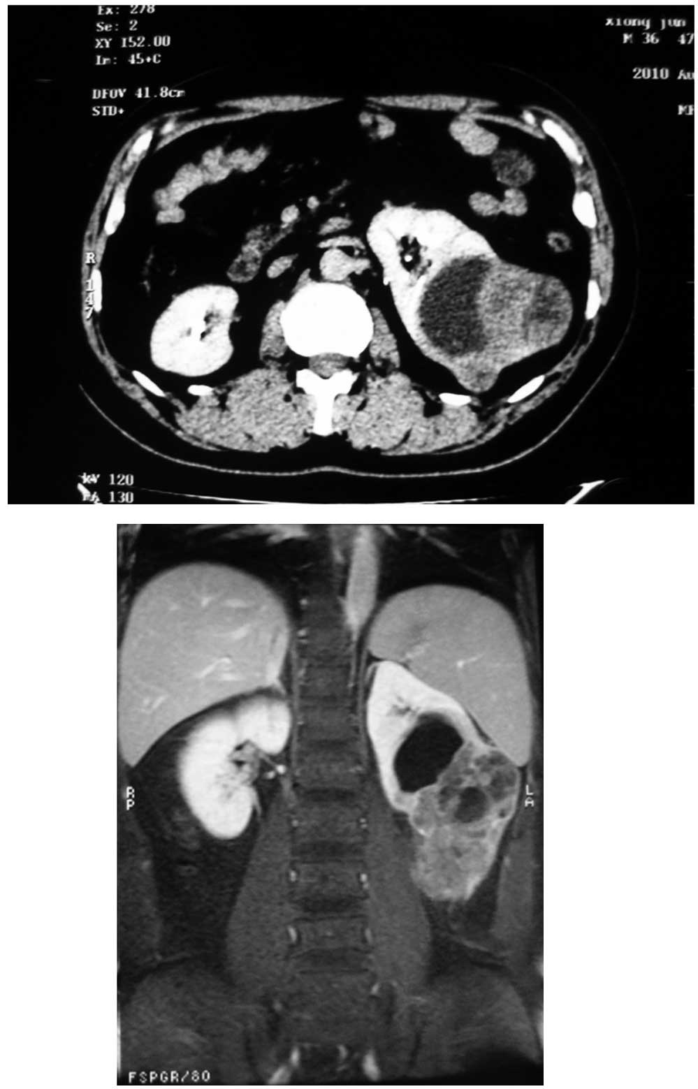
Computed tomography (CT) and magnetic resonance (MR) scan revealed an expansile, solid mass measuring ∼10 cm in the middle lower pole of the left kidney (Fig. 1). The mass was relatively ill-marginated. The patient was admitted for surgical intervention and the lesion was excised under general anesthesia.

Figure 1

Computed tomography and magnetic resonance imaging showing a large solid mass located in the middle and lower pole of the left kidney with hydronephrectasis.

Pathological examination

Specimens from the mass were fixed in 10% formalin, embedded in paraffin, sectioned, and stained with hematoxylin and eosin (H&E) by means of routine procedures. Immunostaining was performed on 4-μm-thick sections using the standard avidin-biotin complex technique. A panel of antibodies (Table I) was used.

Table I

Antibodies and dilutions used in the evaluation of composite metanephric adenoma and Wilms’ tumor of the kidney.

Table I

Antibodies and dilutions used in the evaluation of composite metanephric adenoma and Wilms’ tumor of the kidney.

AntibodyDilutionSourceAntigen retrieval
Vimentin1:20DakoHeat
AE1/AE31:20DakoHeat
CD571:50DakoHeat
WT-11:200DakoHeat
EMA1:100DakoHeat
CK71:200DakoHeat
CK8/181:100DakoHeat
Ki671:40DakoHeat

Immunostaining was performed by an enhancement method based on repetitive microwave heating of slides that were placed into 0.01 M citrate buffer at pH 6.0. Binding of primary antibodies was visualized with an Envision two-step method. Diaminobenzidine was used as the chromogen. Nuclei were stained with Mayer’s hematoxylin. Appropriate positive and negative controls were included.

Results

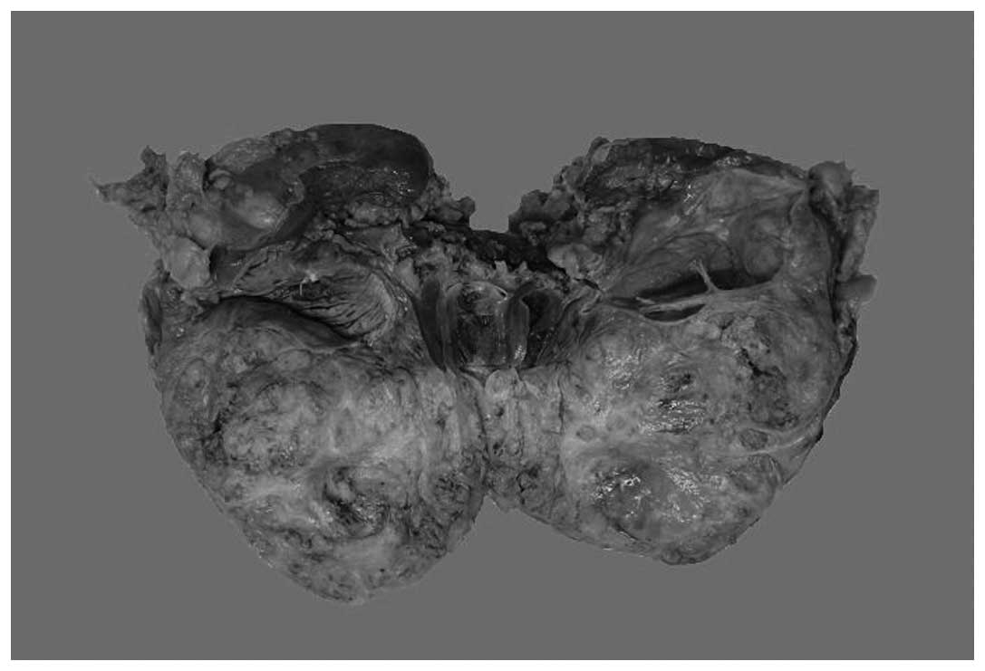
Macroscopically, the specimen was ill-circumscribed and measured 10 cm at its greatest dimension, and hydronephrectasia could be seen in the left kidney due to the occupation of the tumor; the cut surfaces were yellowish gray and solid (Fig. 2). There was no obvious necrosis or hemorrhage. The adjacent renal parenchyma appeared normal. The renal capsule was intact.

Figure 2

The cut surfaces of the tumor were yellowish gray and solid with an ill-circumscribed border.

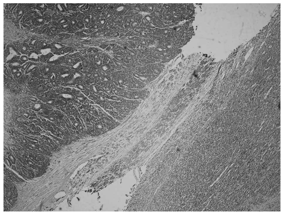
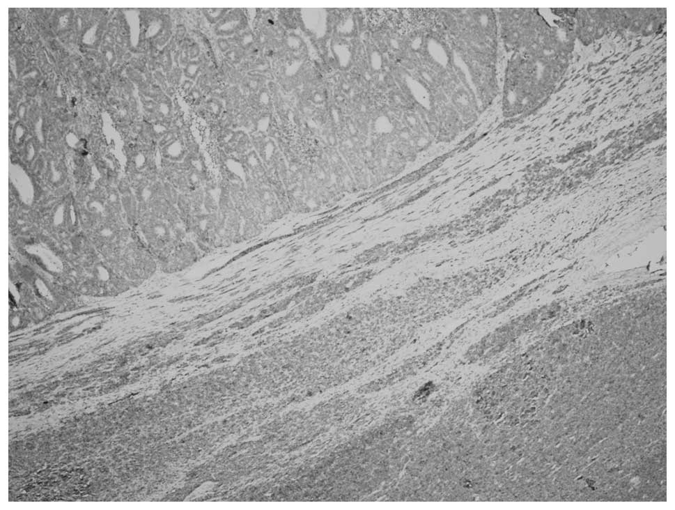
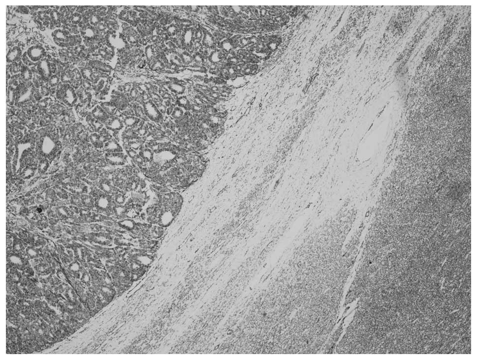
Microscopically, the tumor consisted of two distinct areas of MA and WT that were separated by a band of fibrous stroma 
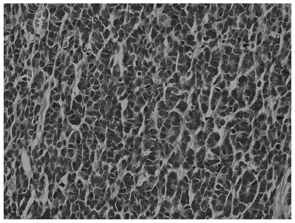
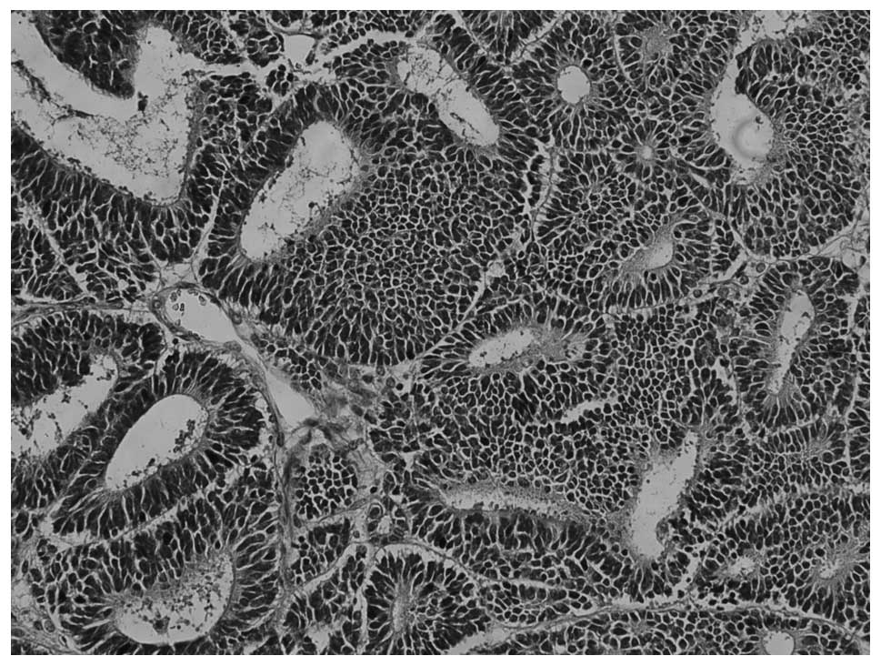
(Fig. 3). MA showed a pushing border with no capsule and an ordered array of small, tightly packed acini and tubules separated by acellular stroma (Fig. 4), but no papillary structure. The tumor cells were small and uniform with round to oval nuclei and scant cytoplasm. Nuclei showed delicate chromatin and inconspicuous nucleoli; mitotic activity was rare to absent.

Figure 3

The composite tumor showing metanephric adenoma (MA; right side) along with Wilms’ tumor (WT; left side) separated by tiny fibrous stroma (hematoxylin and eosin; magnification, ×100).

Figure 4

The metanephric adenoma (MA) area showed ordered array of small, tightly packed acini and tubules. Tumor cells are small and uniform with round to oval nuclei and scant cytoplasm (hematoxylin and eosin; magnification, ×400).

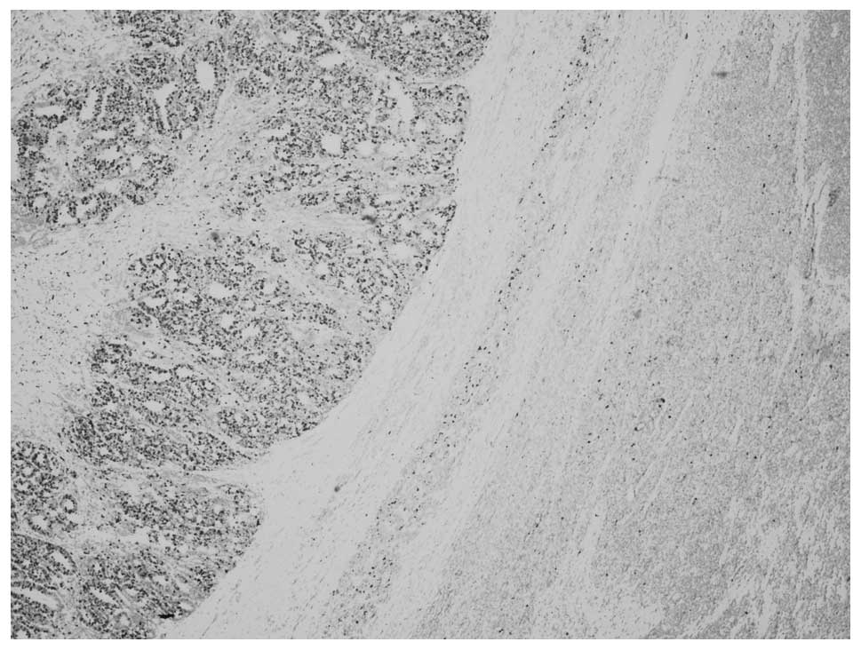
The WT area showed a predominantly epithelial component which was in the form of either poorly formed or well-developed tubules (Fig. 5) The typical blastemal and stromal components were rarely observed.

Figure 5

Wilms’ tumor (WT) area showed predominantly epithelial components which are in the form of poorly formed tubules to well-developed tubular structures. The typical blastemal and stromal components were rare (hematoxylin and eosin; magnification, ×400).

In the MA area, tumor cells showed positive diffuse staining for CD57 (Fig. 6), WT-1 (Fig. 7), focal staining for vimentin, pan CK, CK7 and CK8/18, but negative staining for epithelial membrane antigen (EMA). The Ki67 labeling index (Fig. 8) was 2%. In the WT area, tumor cells stained diffusely for WT-1 (Fig. 7), CK8/18, focally for vimentin, pan CK, EMA, CD57 (Fig. 6), but were negative for CK7. The Ki67 labeling index (Fig. 8) was 50–60%.

Figure 6

Tumor cells showing CD57 expression in the metanephric adenoma area (right side) as compared with negativity in the Wilms’ tumor area (left side) (immunohistochemistry; magnification, ×100).

Figure 7

Tumor cells showing WT-1 expression in the Wilms’ tumor area (left side) as compared with negativity in the metanephric adenoma area (right side) (immunohistochemistry, magnification, ×100).

Figure 8

The Ki67 labeling index was significantly higher in the Wilms’ tumor area (left side) as compared with that of the metanephric adenoma area (right side) (immunohistochemistry; magnification, ×100).

Discussion

Composite renal tumors are rarely reported. The most commonly described association is of Wilms’ tumor and renal cell carcinoma (7). Composite tumors of MA and WT of the kidney are extremely rare (5, 6).

MA is a well-described rare benign renal tumor, predominantly occurring in adult females, and seldom observed in children. WT is the most common malignant renal tumor in children but it is rare in adults. Histologically, MA is composed of tightly packed uniform small epithelial cells in acinar, solid and tubular configurations with small regular nuclei, a high nucleus to cytoplasm ratio, but low mitotic figures.

Wilms’ tumors are typically composed of a mixture of primitive blastemal cells, epithelial cells and mesenchymal elements. In this case, the epithelial component in the WT area was predominant; it was therefore named epithelial-predominant WT (8). Due to overlapping morphological features, histopathological examination of MA often prompts an initial diagnosis of epithelial-predominant WT.

Since MA and epithelial-predominant WT could share microscopical similarities, immunohistochemistry (IHC) played a significant contributory role in the distinction of these two entities. Although CD57 is not particular for diagnosis of MA, it is helpful in the differential diagnosis between MA and epithelial-predominant WT (9, 10). In MA, the epithelial cells are positive for CD57, while the tumor cells in epithelial-predominant WT are negative. The Ki67 labeling index is significantly lower in MA compared with epithelial-predominant WT, which also supports these two areas belonging to distinct lesions (6). The two distinct areas had individual histopathological features and special IHC staining, both of which contributed to the reaffirmation of the histomorphological diagnosis of composite MA and epithelial-predominant WT.

Both MA and epithelial-predominant WT were positive for WT-1, leading to the theory that the two could be linked (9), and MA could even be a more hyperdifferentiated, mature form of WT (11). Since there was no other supporting evidence it was hypothesized that they were two distinct entities.

Another diagnostic challenge is differentiating MA from the solid variant form of papillary renal cell carcinoma (PRCC) based on histologic features alone. IHC is helpful since PRCC is negative for CD57 and WT-1 (12,13).

To date, the genetic basis of MA remains largely unknown since previous reports have given conflicting results (14–16). BRAF mutation has been reported in 90% cases in a series of MA studies but it is rarely detected in other kidney tumors, including WT. Testing for the BRAF mutation could, therefore, serve as a potential diagnostic tool for MA (17).

In summary, a rare case of composite MA and epithelial-predominant WT in an adult kidney is presented. Although there were overlapping morphological features, it was possible to differentiate MA from WT based on the morphologic features and IHC staining. This case also offered additional support to the hypothesis that these two tumors are related.

References

1 

Jones EC, Pins M, Dickersin GR and Young RH: Metanephric adenoma of the kidney. A clinicopathological, immunohistochemical, flow cytometric, cytogenetic, and electron microscopic study of seven cases. Am J Surg Pathol. 19:615–626. 1995. View Article : Google Scholar

2 

Md Zin R, Murch A and Charles A: Pathology, genetics and cytogenetics of Wilms’ tumour. Pathology. 43:302–312. 2011.

3 

Obulareddy SJ, Xin J, Truskinovsky AM, Anderson JK, Franklin J and Dudek AZ: Metanephric adenoma of the kidney: an unusual diagnostic challenge. Rare Tumors. 2:e382010. View Article : Google Scholar : PubMed/NCBI

4 

Portugal R and Barroca H: Clear cell sarcoma, cellular mesoblastic nephroma and metanephric adenoma: cytological features and differential diagnosis with Wilms tumour. Cytopathology. 19:80–85. 2008. View Article : Google Scholar

5 

Davis CJ Jr, Barton JH, Sesterhenn IA and Mostofi FK: Metanephric adenoma. Clinicopathological study of fifty patients Am J Surg Pathol. 19:1101–1114. 1995.PubMed/NCBI

6 

Pasricha S, Gandhi JS, Gupta G, Mehta A and Beg S: Bilateral, multicenteric metanephric adenoma associated with Wilms’ tumor in a child: A rare presentation with important diagnostic and therapeutic implications. Int J Urol. 19:1114–1117. 2012.PubMed/NCBI

7 

Galluzzo ML, Garcia de Davila MT and Vujanic GM: A composite renal tumor: metanephric adenofibroma, Wilms tumor, and renal cell carcinoma: a missing link? Pediatr Dev Pathol. 15:65–70. 2012. View Article : Google Scholar : PubMed/NCBI

8 

Verschuur AC, Vujanic GM, Van Tinteren H, Jones KP, de Kraker J and Sandstedt B: Stromal and epithelial predominant Wilms tumours have an excellent outcome: the SIOP 93 01 experience. Pediatr Blood Cancer. 55:233–238. 2010. View Article : Google Scholar : PubMed/NCBI

9 

Muir TE, Cheville JC and Lager DJ: Metanephric adenoma, nephrogenic rests, and Wilms’ tumor: a histologic and immunophenotypic comparison. Am J Surg Pathol. 25:1290–1296. 2001.

10 

Olgac S, Hutchinson B, Tickoo SK and Reuter VE: Alpha-methylacyl-CoA racemase as a marker in the differential diagnosis of metanephric adenoma. Mod Pathol. 19:218–224. 2006. View Article : Google Scholar : PubMed/NCBI

11 

Argani P: Metanephric neoplasms: the hyperdifferentiated, benign end of the Wilms tumor spectrum? Clin Lab Med. 25:379–392. 2005. View Article : Google Scholar : PubMed/NCBI

12 

Brown JA, Anderl KL, Borell TJ, Qian J, Bostwick DG and Jenkins RB: Simultaneous chromosome 7 and 17 gain and sex chromosome loss provide evidence that renal metanephric adenoma is related to papillary renal cell carcinoma. J Urol. 158:370–374. 1997. View Article : Google Scholar : PubMed/NCBI

13 

Lugli A, Stoffe F, Terracciano L, Dirnhofer S, Mihatsch MJ and Moch H: Differentiated papillary kidney tumors. Differentiation between metanephric adenoma and papillary adenoma. Pathologe. 23:303–307. 2002.(In German).

14 

Burger M, Junker K, Denzinger S, et al: Metanephric adenoma of the kidney: a clinicopathological and molecular study of two cases. J Clin Pathol. 60:832–833. 2007. View Article : Google Scholar : PubMed/NCBI

15 

Brunelli M, Eble JN, Zhang S, Martignoni G and Cheng L: Metanephric adenoma lacks the gains of chromosomes 7 and 17 and loss of Y that are typical of papillary renal cell carcinoma and papillary adenoma. Mod Pathol. 16:1060–1063. 2003. View Article : Google Scholar : PubMed/NCBI

16 

Pesti T, Sukosd F, Jones EC and Kovacs G: Mapping a tumor suppressor gene to chromosome 2p13 in metanephric adenoma by microsatellite allelotyping. Hum Pathol. 32:101–104. 2001. View Article : Google Scholar : PubMed/NCBI

17 

Choueiri TK, Cheville J, Palescandolo E, et al: BRAF mutations in metanephric adenoma of the kidney. Eur Urol. 62:917–922. 2012. View Article : Google Scholar : PubMed/NCBI

Related Articles

  • Abstract
  • View
  • Download
  • Twitter
Copy and paste a formatted citation
Spandidos Publications style
Zhu P, Yan F, Yang Z, Meng L and Ao Q: Composite tumor of metanephric adenoma and Wilms' tumor of the kidney: A case report and review of the literature. Oncol Lett 5: 1311-1314, 2013.
APA
Zhu, P., Yan, F., Yang, Z., Meng, L., & Ao, Q. (2013). Composite tumor of metanephric adenoma and Wilms' tumor of the kidney: A case report and review of the literature. Oncology Letters, 5, 1311-1314. https://doi.org/10.3892/ol.2013.1148
MLA
Zhu, P., Yan, F., Yang, Z., Meng, L., Ao, Q."Composite tumor of metanephric adenoma and Wilms' tumor of the kidney: A case report and review of the literature". Oncology Letters 5.4 (2013): 1311-1314.
Chicago
Zhu, P., Yan, F., Yang, Z., Meng, L., Ao, Q."Composite tumor of metanephric adenoma and Wilms' tumor of the kidney: A case report and review of the literature". Oncology Letters 5, no. 4 (2013): 1311-1314. https://doi.org/10.3892/ol.2013.1148
Copy and paste a formatted citation
x
Spandidos Publications style
Zhu P, Yan F, Yang Z, Meng L and Ao Q: Composite tumor of metanephric adenoma and Wilms' tumor of the kidney: A case report and review of the literature. Oncol Lett 5: 1311-1314, 2013.
APA
Zhu, P., Yan, F., Yang, Z., Meng, L., & Ao, Q. (2013). Composite tumor of metanephric adenoma and Wilms' tumor of the kidney: A case report and review of the literature. Oncology Letters, 5, 1311-1314. https://doi.org/10.3892/ol.2013.1148
MLA
Zhu, P., Yan, F., Yang, Z., Meng, L., Ao, Q."Composite tumor of metanephric adenoma and Wilms' tumor of the kidney: A case report and review of the literature". Oncology Letters 5.4 (2013): 1311-1314.
Chicago
Zhu, P., Yan, F., Yang, Z., Meng, L., Ao, Q."Composite tumor of metanephric adenoma and Wilms' tumor of the kidney: A case report and review of the literature". Oncology Letters 5, no. 4 (2013): 1311-1314. https://doi.org/10.3892/ol.2013.1148
Follow us
  • Twitter
  • LinkedIn
  • Facebook
About
  • Spandidos Publications
  • Careers
  • Cookie Policy
  • Privacy Policy
How can we help?
  • Help
  • Live Chat
  • Contact
  • Email to our Support Team