Spandidos Publications Logo
  • About
    • About Spandidos
    • Aims and Scopes
    • Abstracting and Indexing
    • Editorial Policies
    • Reprints and Permissions
    • Job Opportunities
    • Terms and Conditions
    • Contact
  • Journals
    • All Journals
    • Oncology Letters
      • Oncology Letters
      • Information for Authors
      • Editorial Policies
      • Editorial Board
      • Aims and Scope
      • Abstracting and Indexing
      • Bibliographic Information
      • Archive
    • International Journal of Oncology
      • International Journal of Oncology
      • Information for Authors
      • Editorial Policies
      • Editorial Board
      • Aims and Scope
      • Abstracting and Indexing
      • Bibliographic Information
      • Archive
    • Molecular and Clinical Oncology
      • Molecular and Clinical Oncology
      • Information for Authors
      • Editorial Policies
      • Editorial Board
      • Aims and Scope
      • Abstracting and Indexing
      • Bibliographic Information
      • Archive
    • Experimental and Therapeutic Medicine
      • Experimental and Therapeutic Medicine
      • Information for Authors
      • Editorial Policies
      • Editorial Board
      • Aims and Scope
      • Abstracting and Indexing
      • Bibliographic Information
      • Archive
    • International Journal of Molecular Medicine
      • International Journal of Molecular Medicine
      • Information for Authors
      • Editorial Policies
      • Editorial Board
      • Aims and Scope
      • Abstracting and Indexing
      • Bibliographic Information
      • Archive
    • Biomedical Reports
      • Biomedical Reports
      • Information for Authors
      • Editorial Policies
      • Editorial Board
      • Aims and Scope
      • Abstracting and Indexing
      • Bibliographic Information
      • Archive
    • Oncology Reports
      • Oncology Reports
      • Information for Authors
      • Editorial Policies
      • Editorial Board
      • Aims and Scope
      • Abstracting and Indexing
      • Bibliographic Information
      • Archive
    • Molecular Medicine Reports
      • Molecular Medicine Reports
      • Information for Authors
      • Editorial Policies
      • Editorial Board
      • Aims and Scope
      • Abstracting and Indexing
      • Bibliographic Information
      • Archive
    • World Academy of Sciences Journal
      • World Academy of Sciences Journal
      • Information for Authors
      • Editorial Policies
      • Editorial Board
      • Aims and Scope
      • Abstracting and Indexing
      • Bibliographic Information
      • Archive
    • International Journal of Functional Nutrition
      • International Journal of Functional Nutrition
      • Information for Authors
      • Editorial Policies
      • Editorial Board
      • Aims and Scope
      • Abstracting and Indexing
      • Bibliographic Information
      • Archive
    • International Journal of Epigenetics
      • International Journal of Epigenetics
      • Information for Authors
      • Editorial Policies
      • Editorial Board
      • Aims and Scope
      • Abstracting and Indexing
      • Bibliographic Information
      • Archive
    • Medicine International
      • Medicine International
      • Information for Authors
      • Editorial Policies
      • Editorial Board
      • Aims and Scope
      • Abstracting and Indexing
      • Bibliographic Information
      • Archive
  • Articles
  • Information
    • Information for Authors
    • Information for Reviewers
    • Information for Librarians
    • Information for Advertisers
    • Conferences
  • Language Editing
Spandidos Publications Logo
  • About
    • About Spandidos
    • Aims and Scopes
    • Abstracting and Indexing
    • Editorial Policies
    • Reprints and Permissions
    • Job Opportunities
    • Terms and Conditions
    • Contact
  • Journals
    • All Journals
    • Biomedical Reports
      • Information for Authors
      • Editorial Policies
      • Editorial Board
      • Aims and Scope
      • Abstracting and Indexing
      • Bibliographic Information
      • Archive
    • Experimental and Therapeutic Medicine
      • Information for Authors
      • Editorial Policies
      • Editorial Board
      • Aims and Scope
      • Abstracting and Indexing
      • Bibliographic Information
      • Archive
    • International Journal of Epigenetics
      • Information for Authors
      • Editorial Policies
      • Editorial Board
      • Aims and Scope
      • Abstracting and Indexing
      • Bibliographic Information
      • Archive
    • International Journal of Functional Nutrition
      • Information for Authors
      • Editorial Policies
      • Editorial Board
      • Aims and Scope
      • Abstracting and Indexing
      • Bibliographic Information
      • Archive
    • International Journal of Molecular Medicine
      • Information for Authors
      • Editorial Policies
      • Editorial Board
      • Aims and Scope
      • Abstracting and Indexing
      • Bibliographic Information
      • Archive
    • International Journal of Oncology
      • Information for Authors
      • Editorial Policies
      • Editorial Board
      • Aims and Scope
      • Abstracting and Indexing
      • Bibliographic Information
      • Archive
    • Medicine International
      • Information for Authors
      • Editorial Policies
      • Editorial Board
      • Aims and Scope
      • Abstracting and Indexing
      • Bibliographic Information
      • Archive
    • Molecular and Clinical Oncology
      • Information for Authors
      • Editorial Policies
      • Editorial Board
      • Aims and Scope
      • Abstracting and Indexing
      • Bibliographic Information
      • Archive
    • Molecular Medicine Reports
      • Information for Authors
      • Editorial Policies
      • Editorial Board
      • Aims and Scope
      • Abstracting and Indexing
      • Bibliographic Information
      • Archive
    • Oncology Letters
      • Information for Authors
      • Editorial Policies
      • Editorial Board
      • Aims and Scope
      • Abstracting and Indexing
      • Bibliographic Information
      • Archive
    • Oncology Reports
      • Information for Authors
      • Editorial Policies
      • Editorial Board
      • Aims and Scope
      • Abstracting and Indexing
      • Bibliographic Information
      • Archive
    • World Academy of Sciences Journal
      • Information for Authors
      • Editorial Policies
      • Editorial Board
      • Aims and Scope
      • Abstracting and Indexing
      • Bibliographic Information
      • Archive
  • Articles
  • Information
    • For Authors
    • For Reviewers
    • For Librarians
    • For Advertisers
    • Conferences
  • Language Editing
Login Register Submit
  • This site uses cookies
  • You can change your cookie settings at any time by following the instructions in our Cookie Policy. To find out more, you may read our Privacy Policy.

    I agree
Search articles by DOI, keyword, author or affiliation
Search
Advanced Search
presentation
Oncology Letters
Join Editorial Board Propose a Special Issue
Print ISSN: 1792-1074 Online ISSN: 1792-1082
Journal Cover
April 2013 Volume 5 Issue 4

Full Size Image

Sign up for eToc alerts
Recommend to Library

Journals

International Journal of Molecular Medicine

International Journal of Molecular Medicine

International Journal of Molecular Medicine is an international journal devoted to molecular mechanisms of human disease.

International Journal of Oncology

International Journal of Oncology

International Journal of Oncology is an international journal devoted to oncology research and cancer treatment.

Molecular Medicine Reports

Molecular Medicine Reports

Covers molecular medicine topics such as pharmacology, pathology, genetics, neuroscience, infectious diseases, molecular cardiology, and molecular surgery.

Oncology Reports

Oncology Reports

Oncology Reports is an international journal devoted to fundamental and applied research in Oncology.

Experimental and Therapeutic Medicine

Experimental and Therapeutic Medicine

Experimental and Therapeutic Medicine is an international journal devoted to laboratory and clinical medicine.

Oncology Letters

Oncology Letters

Oncology Letters is an international journal devoted to Experimental and Clinical Oncology.

Biomedical Reports

Biomedical Reports

Explores a wide range of biological and medical fields, including pharmacology, genetics, microbiology, neuroscience, and molecular cardiology.

Molecular and Clinical Oncology

Molecular and Clinical Oncology

International journal addressing all aspects of oncology research, from tumorigenesis and oncogenes to chemotherapy and metastasis.

World Academy of Sciences Journal

World Academy of Sciences Journal

Multidisciplinary open-access journal spanning biochemistry, genetics, neuroscience, environmental health, and synthetic biology.

International Journal of Functional Nutrition

International Journal of Functional Nutrition

Open-access journal combining biochemistry, pharmacology, immunology, and genetics to advance health through functional nutrition.

International Journal of Epigenetics

International Journal of Epigenetics

Publishes open-access research on using epigenetics to advance understanding and treatment of human disease.

Medicine International

Medicine International

An International Open Access Journal Devoted to General Medicine.

Journal Cover
April 2013 Volume 5 Issue 4

Full Size Image

Sign up for eToc alerts
Recommend to Library

  • Article
  • Citations
    • Cite This Article
    • Download Citation
    • Create Citation Alert
    • Remove Citation Alert
    • Cited By
  • Similar Articles
    • Related Articles (in Spandidos Publications)
    • Similar Articles (Google Scholar)
    • Similar Articles (PubMed)
  • Download PDF
  • Download XML
  • View XML
Case Report

Imaging findings of ossifying fibromyxoid tumor with histopathological correlation: A case report

  • Authors:
    • Soshi Ideta
    • Jun Nishio
    • Mikiko Aoki
    • Tetsuro Ishimatsu
    • Kazuki Nabeshima
    • Hiroshi Iwasaki
    • Masatoshi Naito
  • View Affiliations / Copyright

    Affiliations: Department of Orthopaedic Surgery, Faculty of Medicine, Fukuoka University, Fukuoka 814-0180, Japan, Department of Pathology, Faculty of Medicine, Fukuoka University, Fukuoka 814-0180, Japan
  • Pages: 1301-1304
    |
    Published online on: January 31, 2013
       https://doi.org/10.3892/ol.2013.1170
  • Expand metrics +
Metrics: Total Views: 0 (Spandidos Publications: | PMC Statistics: )
Metrics: Total PDF Downloads: 0 (Spandidos Publications: | PMC Statistics: )
Cited By (CrossRef): 0 citations Loading Articles...

This article is mentioned in:


Abstract

Ossifying fibromyxoid tumor (OFMT) is a soft tissue tumor of uncertain lineage that most often arises in the extremities of adults. Imaging findings of this uncommon tumor are rare. We, herein, present a case of OFMT occurring in the left thigh of a 36-year-old male. Radiological examinations revealed a well-circumscribed subcutaneous mass with an incomplete shell of peripheral ossification, suggesting a benign condition. Following complete excision, the mass was histopathologically diagnosed as an OFMT. The patient demonstrated no evidence of local recurrence within 11 months of follow-up. We describe the clinicopathological and radiological features, and review the relevant literature.

Introduction

Ossifying fibromyxoid tumor (OFMT) is a rare, recently identified soft tissue tumor of uncertain histogenesis (1). OFMT predominantly arises in the subcutaneous tissue of extremities, and the median age of OFMT patients is ∼50 years (2–4). Clinically, the tumor often presents as a small, slow-growing, painless, well-defined mass that displays a peripheral incomplete ring of ossification on radiography. Histopathologically, OFMT consists of uniform round, ovoid or spindle-shaped cells arranged in nests and cords, and deposited in a variably fibromyxoid stroma. The biological behavior of this tumor varies. Recently, Graham et al(4) demonstrated that histopathologically malignant OFMTs exist. OFMT may be mistaken for a number of benign and malignant conditions, including myositis ossificans, ossifying hematoma, tumoral calcinosis, extraskeletal chondroma, low-grade fibromyxoid sarcoma, synovial sarcoma and extraskeletal or parosteal osteosarcoma (5). In this study, we describe the imaging findings of an OFMT occurring in the subcutaneous tissue of the thigh. We also review the cytogenetic and molecular cytogenetic features of OFMT, as well as its clinicopathological characteristics.

Case report

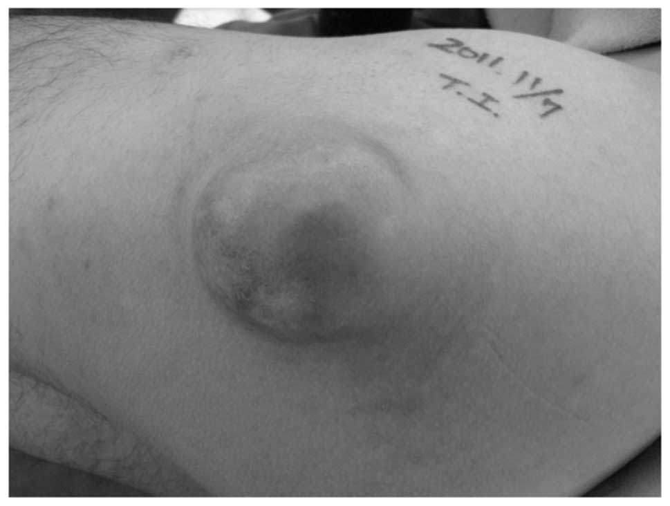
A 36-year-old male presented with a 10-year history of a slow-growing, painless mass in the left proximal thigh. The patient’s medical history was non-contributory. Written informed consent for publication was obtained from the patient. Physical examination revealed a ∼6×5-cm superficial, firm, non-tender mass in the posterolateral aspect of the left proximal thigh (Fig. 1). Neurological and vascular examinations were unremarkable, while the laboratory findings were within normal limits.

Figure 1

Photograph showing the ∼6×5-cm superficial mass in the posterolateral aspect of the left proximal thigh.

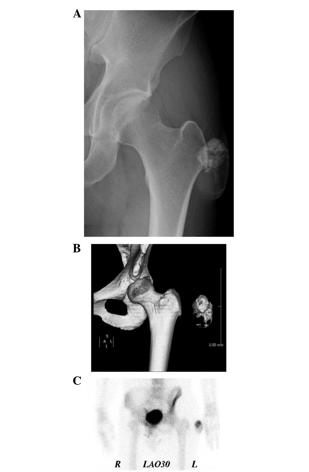
Plain radiographs showed a soft tissue mass with amorphous calcification and extensive foci of ossification (Fig. 2A). Computed tomography (CT) images demonstrated and confirmed an incompletely ossified shell in the lesion (Fig. 2B). Technetium-99m hydroxymethylenediphosphonate bone scintigraphy demonstrated heterogenous uptake in the lateral soft tissue of the left proximal thigh (Fig. 2C). 
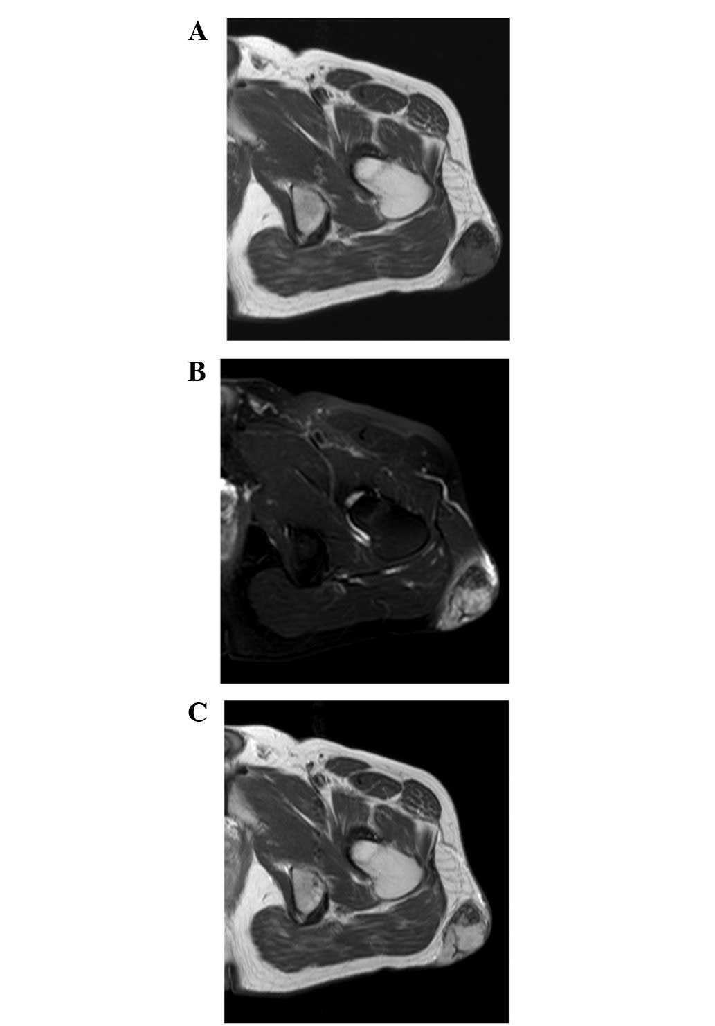
Magnetic resonance imaging (MRI) revealed a well-defined subcutaneous mass. The mass exhibited low to intermediate signal intensity on T1-weighted sequences (Fig. 3A) and heterogeneous high signal intensity, with foci of low signal intensity, on T2-weighted spectral presaturation with inversion recovery sequences (Fig. 3B). A fine linear low signal was observed on both T1- and T2-weighted sequences. Contrast-enhanced T1-weighted sequences demonstrated heterogenous enhancement throughout the mass (Fig. 3C). A benign soft tissue tumor with pronounced ossification was clinically suggested, and the lesion was marginally excised.

Figure 2

(A) Plain radiograph reveals a soft tissue mass with amorphous calcification and extensive foci of ossification. (B) Three-dimensional computed tomography image shows the presence of the lesion and the normal appearance of the proximal femur. (C) Technetium-99m hydroxymethylenediphosphonate bone scintigraphy reveals heterogenous uptake in the lateral soft tissue of the left proximal thigh.

Figure 3

Axial magnetic resonance images of ossifying fibromyxoid tumor in the left proximal thigh. (A) The T1-weighted sequence shows that the mass has low to intermediate signal intensity. (B) The T2-weighted spectral presaturation with inversion recovery sequence shows that the mass has heterogeneous high signal intensity with foci of low signal intensity. (C) The contrast-enhanced T1-weighted sequence shows heterogenous enhancement throughout the mass.

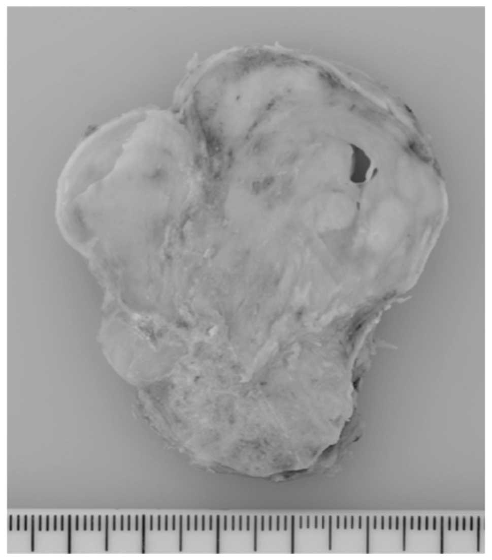
Grossly, the tumor was well circumscribed and covered by a thin fibrous capsule. A cut section revealed that the tumor was gray-white, solid, multinodular and rubbery to firm in consistency (Fig. 4). 
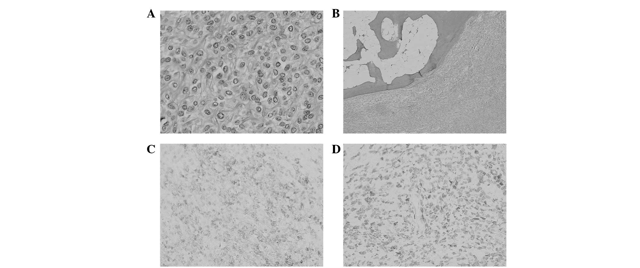
Histopathologically, the tumor was composed of uniform oval, polygonal or spindle-shaped cells, accompanied by an abundant fibromyxoid stroma (Fig. 5A). Ossification-forming bone shells were found in the periphery of the tumor (Fig. 5B). Necrosis and vascular space invasion were not observed. Immunohistochemically, the tumor cells were positive for vimentin, S-100 protein (Fig. 5C) and neuron-specific enolase (Fig. 5D), and focally for smooth muscle actin and CD56. Immunostaining for epithelial membrane antigen, cytokeratin, desmin, glial fibrillary acidic protein, chromogranin A, synaptophysin and CD57 was negative. The MIB-1 labeling index was <1%. Based on these features, the tumor was diagnosed as an OFMT.

Figure 4

Cut section of the ossifying fibromyxoid tumor displaying a well-circumscribed, gray-white, multinodular appearance.

Figure 5

Histopathological and immunohistochemical findings of ossifying fibromyxoid tumor. (A) The tumor is composed of relatively uniform round to oval cells with eosinophilic cytoplasm. (B) A peripheral shell of mature lamellar bone is evident. The tumor cells are immunoreactive for (C) S-100 protein and (D) neuron-specific enolase.

The postoperative course was uneventful, and the patient was doing well with no local recurrence 11 months following surgery.

Discussion

The majority of OFMTs are clinically and histopathologically benign; however, it has been noted that a subset of OFMTs have atypical histopathological features and exhibit correspondingly more aggressive clinical behavior (2,4,6). In 2003, Folpe and Weiss (2) proposed that OFMT may be classified as typical, atypical or malignant on the basis of its cellularity, nuclear grade and mitotic activity. More recently, Graham et al(4) confirmed the existence of malignant OFMT using immunohistochemistry and gene expression profiling. In view of the low to moderate cellularity, low nuclear grade, low mitotic activity and the absence of necrosis or vascular space invasion, our case was regarded as a typical OFMT.

The histogenesis of OFMT remains uncertain. Graham et al(4) recently suggested that this tumor exhibits a scrambled phenotype. Typically, as in our case, the cells are positive for vimentin and express S-100 protein. Atypical or malignant areas are observed to express S-100 protein less frequently than the typical areas (2). Other useful markers are CD10 and EAAT4 (3,4).

To date, a limited number of imaging findings of OFMT have been described in detail in the literature (5,7–10). Plain radiographs typically reveal a non-specific soft tissue mass with an incomplete rim of ossification. Erosion or periosteal reaction of the underlying bone is rarely observed. CT scans clearly demonstrate the presence of surrounding or intralesional ossification (7). The MRI appearances of OFMT are variable. The lesion is isointense to muscle on T1-weighted images and shows intermediate-to-high signal intensity on T2-weighted images. There are high signal intensity areas on T1- and T2-weighted images, suggesting hemorrhage and implying a high degree of vascularity (8,9). In addition, areas of ossification demonstrate low signal intensity on T1- and T2-weighted images. As with our case, the ossific element of OFMT has osteoblastic activity that is detected on bone scintigraphy (5,9,11).

Only seven cases of OFMT have been cytogenetically described (2,12–15). Clonal abnormalities of chromosome band 6p21 are prominent. Notably, a balanced or unbalanced t(6;12) (p21;q24) translocation appears to be characteristic for OFMT. A recent fluorescence in situ hybridization (FISH) study by Graham et al(4) revealed INI-1 deletion in 71% of cases. Most recently, Gebre-Medhin et al(15) demonstrated that PHF1 (at 6p21) is frequently rearranged in OFMT, including atypical and malignant variants. Moreover, PHF1 was fused to EP400 (at 12q24) in one atypical case with the t(6;12) translocation. OFMT is the second neoplasm to be identified, in addition to endometrial stromal tumor, in which PHF1 is involved in fusions with ectopic sequences. A FISH assay for PHF1 rearrangements would therefore be useful for the differential diagnosis of OFMT and its histopathological mimics (16).

In summary, we have described the imaging findings of a typical OFMT with clinical and histopathological correlation. Although rare, OFMT ought to be considered in the differential diagnosis of a well-circumscribed, slow-growing, painless, subcutaneous mass with irregular ossifications and/or calcifications.

Acknowledgements

This study was supported in part by the Ogata Foundation and the Foundation for the Promotion of Medical Science.

References

1 

Enzinger FM, Weiss SW and Liang CY: Ossifying fibromyxoid tumor of soft parts: a clinicopathological analysis of 59 cases. Am J Surg Pathol. 13:817–827. 1989. View Article : Google Scholar : PubMed/NCBI

2 

Folpe AL and Weiss SW: Ossifying fibromyxoid tumor of soft parts: a clinicopathologic study of 70 cases with emphasis on atypical and malignant variants. Am J Surg Pathol. 27:421–431. 2003. View Article : Google Scholar : PubMed/NCBI

3 

Miettinen M, Finnell V and Fetsch JF: Ossifying fibromyxoid tumor of soft parts-a clinicopathologic and immunohistochemical study of 104 cases with long-term follow-up and a critical review of the literature. Am J Surg Pathol. 32:996–1005. 2008. View Article : Google Scholar : PubMed/NCBI

4 

Graham RP, Dry S, Li X, Binder S, Bahrami A, Raimondi SC, Dogan A, Chakraborty S, Souchek JJ and Folpe AL: Ossifying fibromyxoid tumor of soft parts: a clinicopathologic, proteomic, and genomic study. Am J Surg Pathol. 35:1615–1625. 2011. View Article : Google Scholar : PubMed/NCBI

5 

Ogose A, Otsuka H, Morita T, Kobayashi H and Hirata Y: Ossifying fibromyxoid tumor resembling parosteal osteosarcoma. Skeletal Radiol. 27:578–580. 1998. View Article : Google Scholar : PubMed/NCBI

6 

Kilpatrick SE, Ward WG, Mozes M, Miettinen M, Fukunaga M and Fletcher CD: Atypical and malignant variants of ossifying fibromyxoid tumor. Clinicopathologic analysis of six cases. Am J Surg Pathol. 19:1039–1046. 1995. View Article : Google Scholar : PubMed/NCBI

7 

Schaffler G, Raith J, Ranner G, Weybora W and Jeserschek R: Radiographic appearance of an ossifying fibromyxoid tumor of soft parts. Skeletal Radiol. 26:615–618. 1997. View Article : Google Scholar : PubMed/NCBI

8 

Harish S, Polson A, Morris P, Malata C, Griffiths M and Bearcroft PW: Giant atypical ossifying fibromyxoid tumour of the calf. Skeletal Radiol. 35:248–253. 2006. View Article : Google Scholar : PubMed/NCBI

9 

Cha JH, Kwon JW, Cho EY, Lee CS, Yoon YC and Choi SH: Ossifying fibromyxoid tumor invading the spine: a case report and review of the literature. Skeletal Radiol. 37:1137–1140. 2008. View Article : Google Scholar : PubMed/NCBI

10 

Green RAR, Briggs TWR and Tirabosco R: Ossifying fibromyxoid tumour, an unusual cause of rim ossification: imaging features and correlation with histopathology. Eur J Radiol Extra. 70:e37–e40. 2009. View Article : Google Scholar

11 

Raith J, Ranner G, Schaffler G, Gröll R, Lindbichler F, Fritz K and Weybora W: Bone scan in ossifying fibromyxoid tumor of soft parts. Clin Nucl Med. 23:262–264. 1998. View Article : Google Scholar : PubMed/NCBI

12 

Sovani V, Velagaleti GV, Filipowicz E, Gatalica Z and Knisely AS: Ossifying fibromyxoid tumor of soft parts: report of a case with novel cytogenetic findings. Cancer Genet Cytogenet. 127:1–6. 2001. View Article : Google Scholar : PubMed/NCBI

13 

Nishio J, Iwasaki H, Ohjimi Y, Ishiguro M, Isayama T, Naito M, Okabayashi H, Kaneko Y and Kikuchi M: Ossifying fibromyxoid tumor of soft parts. Cytogenetic findings. Cancer Genet Cytogenet. 133:124–128. 2002. View Article : Google Scholar : PubMed/NCBI

14 

Kawashima H, Ogose A, Umezu H, Hotta T, Tohyama T, Tsuchiya M and Endo N: Ossifying fibromyxoid tumor of soft parts with clonal chromosomal aberrations. Cancer Genet Cytogenet. 176:156–160. 2007. View Article : Google Scholar : PubMed/NCBI

15 

Gebre-Medhin S, Nord KH, Möller E, Mandahl N, Magnusson L, Nilsson J, Jo VY, Vult von Steyern F, Brosjö O, Larsson O, Domanski HA, Sciot R, Debiec-Rychter M, Fletcher CDM and Mertens F: Recurrent rearrangement of the PHF1 gene in ossifying fibromyxoid tumors. Am J Pathol. 181:1069–1077. 2012. View Article : Google Scholar : PubMed/NCBI

16 

Nishio J: Updates on the cytogenetics and molecular cytogenetics of benign and intermediate soft tissue tumors (Review). Oncol Lett. 5:12–18. 2013.PubMed/NCBI

Related Articles

  • Abstract
  • View
  • Download
  • Twitter
Copy and paste a formatted citation
Spandidos Publications style
Ideta S, Nishio J, Aoki M, Ishimatsu T, Nabeshima K, Iwasaki H and Naito M: Imaging findings of ossifying fibromyxoid tumor with histopathological correlation: A case report. Oncol Lett 5: 1301-1304, 2013.
APA
Ideta, S., Nishio, J., Aoki, M., Ishimatsu, T., Nabeshima, K., Iwasaki, H., & Naito, M. (2013). Imaging findings of ossifying fibromyxoid tumor with histopathological correlation: A case report. Oncology Letters, 5, 1301-1304. https://doi.org/10.3892/ol.2013.1170
MLA
Ideta, S., Nishio, J., Aoki, M., Ishimatsu, T., Nabeshima, K., Iwasaki, H., Naito, M."Imaging findings of ossifying fibromyxoid tumor with histopathological correlation: A case report". Oncology Letters 5.4 (2013): 1301-1304.
Chicago
Ideta, S., Nishio, J., Aoki, M., Ishimatsu, T., Nabeshima, K., Iwasaki, H., Naito, M."Imaging findings of ossifying fibromyxoid tumor with histopathological correlation: A case report". Oncology Letters 5, no. 4 (2013): 1301-1304. https://doi.org/10.3892/ol.2013.1170
Copy and paste a formatted citation
x
Spandidos Publications style
Ideta S, Nishio J, Aoki M, Ishimatsu T, Nabeshima K, Iwasaki H and Naito M: Imaging findings of ossifying fibromyxoid tumor with histopathological correlation: A case report. Oncol Lett 5: 1301-1304, 2013.
APA
Ideta, S., Nishio, J., Aoki, M., Ishimatsu, T., Nabeshima, K., Iwasaki, H., & Naito, M. (2013). Imaging findings of ossifying fibromyxoid tumor with histopathological correlation: A case report. Oncology Letters, 5, 1301-1304. https://doi.org/10.3892/ol.2013.1170
MLA
Ideta, S., Nishio, J., Aoki, M., Ishimatsu, T., Nabeshima, K., Iwasaki, H., Naito, M."Imaging findings of ossifying fibromyxoid tumor with histopathological correlation: A case report". Oncology Letters 5.4 (2013): 1301-1304.
Chicago
Ideta, S., Nishio, J., Aoki, M., Ishimatsu, T., Nabeshima, K., Iwasaki, H., Naito, M."Imaging findings of ossifying fibromyxoid tumor with histopathological correlation: A case report". Oncology Letters 5, no. 4 (2013): 1301-1304. https://doi.org/10.3892/ol.2013.1170
Follow us
  • Twitter
  • LinkedIn
  • Facebook
About
  • Spandidos Publications
  • Careers
  • Cookie Policy
  • Privacy Policy
How can we help?
  • Help
  • Live Chat
  • Contact
  • Email to our Support Team