Introduction
Lung cancer remains the leading cause of cancer
mortality in a number of countries worldwide and non-small cell
lung cancer (NSCLC) accounts for 80% of all lung malignancies
(1). Lung adenocarcimoma is one of
the most common subtypes of NSCLC, with high mortality rates and
poor prognosis (2). The majority of
lung adenocarcimoma cases present with a small primary tumor with
an increased tendency to metastasize to regional lymph nodes and
distant organs. Although significant advances in the diagnosis and
therapy of lung cancer have been made in recent decades, the
survival rate of this malignant disease has only minimally
improved. Lung cancer is a multi-step process with morphological
progression involving multiple molecular events (2). Thus, the identification of molecular
and biological alterations that occur during carcinogenesis and
progression may facilitate the investigation of the pathology of
the disease and generate new markers to more accurately predict and
monitor patients’ clinical outcome and therapy, subsequently aiding
to individualize the treatment of cancer in patients.
Circulating tumor cells (CTCs), which are derived
from primary sites or metastases, are capable of circulating freely
in the peripheral blood (PB) of cancer patients (3). CTCs have been isolated and
characterized in a variety of human solid tumors, including breast
cancer (4), melanoma (5), gastrointestinal cancer (6), lung cancer (7) and prostate cancer (8). Evidence from previous studies
highlighted that the presence of CTCs may reflect the tumor burden
and is associated with tumor relapse and progression (5,9–11).
Improvements in molecular technology have enabled the detection of
rare CTCs in PB samples using quantification methods, including
real-time PCR techniques. The main advantage of this approach lies
in its sensitivity, which is considered to be higher than the
reported sensitivity of immune-mediated detection, including
immunocytochemistry (12). Given
the heterogeneity of metastatic tumor cells, it is unlikely that
one marker suitable for the detection of CTCs in all cancer
patients exists. Therefore, we suggest that a multimarker assay is
likely to provide an improved strategy compared with single marker
assays and real-time reverse transcription PCR (RT-PCR) is easily
adaptable to multimarker assays (13).
Survivin is a member of the family of inhibitors of
the apoptotic proteins and has been implicated in the regulation of
cell survival and mitosis in cancer (14–16).
High levels of survivin expression have been detected in cancer
cells, with low levels of expression detected in the majority of
normal differentiated adult tissues (14). Previous studies suggest that the
overexpression of survivin is correlated with advanced disease,
accelerated time to recurrence, reduced survival and resistance to
therapy (17). Numerous studies
have demonstrated that the detection of survivin expression in CTCs
is significantly correlated with disease diagnosis and clinical
prognosis (18–20). The expression of the human
telomerase reverse transcriptase (hTERT) gene functions as the
major limiting factor for telomerase activity, which is important
in cellular immortalization, tumorigenesis and the progression of
cancer (21). The telomerase enzyme
complex consists of two major subunits and its expression is mainly
regulated by the catalytic subunit, hTERT (22). The cancer-related gene, hTERT, has
been used for the detection of CTCs in the PB of breast and gastric
cancer patients (20,23). Thyroid transcription factor 1
(TTF-1), which is a 38-kDa nuclear protein member of the NKx2
family of homodomain transcription factors, is selectively
expressed in the lung, thyroid and diencephalons (24). It has been used to identify primary
lung adenocarcinoma or thyroid tumors (25,26).
Epithelial tumors usually retain the keratin expression profile of
their normal epithelial origin. Cytokeratin-7 (CK-7) is a type II
cytoskeletal keratin that is frequently expressed in the epithelial
tissue lining the cavities (27).
It has been demonstrated that CK-7 is frequently expressed in a
variety of human tumors, including lung (27), gallbladder (28), breast (29), gastrointestinal (30) and urinary tract carcinoma (31). A previous study demonstrated that
CK-7 may serve as a potential marker for the detection of CTCs in
lung cancer (32).
In this study, we aimed to develop a real-time
RT-PCR assay to investigate the expression of four epithelial or
cancer-related mRNA markers, survivin, hTERT, CK-7 and TTF-1, in
order to detect levels of CTCs in PB samples of advanced lung
adenocarcinoma patients as well as from healthy controls. We
additionally evaluated the efficacy of each individual circulating
mRNA marker and all four mRNA markers in combination. Furthermore,
we investigated the correlation between the expression levels of
these four mRNA markers with the clinicopathological features of
advanced lung adenocarcinoma patients. Lastly, we analyzed the
correlation between CTC status with patients’ disease
progression.
Materials and methods
Patient and control sample selection
Sixty-eight lung adenocarcinoma patients undergoing
treatment at The Third Affiliated Hospital of Harbin Medical
University (Harbin, China), between January and June 2011, were
enrolled into the study and underwent complete tumor evaluation.
All patients provided written informed consent for the analysis.
The study was approved by the ethics committee of the Third
Affiliated Hospital of Harbin Medical University, Harbin, China.
Clinical stages were determined according to the criteria of the
American Joint Commission on Cancer (33) and the pathological type was
diagnosed as lung adenocarcinoma. In addition, PB samples were
obtained from 30 healthy patients who exhibited no evidence of any
clinically detectable disease at the time of blood withdrawal.
Processing of blood samples
To prevent the contamination of epithelial cells, a
catheter was used and the first 5 ml of blood was discarded.
Following this, a 5-ml sample of PB was obtained from patients
prior to therapy. Sample processing was performed within 2 h of
blood withdrawal. Mononuclear cells were enriched using density
gradient centrifugation, which removed red blood cells and
serum.
Cell culture and processing blood-spiking
samples
The lung adenocarcinoma cell line A549 was
maintained in RPMI-1640 medium (Invitrogen, Carlsbad, CA, USA) and
supplemented with 10% fetal bovine serum (Hyclone, Logan, UT, USA),
penicillin (100 U/ml) and streptomycin (100 U/ml) at 37°C in a 5%
CO2 air environment. For blood-spiking experiments, A549
cells were serially diluted in PBS and mixed with a 5-ml sample of
PB which was obtained from one healthy patient to form
concentrations of tumor cells of 1, 10, 100, 1,000 and 10,000 cells
per 5-ml blood sample.
Total RNA extraction and reverse
transcription
Total RNA from mononuclear cells of the PB were
extracted using TRIzol reagent (Invitrogen). RNA integrity was
verified electrophoretically and quantified using UV
spectrophotometry. The 260/280 ratio was 1.8–1.9. First strand cDNA
was synthesized from total RNA using a first strand cDNA synthesis
kit (Promega Corporation, Madison, WI, USA). The reverse
transcription was performed in a reaction mixture consisting of AMV
reverse transcriptase (high concentration), MgCl2 (25
mM), reverse transcription 10X buffer, dNTP mixture (10 mM),
recombinant RNasin ribonuclease inhibitor and
oligo(dT)15 primer. The reaction mixture, including 2
μg RNA, was incubated at 70°C for 10 min, spun briefly and
placed on ice. The 20-μl reaction mixture was then incubated
at 42°C for 45 min, heated to 95°C for 5 min and the resulting cDNA
was stored at −20°C.
Real-time PCR
Primers were designed for survivin, hTERT, CK-7 and
TTF-1 using Premier 5.0 software (Palo Alto, CA, USA). The primers
and Taqman probes were positioned to span exon-intron boundaries in
order to reduce the detection of genomic DNA. The primer sequences
and probes of each gene are shown in Table I. Real-time PCR and data collection
were performed with a MiniOpticon™ system (Bio-Rad, CDF-3120,
Hercules, CA, USA). The housekeeping gene, GAPDH, was used as an
internal control to normalize the expression levels of survivin,
hTERT, CK-7 and TTF-1. For the amplification of a total volume of
25 μl, each reaction mixture contained 12.5 μl
PremixExTaq (DRR039S, Takara Bio, Inc., Shiga, Japan), 0.6
μl forward primers (12.5 μM), 0.6 μl reverse
primers (12.5 μM), 0.3 μl Taqman probe (5 μM),
2 μl cDNA and 9 μl ddH2O. Amplification
conditions are shown in Table II.
For all samples, positive, negative and no template controls were
performed. To ensure the reproducibility of results, all genes were
tested in triplicate and the real-time PCR analyses were performed
blinded for the identity and clinical outcome of the patients.
 | Table IList of all primers used for PCR. |
Table I
List of all primers used for PCR.
| Primer | 5′-3′ sequence | Size of product
(bp) |
|---|
| Survivin (sense) |
AAGAACTGGCCCTTCTTGGA | 253 |
| Survivin
(antisense) |
CAACCGGACGAATGCTTTT | |
| Survivin Taqman
probe |
CCAGATGACGACCCCATTGGGCCGG | |
| hTERT (sense) |
TACGTCGTGGGAGCCAGAAC | 86 |
| hTERT
(antisense) |
TTCCGCAGAGAAAAGAGGGCCGA | |
| hTERT Taqman
probe |
TTCCGCAGAGAAAAGAGGGCCGA | |
| CK-7 (sense) |
GACATCGAGATCGCCACCTAC | 162 |
| CK-7
(antisense) |
ATTGCTGCCCATGGTTCCC | |
| CK-7 Taqman
probe |
AATGCCACCGCCACTGCTACTGCC | |
| TTF-1 (sense) |
CTTCGCCTTCCCCCTCTCC | 156 |
| TTF-1
(antisense) |
CCCTCCATGCCCACTTTCTTG | |
| TTF-1 Taqman
probe |
TCTTCCTTCCTCCTCCAGCCGCCG | |
| GAPDH (sense) |
GAAGGTGAAGGTCGGAGTC | 225 |
| GAPDH
(antisense) |
GAAGATGGTGATGGGATTTC | |
| GAPDH Taqman
probe |
CAAGCTTCCCGTTCTCAGCC | |
| Table IIReal-time PCR amplification
conditions. |
Table II
Real-time PCR amplification
conditions.
| Marker |
Pre-denaturation | Denaturation | Annealing | Extension | Cycles |
|---|
| Survivin | 95°C (2 min) | 95°C (5 sec) | 55°C (20 sec) | 72°C (20 sec) | 40 |
| hTERT | 95°C (5 min) | 95°C (20 sec) | 56°C (30 sec) | 72°C (30 sec) | 40 |
| CK-7 | 95°C (2 min) | 95°C (15 sec) | 58°C (30 sec) | 72°C (30 sec) | 40 |
| TTF-1 | 95°C (2 min) | 95°C (15 sec) | 58°C (30 sec) | 72°C (30 sec) | 40 |
Data analysis
Based on the three replications, mean crossing
points (the beginning of the PCR exponential phase) were used in
the calculations and the concentration of each tumor mRNA was
normalized against that of GAPDH in each sample. If the mean Ct
(threshold cycle) value for a gene of interest was ≥40, the gene
expression was considered to be undetectable. Based on previous
studies (11,34), the highest value of each marker in
healthy patients was determined as a cut-off value, which was used
to determine whether a patient’s PB sample was positive or not. For
one marker, gene expression was regarded as positive when it was
greater than the cut-off value. A PB sample was defined as
CTC-positive (CTC+) if any of the four markers tested
positive.
Statistical analysis
All data were analyzed using the SPSS 17.0 software
(Chicago, IL, USA). Chi-square analysis and Fisher’s exact test
were used to analyze the correlation between the positive
expression of the four markers and clinicopathological
characteristics among lung adnocarcinoma patients and healthy
patients. Bivariate correlations between variables were calculated
by Spearman’s rank correlation coefficients. Patients’ survival
curves were plotted by the Kaplan-Meier method and differences in
survival were compared using the log-rank test. P<0.05 was
considered to indicate a statistically significant result.
Real-time PCR was calculated using linear regression and Pearson’s
correlation.
Results
Sensitivity and specificity tests of
real-time PCR
To confirm the sensitivity and specificity of our
real-time RT-PCR assays, blood-spiking tests were performed to
examine the number of detectable cells within PB samples. Serial
numbers of human lung adenocarcinoma A549 cells were subjected to
RNA extraction and real-time RT-PCR procedures. In our assay, the
quantification of survivin, hTERT, CK-7 and TTF-1 was linearly
correlated with the loading numbers of A549 cells in PB samples and
the highest detection sensitivity observed was one A549 cell in 5
ml of PB.
Standard curves for the four markers were calculated
with MiniOpticon system software (Fig.
1). The corresponding real-time PCR efficiency (E) of one cycle
in the exponential phase was calculated according to the equation:
E=10[−slope]−1 (35). Investigated
transcripts demonstrated high real-time PCR efficiency rates which
were detected using the Taqman probe. The efficiency of survivin,
hTERT, CK-7 and TTF-1 was 85.6, 101, 108 and 102%, respectively. As
shown in Fig. 1, the investigated
concentration from 1 to 10000 cells/5 ml cDNA input appeared to
have high linearity (Pearson correlation coefficient
r>0.95).
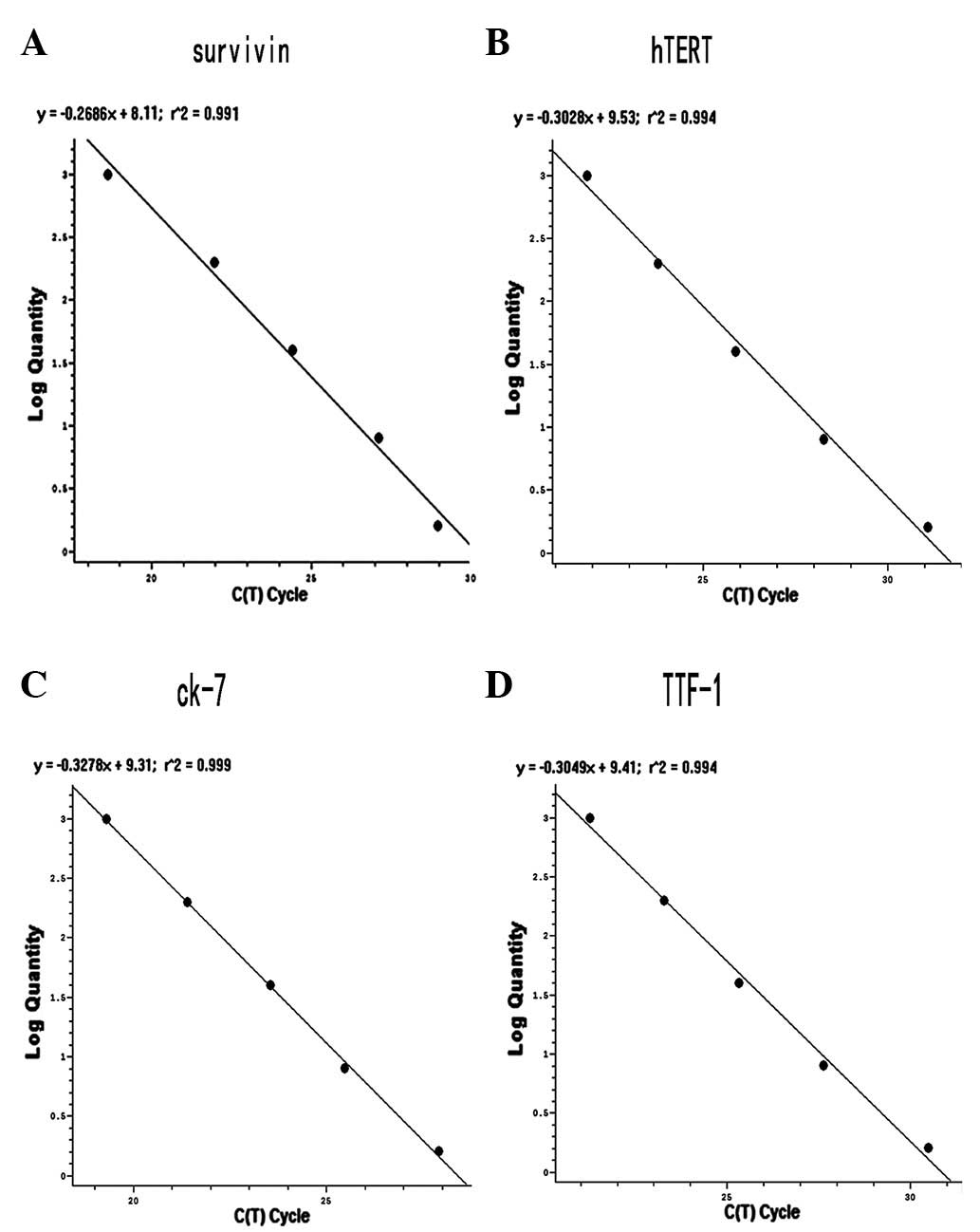 | Figure 1Standard curves for (A) survivin, (B)
hTERT, (C) CK-7 and (D) TTF-1 estimation. Each curve was
constructed using data from five external standards by plotting the
Ct value against the input quantity of A549 cells (the
concentration is 10,000 cells/5 ml, 1,000 cells/5 ml, 100 cells/5
ml, 10 cell/5 ml and 1 cell/5 ml. The concentration is represented
from top to bottom in each figure) of the four markers. hTERT,
human telomerase reverse transcriptase; TTF-1, thyroid
transcription factor 1; CK-7, cytokeratin-7; Ct, threshold
cycle. |
Relative expression of survivin, hTERT,
CK-7 and TTF-1 mRNA in the PB of advanced lung adenocarcinoma
patients
In our assay, the mRNA levels of these four markers
were slightly amplified in PB samples of 30 healthy controls. To
define a cut-off value for the normalization of mRNA expression of
the four markers in the peripherial blood, the relative mRNA
expression levels of each marker were examined in healthy patients.
The highest values of survivin, hTERT, CK-7 and TTF-1 mRNA
expression were 0.279, 0.0406, 0.094 and 1.182, respectively, which
were equal to their expression levels in 0.4–11.8 A549 cells.
Subsequently, four gene markers were compared with the highest
value to determine the expression levels in PB samples of 68 lung
adenocarcinoma patients. The sensitivity of survivin, hTERT, CK-7
and TTF-1 mRNA in patients was 41.18, 61.76, 41.18 and 35.29%,
respectively. The sensitivity of the four markers combined was
82.35%. Compared with single marker detection, the sensitivity of
the four markers in combination was significantly higher (Table III).
| Table IIIPositive detection rates for four
gene markers in the peripheral blood of patients with advanced lung
adenocarcinoma (n=68). |
Table III
Positive detection rates for four
gene markers in the peripheral blood of patients with advanced lung
adenocarcinoma (n=68).
| Survivin | hTERT | CK-7 | TTF-1 | Combined |
|---|
| Above cut-off
(n) | 28 | 42 | 28 | 24 | 56 |
| Below cut-off
(n) | 40 | 26 | 40 | 44 | 12 |
| Positive rate
(%) | 41.18 | 61.76 | 41.18 | 35.29 | 82.35 |
Correlation of the mRNA expression levels
of four markers with the clinicopathological features of advanced
lung adenocarcinoma patients
Real-time PCR determination of the four marker mRNA
expression levels was statistically analyzed to identify an
association with the clinicopathological features of advanced lung
adenocarcinoma patients. The characteristics of patients are
summarized in Table IV. Survivin,
hTERT and TTF-1 mRNA expression was significantly correlated with N
classification (P=0.023, P=0.007 and P=0.007, respectively).
Survivin, hTERT, CK-7 and TTF-1 mRNA expression was significantly
correlated with distant metastases (P=0.031, P=0.001, P=0.031 and
P=0.019, respectively). However, there was no significant
association between the mRNA expression levels of the four markers
and patients’ age or smoking (Table
IV). Spearman correlation analysis demonstrated that high
expression levels of survivin, hTERT and TTF-1 mRNA are positively
correlated with lymph node classification, and survivin, hTERT,
CK-7 and TTF-1 mRNA are positively correlated with distant
metastasis (all r>0, P<0.05, data not shown).
| Table IVCorrelation between
clinicopathological features of advanced lung adenocarcinoma
patients and the expression of mRNA markers. |
Table IV
Correlation between
clinicopathological features of advanced lung adenocarcinoma
patients and the expression of mRNA markers.
| Survivin (%) | | hTERT (%) | | CK-7 (%) | | TTF-1 (%) | |
|---|
|
|
|
|
|---|
| Characteristic | Positive | Negative | P-value | Positive | Negative | P-value | Positive | Negative | P-value | Positive | Negative | P-value |
|---|
| Age (years) | | | | | | | | | | | | |
| ≤50 | 6 (60.0) | 4 (40.0) | | 4 (40.0) | 6 (60.0) | | 6 (60.0) | 4 (60.0) | | 6 (60.0) | 4 (60.0) | |
| >50 | 22 (37.9) | 36 (62.1) | 0.190a | 38 (65.5) | 20 (34.5) | 0.125a | 22 (37.9) | 36 (62.1) | 0.190a | 18 (31.0) | 40 (69.0) | 0.077a |
| Smoking | | | | | | | | | | | | |
| No | 16 (44.4) | 20 (55.6) | | 20 (55.6) | 16 (44.4) | | 12 (33.3) | 24 (66.7) | | 14 (38.9) | 22 (61.1) | |
| Yes | 12 (37.5) | 20 (62.5) | 0.635 | 22 (68.8) | 10 (31.3) | 0.264 | 16 (50.0) | 16 (50.0) | 0.163 | 10 (31.3) | 22 (68.8) | 0.511 |
| N
classification | | | | | | | | | | | | |
| N0 | 7 (25.0) | 21 (75.0) | | 12 (42.9) | 16 (57.1) | | 8 (28.6) | 20 (71.4) | | 4 (14.3) | 24 (85.7) | |
| N1–3 | 21 (52.5) | 19 (47.5) | 0.023 | 30 (75.0) | 10 (25.0) | 0.007 | 20 (50.0) | 20 (50.0) | 0.077 | 20 (50.0) | 20 (50.0) | 0.002 |
| Distant
metastases | | | | | | | | | | | | |
| No | 8 (26.7) | 22 (73.3) | | 12 (40.0) | 18 (60.0) | | 8 (26.7) | 22 (73.3) | | 6 (20.0) | 24 (80.0) | |
| Yes | 20 (52.6) | 18 (47.4) | 0.031 | 30 (78.9) | 8 (21.1) | 0.001 | 20 (52.6) | 18 (47.4) | 0.031 | 18 (47.4) | 20 (52.6) | 0.019 |
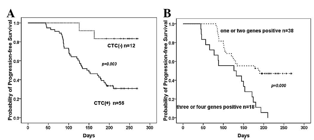
Disease progression and its association
with CTC status
To evaluate whether the mRNA expression levels of
the four markers in combination is associated with disease
progression, we defined the PB sample from advanced lung
adenocacinoma patients as CTC+ if any of the four
markers tested positive. The rate of disease progression in the
CTC+ group was 67.86% (38/56), while the rate of disease
progression in the CTC− group was 16.67% (2/12). There
was a significant difference between the two groups (P=0.001),
which was positively correlated with tumor progression (r=0.397,
P<0.001). Meanwhile, we stratified the CTC+ group to
a 1–2 marker mRNA overexpression group and a 3–4 marker mRNA
overexpression group. Disease progression rate of the 3–4 marker
mRNA overexpression group was 100% (18/18), while the 1–2 marker
mRNA expression group was 52.63% (20/38). There was a significant
difference between the two groups (P=0.000), which was positively
correlated with tumor progression (r=0.474, P<0.000).
The median progression time for the 68 cases was 150
days, with a range between 45 and 295 days. As shown in Fig. 2, compared with the CTC−
group, the CTC+ group had a shorter disease progression-free
survival (P=0.003). In addition, in the 3–4 marker overexpression
group, a shorter disease progression-free survival time was
observed compared with the 1–2 marker expression group
(P=0.000).
Discussion
In this study, we developed a real-time RT-PCR assay
to detect CTCs in advanced lung adenocarcinoma patients. To the
best of our knowledge, this is the first time that survivin, hTERT,
CK-7 and TTF-1 tumor-related mRNA markers have been used in
combination to detect CTCs of advanced lung adenocarcinoma
patients. This assay was designed to use Taqman probe to improve
the specificity of the amplification products. In addition, the
sensitivity demonstrated that target mRNAs are able to be detected
at a sensitivity of one A549 cell in 5 ml of PB, which is
sufficient to detect low levels of survivin, hTERT, CK-7 and TTF-1
mRNAs. Our present study demonstrates that the positive detection
rates of survivin, hTERT, CK-7 and TTF-1 in the PB of advanced lung
adenocarcinoma patients were 41.18, 61.76, 41.18 and 35.29%,
respectively. The sensitivity of this multimarker combination was
82.35%, significantly higher than that of any single marker.
Overexpression of survivin, hTERT, CK-7 and TTF-1 was significantly
associated with distant metastasis and overexpression of survivin,
hTERT and TTF-1 was significantly associated with N classification.
In addition, the CTC+ group was inversely correlated
with the disease progression of advanced lung adenocarcinoma
patients.
Compared with the sampling of lymph nodes and bone
marrow, PB collection is a noninvasive approach that may be
conducted throughout the course of disease. A number of
tumor-associated or epithelial-specific genes are used in the
detection of CTCs of different types of cancer, including CK, Her2,
CEA, MUC1, EpCAM, EGFR, hTERT, survivin, c-met, FN1 and several
other mRNA markers (4,6,11,20).
In our study, the tumor-associated genes survivin and hTERT,
epithelial-specific gene CK-7 and lung or thyroid
epithelial-specific TTF-1 were selected, and the positive detection
rate of the four gene mRNAs ranged between 35.29 and 61.76%, which
is similar to previous studies. These studies have demonstrated
that the detection rate of circulating tumor-related mRNAs ranged
between 30 and 60% in the PB of cancer patients (11,20).
Since genes are expressed heterogeneously and the alternative
expression of genes continuously occurs in tumor progression, no
tumor marker was identified to be consistently and specifically
expressed in all tumor cases. Single marker detection may limit the
reliability of the assay. Consequently, we combined the four
markers to detect levels of CTCs in advanced lung adenocarcinoma
patients. The sensitivity of the four markers in combination was
82.35%, an increase of 10.35% compared with results from the study
by Sher et al, detecting a combination of four marker genes
(36). We demonstrated that,
compared with single marker detection, combined use of the four
mRNA markers is capable of improving the sensitivity of CTC
detection in the PB of patients with advanced lung adenocarcinoma.
These results suggest that the use of multiple markers is able to
compensate for tumor cell heterogeneity in marker expression, low
mRNA levels and the rarity of CTCs in the PB.
Furthermore, we have analyzed the correlation
between these four mRNA markers with the clinicopathological
features of advanced lung adenocarcinoma. We identified that the
over-expression of survivin, hTERT and TTF-1 mRNA is positively
correlated with lymph node classification, and overexpression of
survivin, hTERT, CK-7 and TTF-1 mRNA is positively correlated with
distant metastasis. The present study strongly suggests that the
four gene mRNA marker combination may provide a valuable tool to
identify subsets of advanced lung adenocarcinoma patients with more
aggressive tumors, which have a high risk of metastasis and
recurrence. These results are consistent with the majority of other
studies of lung and breast cancer (7,11,20).
Following 10 months of follow-up, disease progression-free survival
was also significantly shorter in patients with CTC+
compared with those with CTC−, and with the increased
number of genes expressed, and increased risk of disease
progression, the progression-free survival shortened. These results
suggest that survivin, hTERT, CK-7 and TTF-1 mRNA are important in
lung adenocarcinoma development and analysis of the four marker
genes may provide valuable prediction information of disease
progression in patients. In terms of overall survival, the
follow-up period of the present study was only 10 months, which is
too short a time to assess the values of the investigated CTC
markers as predictors of overall survival. A longer observation
time with more serial monitoring is necessary to validate the
potential usefulness of these four markers in combination as an
overall predictor of survival.
In conclusion, we used quantitative real-time RT-PCR
to detect survivin, hTERT, CK-7 and TTF-1 mRNA expression levels in
PB samples of advanced lung adenocarcinoma patients. We identified
four mRNA markers that were capable of significantly improving the
sensitivity of detecting CTCs compared with single marker assays.
Multiple marker expression is positively correlated with N
classification and distant metastasis. Multiple marker-positive
CTCs are a useful surrogate predictor of disease progression.
However, it needs to be studied in larger patient cohorts including
early stage patients to precisely define the clinical relevance of
the four mRNA markers.
Abbreviations:
|
CTCs
|
circulating tumor cells
|
|
PB
|
peripheral blood
|
Acknowledgements
This study was supported by grants
from the National Natural Science Foundation of China (Nos.
30973417 and 81101758), grants from the Research Fund for the
Doctoral Program of China (No. 20072307110020), as well as grants
from the Postdoctoral Science Foundation of China (No.
2012M511514). The authors would like to thank Professor Ji-Lai Liu
and Professor Hai-feng Duan from the Beijing Institute of Radiation
Medicine for supplying laboratory materials and their technical
support.
References
|
1
|
Parkin DM, Pisani P and Ferlay J: Global
cancer statistics. CA Cancer J Clin. 49:33–64. 1999. View Article : Google Scholar
|
|
2
|
Hoffman PC, Mauer AM and Vokes EE: Lung
cancer. Lancet. 355:479–485. 2000. View Article : Google Scholar : PubMed/NCBI
|
|
3
|
Allard WJ, Matera J, Miller MC, et al:
Tumor cells circulate in the peripheral blood of all major
carcinomas but not in healthy subjects or patients with
nonmalignant diseases. Clin Cancer Res. 10:6897–6904. 2004.
View Article : Google Scholar : PubMed/NCBI
|
|
4
|
Tewes M, Aktas B, Welt A, et al: Molecular
profiling and predictive value of circulating tumor cells in
patients with metastatic breast cancer: an option for monitoring
response to breast cancer related therapies. Breast Cancer Res
Treat. 115:581–590. 2009. View Article : Google Scholar
|
|
5
|
Schuster R, Bechrakis NE, Stroux A, et al:
Circulating tumor cells as prognostic factor for distant metastases
and survival in patients with primary uveal melanoma. Clin Cancer
Res. 13:1171–1178. 2007. View Article : Google Scholar : PubMed/NCBI
|
|
6
|
Iinuma H, Okinaga K, Egami H, et al:
Usefulness and clinical significance of quantitative real-time
RT-PCR to detect isolated tumor cells in the peripheral blood and
tumor drainage blood of patients with colorectal cancer. Int J
Oncol. 28:297–306. 2006.PubMed/NCBI
|
|
7
|
Devriese LA, Bosma AJ, van de Heuvel MM,
Heemsbergen W, Voest EE and Schellens JH: Circulating tumor cell
detection in advanced non-small cell lung cancer patients by
multi-marker QPCR analysis. Lung Cancer. 75:242–247. 2012.
View Article : Google Scholar : PubMed/NCBI
|
|
8
|
Moreno JG, Miller MC, Gross S, Allard WJ,
Gomella LG and Terstappen LW: Circulating tumor cells predict
survival in patients with metastatic prostate cancer. Urology.
65:713–718. 2005. View Article : Google Scholar : PubMed/NCBI
|
|
9
|
Cristofanilli M and Mendelsohn J:
Circulating tumor cells in breast cancer: Advanced tools for
‘tailored’ therapy? Proc Natl Acad Sci USA. 103:17073–17074.
2006.
|
|
10
|
Nagrath S, Sequist LV, Maheswaran S, et
al: Isolation of rare circulating tumour cells in cancer patients
by microchip technology. Nature. 450:1235–1239. 2007. View Article : Google Scholar : PubMed/NCBI
|
|
11
|
Yoon SO, Kim YT, Jung KC, Jeon YK, Kim BH
and Kim CW: TTF-1 mRNA-positive circulating tumor cells in the
peripheral blood predict poor prognosis in surgically resected
non-small cell lung cancer patients. Lung Cancer. 71:209–216. 2011.
View Article : Google Scholar : PubMed/NCBI
|
|
12
|
Zieglschmid V, Hollmann C and Böcher O:
Detection of disseminated tumor cells in peripheral blood. Crit Rev
Clin Lab Sci. 42:155–196. 2005. View Article : Google Scholar : PubMed/NCBI
|
|
13
|
Sieuwerts AM, Kraan J, Bolt-de Vries J, et
al: Molecular characterization of circulating tumor cells in large
quantities of contaminating leukocytes by a multiplex real-time
PCR. Breast Cancer Res Treat. 118:455–468. 2009. View Article : Google Scholar : PubMed/NCBI
|
|
14
|
Ambrosini G, Adida C and Altieri DC: A
novel anti-apoptosis gene, survivin, expressed in cancer and
lymphoma. Nat Med. 3:917–921. 1997. View Article : Google Scholar : PubMed/NCBI
|
|
15
|
Blanc-Brude OP, Mesri M, Wall NR, Plescia
J, Dohi T and Altieri DC: Therapeutic targeting of the survivin
pathway in cancer: initiation of mitochondrial apoptosis and
suppression of tumor-associated angiogenesis. Clin Cancer Res.
9:2683–2692. 2003.PubMed/NCBI
|
|
16
|
Pennati M, Folini M and Zaffaroni N:
Targeting survivin in cancer therapy: fulfilled promises and open
questions. Carcinogenesis. 28:1133–1139. 2007. View Article : Google Scholar : PubMed/NCBI
|
|
17
|
Adida C, Berrebi D, Peuchmaur M,
Reyes-Mugica M and Altieri DC: Anti-apoptosis gene, survivin, and
prognosis of neuroblastoma. Lancet. 351:882–883. 1998. View Article : Google Scholar : PubMed/NCBI
|
|
18
|
Bertazza L, Mocellin S, Marchet A, et al:
Survivin gene levels in the peripheral blood of patients with
gastric cancer independently predict survival. J Transl Med.
7:1112009. View Article : Google Scholar : PubMed/NCBI
|
|
19
|
Cao W, Yang W, Li H, et al: Using
detection of survivin-expressing circulating tumor cells in
peripheral blood to predict tumor recurrence following curative
resection of gastric cancer. J Surg Oncol. 103:110–115. 2011.
View Article : Google Scholar
|
|
20
|
Shen C, Hu L, Xia L and Li Y: The
detection of circulating tumor cells of breast cancer patients by
using multimarker (Survivin, hTERT and hMAM) quantitative real-time
PCR. Clin Biochem. 42:194–200. 2009. View Article : Google Scholar : PubMed/NCBI
|
|
21
|
Daniel M, Peek GW and Tollefsbol TO:
Regulation of the human catalytic subunit of telomerase (hTERT).
Gene. 498:135–146. 2012. View Article : Google Scholar : PubMed/NCBI
|
|
22
|
Miura N, Horikawa I, Nishimoto A, et al:
Progressive telomere shortening and telomerase reactivation during
hepatocellular carcinogenesis. Cancer Genet Cytogenet. 93:56–62.
1997. View Article : Google Scholar : PubMed/NCBI
|
|
23
|
Wu CH, Lin SR, Hsieh JS, et al: Molecular
detection of disseminated tumor cells in the peripheral blood of
patients with gastric cancer: evaluation of their prognostic
significance. Dis Markers. 22:103–109. 2006. View Article : Google Scholar : PubMed/NCBI
|
|
24
|
Chang YL, Lee YC, Liao WY and Wu CT: The
utility and limitation of thyroid transcription factor-1 protein in
primary and metastatic pulmonary neoplasms. Lung Cancer.
44:149–157. 2004. View Article : Google Scholar : PubMed/NCBI
|
|
25
|
Lazzaro D, Price M, de Felice M and Di
Lauro R: The transcription factor TTF-1 is expressed at the onset
of thyroid and lung morphogenesis and in restricted regions of the
foetal brain. Development. 113:1093–1104. 1991.PubMed/NCBI
|
|
26
|
Pelosi G, Fraggetta F, Pasini F, et al:
Immunoreactivity for thyroid transcription factor-1 in stage I
non-small cell carcinomas of the lung. Am J Surg Pathol.
25:363–372. 2001. View Article : Google Scholar : PubMed/NCBI
|
|
27
|
Wicha MS: Cancer stem cells and
metastasis: lethal seeds. Clin Cancer Res. 12:5606–5607. 2006.
View Article : Google Scholar : PubMed/NCBI
|
|
28
|
Kalekou H and Miliaras D: Cytokeratin 7
and 20 expression in gallbladder carcinoma. Pol J Pathol. 62:25–30.
2011.PubMed/NCBI
|
|
29
|
Kawaguchi K and Shin SJ:
Immunohistochemical staining characteristics of low-grade
adenosquamous carcinoma of the breast. Am J Surg Pathol.
36:1009–1020. 2012. View Article : Google Scholar : PubMed/NCBI
|
|
30
|
Coban S, Ormeci N, Savaş B, et al:
Evaluation of Barrett’s esophagus with CK7, CK20, p53, Ki67, and
cyclooxygenase expressions using chromoendoscopical examination.
Dis Esophagus. May 16–2012.(Epub ahead of print).
|
|
31
|
Ross H, Martignoni G and Argani P: Renal
cell carcinoma with clear cell and papillary features. Arch Pathol
Lab Med. 136:391–399. 2012. View Article : Google Scholar : PubMed/NCBI
|
|
32
|
Xi L, Nicastri DG, El-Hefnawy T, Hughes
SJ, Luketich JD and Godfrey TE: Optimal markers for real-time
quantitative reverse transcription PCR detection of circulating
tumor cells from melanoma, breast, colon, esophageal, head and
neck, and lung cancers. Clin Chem. 53:1206–1215. 2007. View Article : Google Scholar : PubMed/NCBI
|
|
33
|
Greene FL, Page DL, Fleming ID, Fritz AG,
Balch CM, Haller DG and Morrow M: AJCC Cancer Staging Handbook. 6th
edition. Springer-Verlag; New York: pp. 170–171. 2001
|
|
34
|
Tjensvoll K, Oltedal S, Farmen RK, et al:
Disseminated tumor cells in bone marrow assessed by TWIST1,
cytokeratin 19, and mammaglobin A mRNA predict clinical outcome in
operable breast cancer patients. Clin Breast Cancer. 10:378–384.
2010. View Article : Google Scholar : PubMed/NCBI
|
|
35
|
Bustin SA, Benes V, Garson JA, et al: The
MIQE guidelines: minimum information for publication of
quantitative real-time PCR experiments. Clin Chem. 55:611–622.
2009. View Article : Google Scholar : PubMed/NCBI
|
|
36
|
Sher YP, Shih JY, Yang PC, et al:
Prognosis of non-small cell lung cancer patients by detecting
circulating cancer cells in the peripheral blood with multiple
marker genes. Clin Cancer Res. 11:173–179. 2005.PubMed/NCBI
|