Introduction
Leiomyoma is a benign tumour commonly encountered in
the genitourinary and gastrointestinal organs in adults (1). Cutaneous leiomyomas are rare benign
tumors arising from the arrector pili muscle of hair follicles,
ranging in number from a few to several hundred (2,3). The
skin is the second most common location for leiomyoma after the
uterus, hosting ∼5% of all leiomyomas (4). According to their site of origin,
leiomyomas may be classified into three types: i) piloleiomyomas,
ii) angioleiomyomas and iii) dartoic leiomyomas. Piloleiomyomas are
derived from the arrector pili muscle of hair follicles, whereas
angioleiomyomas include those originating from the vascular smooth
muscle and dartoic leiomyomas consist of those originating from the
smooth muscle of genital skin (5).
Cutaneous leiomyomas are more likely to occur in adults than in
children, and often arise in the fifth and sixth decades of life
(6). These lesions may be
hereditary or sporadic (7).
Case report
In the present study, we describe a case of a
10-year-old female submitted to the Dermatology Department of Recep
Tayyip Erdoğan University Medical Faculty, Rize, Turkey, with a
two-month history of a lesion in the right scapular and lumbar
regions. During the second month, the mass was observed to have
increased in size and become painful. On clinical examination,
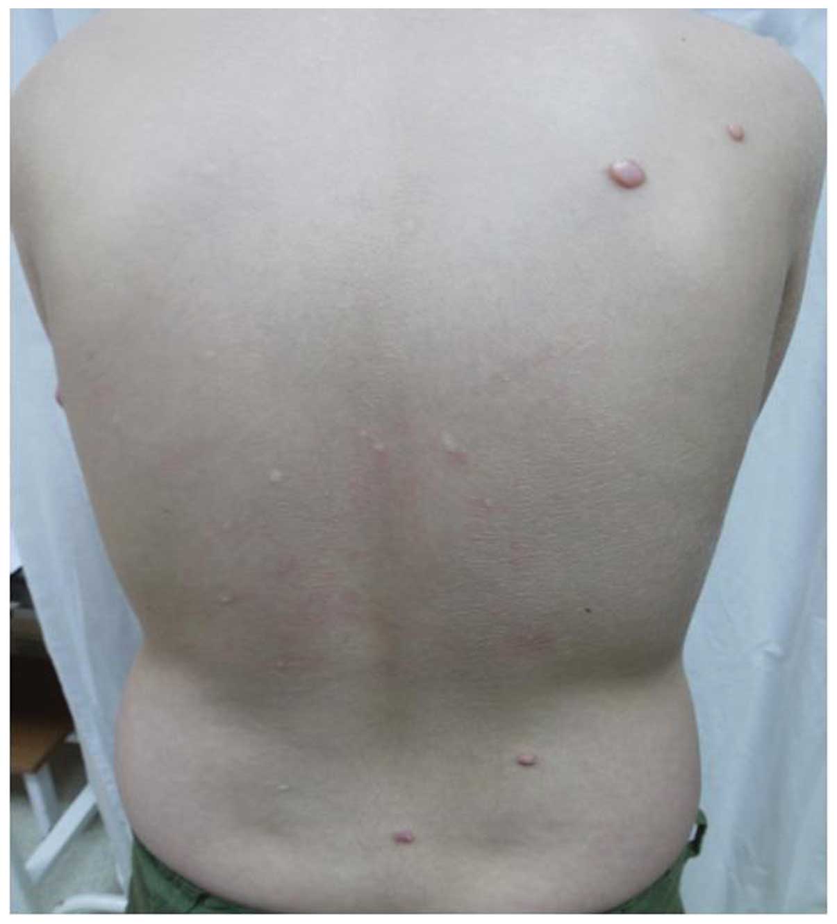
multiple firm red-brown masses were observed on the back of the
patient, the largest of which measured ∼10×15 mm and was located in
the left scapular region (Fig. 1).
Thorough clinical examination did not reveal any evidence of tumors
located elsewhere or any pertinent past clinical history. No
history of significant or hereditary diseases in the family were
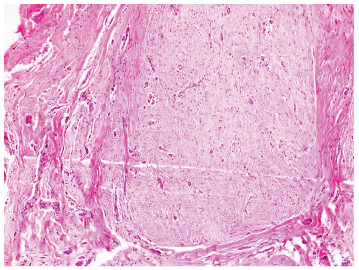
reported. A punch biopsy was performed by the clinician. Spindle
cells with an eosinophilic cytoplasm were observed under high-power
examination (Fig. 2).
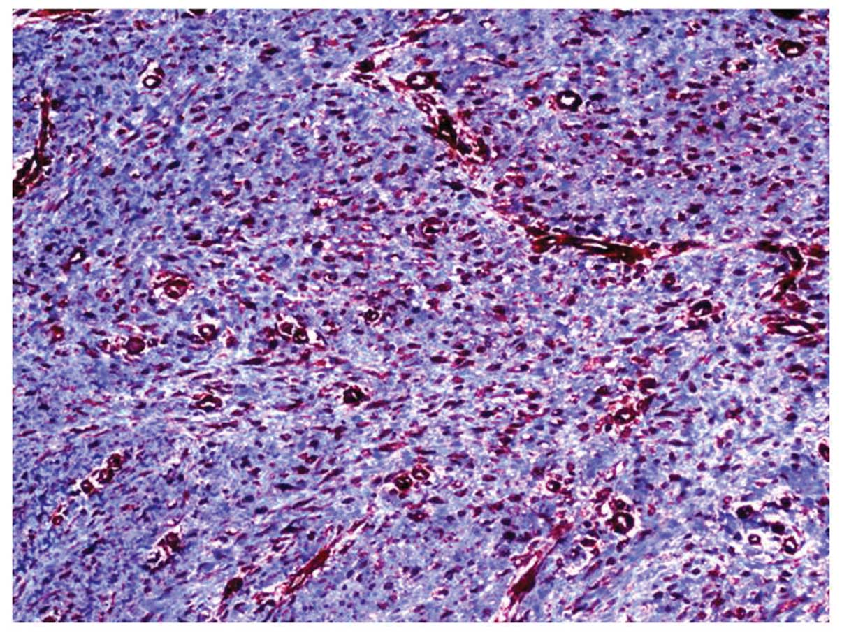
Immunohistochemical studies were performed and the cells stained
strongly positive for smooth muscle actin (SMA) (Fig. 3). As a result, the patient was
diagnosed with pilar leiomyoma.
This study was approved by the ethics committee of
the University of Rize, Turkey. The patient consented to the
publication of this study.
Discussion
The anatomical distribution of cutaneous leiomyomas
is extensive. Leiomyomas may present clinically as either solitary
or multiple lesions that have a skin-colored or red surface, and
are most commonly located in the extremities (7). Solitary and multiple pilar leiomyomas
arise from arrector pili muscles (8). Pilar leiomyomas are the most common
type of leiomyoma and range from 2 to 20 mm in diameter. When
multiple leiomyomas exist, these typically consist of red-brown
grouped papules, commonly located on the trunk or the extremities
(9). Cutaneous leiomyomas may be
asymptomatic, but are typically extremely painful (3). The pain experienced may be spontaneous
or as a result of exposure to cold, pressure or emotional stress
(3,10). The diagnosis of cutaneous leiomyomas
may be accomplished by microscopic examination of a hematoxylin and
eosin-stained biopsy of the papule or nodule (10). Tumors in each classification have
distinct clinical and/or histologic characteristics (4). Pilar leiomyomas are non-capsulated,
circumscribed dermal tumors composed of numerous fascicles of
smooth muscle in an interlacing and whorled arrangement (9).
While solitary lesions may be easily treated by
surgical excision, multiple lesions covering large areas are more
difficult to treat (3). Our patient
was referred to a plastic surgeon for surgical treatment.
In conclusion, although cutaneous leiomyoma is a
rare disorder we identified a case of pilar leiomyoma in a young
female. In the present case, a careful clinical assessment led to
the correct diagnosis and therapy. Although cutaneous leiomyomas
occur more frequently in adults, we suggest that leiomyoma ought to
be considered in the differential diagnosis of any cutaneous or
mucosal mass in children.
References
|
1
|
Yamato M, Nishimura G, Koguchi Y and
Saotome K: Calcified leiomyoma of deep soft tissue in a child.
Pediatr Radiol. 29:135–137. 1999. View Article : Google Scholar : PubMed/NCBI
|
|
2
|
Martinez-Mir A, Gordon D, Horev L,
Klapholz L, Ott J, Christiano AM and Zlotogorski A: Multiple
cutaneous and uterine leiomyomas: refinement of the genetic locus
for multiple cutaneous and uterine leiomyomas on chromosome
1q42.3–43. J Invest Dermatol. 118:876–880. 2002.PubMed/NCBI
|
|
3
|
Christenson LJ, Smith K and Arpey CJ:
Treatment of multiple cutaneous leiomyomas with CO2
laser ablation. Dermatol Surg. 26:319–322. 2000. View Article : Google Scholar : PubMed/NCBI
|
|
4
|
Malhotra P, Walia H, Singh A and Ramesh V:
Leiomyoma cutis: a clinicopathological series of 37 cases. Indian J
Dermatol. 55:337–341. 2010. View Article : Google Scholar : PubMed/NCBI
|
|
5
|
Badeloe S, van Geel M, van Steensel MA,
Bastida J, Ferrando J, Steijlen PM, Frank J and Poblete-Gutiérrez
P: Diffuse and segmental variants of cutaneous leiomyomatosis:
novel mutations in the fumarate hydratase gene and review of the
literature. Exp Dermatol. 15:735–741. 2006. View Article : Google Scholar : PubMed/NCBI
|
|
6
|
Robati RM, Barikbin B, Kavand S,
Sarrafi-Rad N and Moradloo M: Solitary cutaneous leiomyoma in an
infant. Pediatr Dermatol. 26:621–623. 2009. View Article : Google Scholar : PubMed/NCBI
|
|
7
|
Fons ME, Bachhuber T and Plaza JA:
Cutaneous leiomyosarcoma originating in a symplastic pilar
leiomyoma: a rare occurrence and potential diagnostic pitfall. J
Cutan Pathol. 38:49–53. 2011. View Article : Google Scholar : PubMed/NCBI
|
|
8
|
Akay BN, Boyvat A, Heper AO and Unlu E:
Congenital pilar leiomyoma. J Am Acad Dermatol. 59(5 Suppl):
S102–S104. 2008. View Article : Google Scholar : PubMed/NCBI
|
|
9
|
Matthews JH, Pichardo RO, Hitchcock MG and
Leshin B: Cutaneous leiomyoma with cytologic atypia, akin to
uterine symplastic leiomyoma. Dermatol Surg. 30:1249–1251.
2004.PubMed/NCBI
|
|
10
|
Stewart L, Glenn G and Toro JR: Cutaneous
leiomyomas: a clinical marker of risk for hereditary leiomyomatosis
and renal cell cancer. Dermatol Nurs. 18:335–341. 2006.PubMed/NCBI
|