Introduction
Gastric cancer (GC) is an extremely common disease
worldwide and the second most common cause of cancer-related
mortality (1). GC is considered to
be a disease of the middle aged and elderly. However, a previous
study revealed that 2 to 15% of patients with GC are younger than
40 years of age (defined as young GC patients), and that the
relative proportion of young GC patients is higher than that of
older GC patients (2). Despite the
advances in chemotherapy and surgical techniques, the overall
5-year survival rate of GC patients in China remains low (3). At present, clinical staging and
histopathological criteria are the only parameters used to stratify
patients. However, the current staging classifications do not
produce accurate predictions of patient outcomes. Molecular
biomarkers may account for this diversity and several prognostic
factors have been identified (4).
However, none of these methods have been proven to be robust enough
to be incorporated into routine practice.
S100A4 belongs to the S100 family, which is involved
in the regulation of a wide range of intracellular and
extracellular biological functions, including cell motility,
differentiation and contractility, and is classified as a
metastasis-related gene (5). A
number of studies have suggested that S100A4 overexpression is
correlated with poor clinical outcomes in various human cancers,
such as bladder (6), colorectal
(7), ovarian (8) and esophageal carcinoma (9). In the present study,
immunohistochemistry (IHC) and RT-PCR were used to analyze the
expression of S100A4 in 85 clinicopathologically characterized
young GC patients.
Patients and methods
Patient population
A total of 85 young GC patients treated at Tianjin
Medical University Cancer Hospital and Institute of China (Tianjin,
China) between January 2001 and December 2006 were included in the
study at the time of primary surgery for GC. The study was approved
by the Ethics Committee of the hospital and informed consent was
obtained from the patients. Patients with any of the following
conditions were excluded from the present study: older than 40
years old, histology other than adenocarcinoma, preoperative
chemoradiotherapy, mortality not caused by cancer and unknown stage
of disease. The study population included 85 patients in
tumor-node-metastasis (TNM) stages I–IV who had undergone surgery.
Paraffin sections of tissues from these patients were prepared. The
presence or absence of distant metastases was determined through
radiological examination. Primary tumor sections were re-evaluated
by an experienced pathologist who was blinded to the patients’
survival or other clinical variables to ensure consistent staging
and grading. Representative sections for each tumor were identified
and prepared for subsequent IHC analysis.
IHC and scoring
IHC staining was performed based on a previously
described method (10) to identify
changes in the protein expression of the primary GC, metastatic LN
and their normal counterpart tissues. In brief, the slides were
placed in an oven at 60°C for 2 h, deparaffinized with xylene and
then rehydrated. The sections were submerged in an EDTA antigenic
retrieval buffer, placed in a microwave for antigen retrieval,
treated with 3% hydrogen peroxide in methanol to quench endogenous
peroxidase activity and then incubated with 1% bovine serum albumin
to block non-specific binding. The sections were incubated with
mouse anti-S100A4 (Santa Cruz Biotechnology, Santa Cruz, CA, USA)
overnight at 4°C. Normal goat serum was used as the negative
control. The tissue sections were washed, treated with secondary
antibody, counterstained with hematoxylin, dehydrated and then
mounted.
S100A4 was stained yellow-brown in the cytoplasm and
nucleus. The degree of immunostaining was reviewed and scored
independently by two observers based on the staining intensity and
percentage of positive cells. The intensity grading scale was
according to the following criteria: 0 (no staining), 1 (weak
staining, light yellow), 2 (moderate staining yellow-brown) and 3
(strong staining, brown). Moderate and strong staining indicated
tumors with high S100A4 expression, while no and weak staining
indicated low S100A4 expression.
RT-PCR
Total RNA was extracted from the primary GC,
metastatic LN and their normal counterpart tissues using TRIzol
reagent (Takara Biotech, Dalian Co., Ltd., Otsu, Japan). Reverse
transcriptase reactions were performed using 1 mg of total RNA and
PrimeScript™ RT reagent kit (Takara Biotech) followed by PCR
amplification (TC-412 thermal cycler; Techne, Stone, Staffordshire,
UK) with specific primers. PCR amplification was performed as
follows: 30 cycles of 94°C for 5 min, 58°C for 30 sec and 72°C for
30 sec, with a final extension of 72°C for 10 min. After the PCR
products were electrophoresed on 1.5% agarose gels, they were
arrayed using a Bio-Rad scanner system (Hercules, CA, USA) and
analyzed with Quantity One software. The specific primers for the
S100A4 gene were S100A4-sense (5′-GATGTGATGGTGTCCAccTT-3′) and
S100A4-antisense (5′-ATTTCTTCCTGGGCTGCTTA-3′) whose target was a
277-bp fragment. Primer pairs specific to the β-actin gene
(β-actin-sense, 5′-CCAGATCAtGTTTGAGACCT-3′; β-actin-antisense,
5′-TTGAAGGTAGTTTCGTGGAT-3′; PCR product, 480 bp) served as the
internal standard.
Postoperative follow-up
Following surgery, each patient was scheduled for a
follow-up examination every 4 months in the first year,
semi-annually in the second year and annually thereafter. More
frequent examinations were scheduled if clinically indicated. The
cause of mortality was registered and classified as mortality due
to GC, other causes or unknown causes. For overall survival, the
median follow-up of the surviving patients was 15 months (range,
2–81 months).
Statistical analysis
The associations between S100A4 staining and
clinicopathological variables were tested using the two-tailed
Fisher’s exact test or linear-by-linear association Chi-squared
test. The Kaplan-Meier method was used to calculate the survival
functions and differences were assessed with the log-rank test.
Multivariate analysis was performed using the Cox proportional
hazards regression model. Overall and disease-specific survival
were determined from the time of surgery until mortality. P<0.05
was considered to indicate statistically significant differences.
All reported P-values were two-sided and all analyses were
performed using the Statistical Package for Social Sciences,
version 19.0, for Windows (SPSS Inc., Chicago, IL, USA).
Results
Expression of S100A4
Expression of S100A4 in the gastric
mucosa
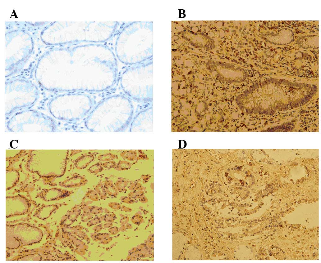
S100A4 protein was detected in 18 (21.18%) out of
the 85 non-tumor mucosal samples obtained from young patients. The
majority of the samples expressed the protein at a low level. The
expression was negligible or negative in the normal gastric mucosa
but moderate to strong in lymphocytes and smooth muscles cells.
Among the 85 GC tissue specimens from young patients screened for
S100A4 protein expression, 53 (62.35%) exhibited S100A4
overexpression, in which immunostaining was observed in the
cytoplasm or the nucleus of the tumor cells (Figs. 1–4).
Statistically, S100A4 overexpression was closely associated with
the tumor size (P=0.017), Lauren classification (P=0.002),
histological classification (P=0.010), histological differentiation
(P=0.000), Borrmann classification (P=0.020), TNM stage (P=0.000),
LN metastasis (P=0.000) and distant metastasis (P=0.024). However,
no significant correlation was observed between S100A4
overexpression and other clinicopathological parameters, including
age, gender, tumor location, surgery type and Heliobacter
pylori infection (Table I).
 | Table IAssociation between S100A4 expression
and clinicopathological characteristics. |
Table I
Association between S100A4 expression
and clinicopathological characteristics.
| S100A4
| | |
|---|
| Characteristics | Low | High | χ2 | P-value |
|---|
| Age (years) | | | 0.134 | 0.935 |
| <30 | 6 | 11 | | |
| 31–35 | 9 | 16 | | |
| 36–40 | 17 | 26 | | |
| Gender | | | 1.011 | 0.315 |
| Male | 15 | 19 | | |
| Female | 17 | 34 | | |
| Tumor location | | | 0.894 | 0.640 |
| Proximal | 5 | 11 | | |
| Middle | 10 | 12 | | |
| Distal | 17 | 30 | | |
| Tumor size (cm) | | | 5.647 | 0.017 |
| <5 | 18 | 16 | | |
| ≥5 | 14 | 37 | | |
| Surgery | | | 3.348 | 0.341 |
| Distal subtotal
gastrectomy | 17 | 28 | | |
| Proximal subtotal
gastrectomy | 5 | 9 | | |
| Total
gastrectomy | 9 | 9 | | |
| Other | 1 | 7 | | |
| Lauren
classification | | | 9.465 | 0.002 |
| Intestinal | 17 | 11 | | |
| Diffuse | 15 | 42 | | |
| Histological
classification | | | 13.326 | 0.010 |
| Tubular
adenocarcinoma | 14 | 10 | | |
| Papillary
adenocarcinoma | 5 | 3 | | |
| Mucinous
adenocarcinoma | 7 | 11 | | |
| Poorly
differentiated adenocarcinoma | 6 | 25 | | |
| Other | 0 | 4 | | |
| Histological
differentiation | | | 18.501 | 0.000 |
| Well | 8 | 1 | | |
| Moderately | 7 | 3 | | |
| Poorly | 17 | 49 | | |
| Borrmann
classification (type) | | | 9.890 | 0.020 |
| I | 6 | 1 | | |
| II | 11 | 13 | | |
| III | 11 | 27 | | |
| IV | 4 | 12 | | |
|
Tumor-node-metastasis stage | | | 19.178 | 0.000 |
| I | 9 | 1 | | |
| II | 4 | 1 | | |
| III | 8 | 17 | | |
| IV | 11 | 34 | | |
| Lymph node
metastasis | | | 27.400 | 0.000 |
| N0 | 12 | 2 | | |
| N1 | 7 | 4 | | |
| N2 | 10 | 19 | | |
| N3 | 3 | 28 | | |
| Distant
metastasis | | | 5.070 | 0.024 |
| M0 | 31 | 46 | | |
| M1 | 0 | 8 | | |
| H.
pylori | | | 3.108 | 0.078 |
| (+) | 13 | 12 | | |
| (−) | 19 | 41 | | |
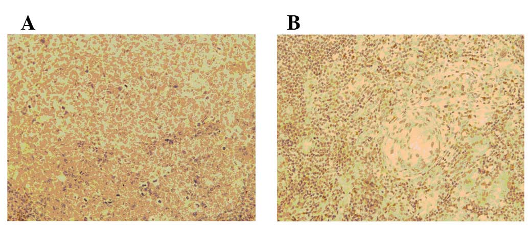
Expression of S100A4 in the LN
The S100A4 protein was present in non-metastatic LNs
at low levels (Fig. 2). The S100A4
protein level in the metastatic LNs was significantly higher than
that of the non-metastatic LNs. S100A4 overexpression was closely
correlated with LN staging (P=0.000; Table II). No significant difference in
S100A4 expression was observed between the GC tissues and
metastatic LNs (P=0.896).
| Table IIAssociation between S100A4 expression
and LN metastasis. |
Table II
Association between S100A4 expression
and LN metastasis.
| S100A4
| | |
|---|
|
Characteristics | Low | High | χ2 | P-value |
|---|
| LN metastasis | | | 14.409 | 0.000 |
| Negative | 45 | 17 | | |
| Positive | 24 | 38 | | |
| No. of LN
metastases | | | 14.409 | 0.000 |
| 1–6 | 13 | 2 | | |
| 7–15 | 7 | 15 | | |
| >15 | 4 | 21 | | |
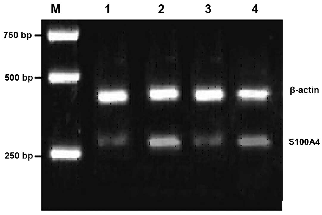
Expression of S100A4 mRNA
S100A4 expression was detected using IHC. The
relative expression level of β-actin/S100A4 in the primary GC
(0.4493±0.0453) was higher than that in the normal tissues
(0.1145±0.1000). In addition, the relative expression level of the
metastatic LNs (0.5491±0.0197) was higher than that in the normal
LN tissues (0.1558±0.0318). The difference between each group was
statistically significant (P<0.05; Fig. 3).
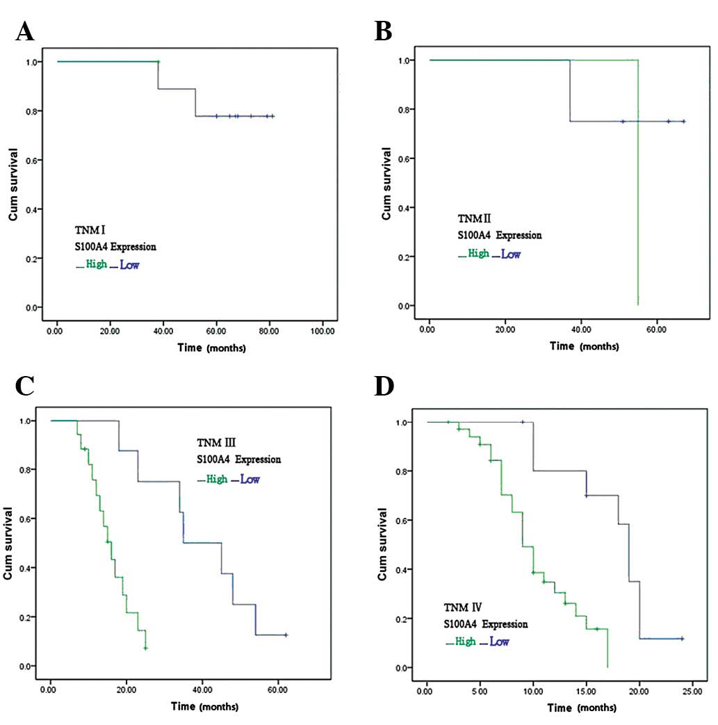
Correlation between S100A4 expression
and patient prognosis
In stage I–IV tumors, the 1- or 3-year survival rate
of the patients with high S100A4 expression was significantly lower
than that in the patients with low S100A4 expression (P<0.05;
Fig. 4).
Multivariate analysis of
clinicopathological parameters and prognosis
The factors with possible prognostic effects on
young GC patients were analyzed by Cox regression. The results
showed that patient age (P=0.035), tumor size (P=0.002), TNM stage
(P=0.001) and S100A4 upregulation (P=0.000) were independent
prognostic indicators for the disease (Table III).
| Table IIIMultivariate Cox regression survival
analysis of clinicopathological parameters and S100A4. |
Table III
Multivariate Cox regression survival
analysis of clinicopathological parameters and S100A4.
| Parameters | B | SE | Wald | Exp (B) | P-value |
|---|
| Age | 0.072 | 0.034 | 4.467 | 0.930 | 0.035 |
| Gender | 0.350 | 0.361 | 0.937 | 1.419 | 0.333 |
| Location | 0.204 | 0.246 | 0.684 | 0.816 | 0.408 |
| Size | 1.474 | 0.471 | 9.799 | 4.369 | 0.002 |
| Operation | 0.221 | 0.245 | 0.816 | 1.248 | 0.366 |
| Lauren | 0.417 | 0.440 | 0.897 | 1.517 | 0.344 |
| Histology | 0.069 | 0.178 | 0.151 | 0.933 | 0.698 |
|
Differentiaiton | 0.560 | 0.413 | 1.834 | 1.750 | 0.176 |
| Borrmann | 0.116 | 0.377 | 0.095 | 0.890 | 0.758 |
| TNM | 1.510 | 0.441 | 11.711 | 4.525 | 0.001 |
| Metastasis | 1.391 | 0.896 | 2.414 | 4.021 | 0.120 |
| H.
pylori | 0.315 | 0.332 | 0.899 | 1.370 | 0.343 |
| Expression | 2.315 | 0.507 | 20.864 | 10.129 | 0.000 |
Discussion
A number of studies on the clinicopathological and
molecular biological features of GC in the elderly have been
performed. However, only a few studies with a small sample size
have analyzed GC in young patients (11). Previous studies have associated
S100A4 protein expression with survival in several tumor types,
including bladder (6), colorectal
(7), ovarian (8) and esophageal carcinoma (9). S100A4 protein expression has also been
revealed to have prognostic significance in GC (12–14).
In a study of 436 cases, Wang et al(12) demonstrated that IHC staining for
S100A4 is associated with LN metastasis and poor prognosis in GC
patients. The present study demonstrates the prognostic
significance of S100A4 overexpression in the tumor cells of young
GC patients for the first time. Multivariate analysis showed that
S100A4 overexpression was associated with patient outcome. The
observed results may translate into clinically important
differences.
In certain studies (11,15), a
female predominance was observed among young GC patients. However,
a higher proportion of male patients has been noted in elderly GC
patients (16).
In the present study, the female-to-male ratio was
1.5:1 in the young GC patients. The reason for the higher number of
female patients is not yet known. In the present study, no
significant difference in S100A4 expression was observed between
the male and female patients (P>0.05).
Infection caused by H. pylori is considered
to be an important epidemiological risk factor for GC patients of
all ages (17,18). Moreover, certain epidemiological
data have revealed an association between H. pylori
infection and an increased risk of GC presenting at a young age
(19,20). However, in the present study, no
significant difference in S100A4 expression was observed in the
patients with and without H. pylori infection. In addition,
H. pylori positivity was infrequently observed among the
young GC patients.
Chung et al(15) reported that the incidence of primary
lesions in the upper third of the stomach is higher in young
patients than in elderly patients. In the present study, the same
results were reported, although no significant difference was
observed.
Similar to the results of previous studies (11,15),
the histology in young patients was observed to be more poorly
differentiated.
Gastrectomy in combination with lymphadenectomy is
the only potentially curative treatment for localized gastric
carcinomas, and curative resection offers the only chance of
long-term survival. However, radical resection is not common in
young GC patients due to clinicopathological characteristics.
Numerous studies have reported that the curative resection rate in
young patients with GC is low. In the present study, the type of
surgery showed no significant effect on S100A4 expression.
In the present study, quantitative RT-PCR revealed
the presence of S100A4 mRNA in non-tumor mucosal samples from young
patients. The non-metastatic LNs expressed S100A4 mRNA at low
levels. The levels of S100A4 mRNA in the GC tissues and metastatic
LNs were significantly higher compared with those in the matched
normal gastric mucosa and non-metastatic LNs, respectively.
Similarly, S100A4 expression was also detected using IHC. IHC
showed that the S100A4 protein was associated with GC cells and
other non-parenchymal cell types including lymphocytes. The
proportion of specimens with epithelial cells stained for S100A4
increased between the GC tissues and metastatic LNs. However, no
significant difference was observed. By contrast, the proportion of
lymphocytes stained for S100A4 remained at a consistently high
level in the normal gastric tissues, GC tissues and metastatic LNs.
Significant differences in the expression levels of the S100A4
protein were observed among the TNM stages of GC, as well as
between metastatic GC and non-metastatic GC. The results of the
present study are consistent with the results of previous studies
(12–14), in which S100A4 expression detected
via IHC was increased in malignant relative to non-malignant GC
specimens. Moreover, the deeper and more invasive regions of the
specimens in these studies were enhanced in immunohistochemically
detectable S100A4. This suggests that high levels of S100A4 protein
are associated with the invading regions of the tumors. S100A4
overexpression was more frequently observed in the metastatic LNs
than in the primary GC tissues from young patients. However, no
significant difference was observed.
Previous studies reported that S100A4 overexpression
is closely correlated with several factors for GC aggressiveness,
such as LN metastasis, distant metastasis and TNM stage (12–14).
In the present study, similar results were observed in the young GC
patients. These data further support a significant correlation
between S100A4 overexpression and GC progression, indicating the
putative role of S100A4 in tumor cell aggressiveness.
In conclusion, S100A4 overexpression is an
independent predictor of adverse prognosis in young GC patients.
Thus, it may be used as a biomarker in future clinical studies.
Acknowledgements
This work was supported by the China
National Natural Science Fund Committee (Grant No. 30971421) and
Tianjin Natural Science Fund Committee (Grant No.
10JCYBJC10200).
References
|
1
|
Kamangar F, Dores GM and Anderson WF:
Patterns of cancer incidence, mortality, and prevalence across five
continents: defining priorities to reduce cancer disparities in
different geographic regions of the world. J Clin Oncol.
24:2137–2150. 2006. View Article : Google Scholar
|
|
2
|
Simsa J, Leffler J, Hoch J, et al: Gastric
cancer in young patients - is there any hope for them. Acta Chir
Belg. 104:673–676. 2004.PubMed/NCBI
|
|
3
|
Chen CY, Wu CW, Lo SS, et al: Peritoneal
carcinomatosis and lymph node metastasis are prognostic indicators
in patients with Borrmann type IV gastric carcinoma.
Hepatogastroenterology. 49:874–877. 2002.PubMed/NCBI
|
|
4
|
Fareed KR, Kaye P, Soomro IN, et al:
Biomarkers of response to therapy in oesophago-gastric cancer. Gut.
58:127–143. 2009. View Article : Google Scholar : PubMed/NCBI
|
|
5
|
Garrett SC, Varney KM, Weber DJ and
Bresnick AR: S100A4, a mediator of metastasis. J Biol Chem.
281:677–680. 2006. View Article : Google Scholar : PubMed/NCBI
|
|
6
|
Matsumoto K, Irie A, Satoh T, et al:
Expression of S100A2 and S100A4 predicts for disease progression
and patient survival in bladder cancer. Urology. 70:602–607. 2007.
View Article : Google Scholar : PubMed/NCBI
|
|
7
|
Sack U, Walther W, Scudiero D, et al:
Novel effect of antihelminthic Niclosamide on S100A4-mediated
metastatic progression in colon cancer. J Natl Cancer Inst.
103:1018–1036. 2011. View Article : Google Scholar : PubMed/NCBI
|
|
8
|
Kikuchi N, Horiuchi A, Osada R, et al:
Nuclear expression of S100A4 is associated with aggressive behavior
of epithelial ovarian carcinoma: an important autocrine/paracrine
factor in tumor progression. Cancer Sci. 97:1061–1069. 2006.
View Article : Google Scholar
|
|
9
|
Lee OJ, Hong SM, Razvi MH, et al:
Expression of calcium-binding proteins S100A2 and S100A4 in
Barrett’s adenocarcinomas. Neoplasia. 8:843–850. 2006.PubMed/NCBI
|
|
10
|
Flatmark K, Pedersen KB, Nesland JM, et
al: Nuclear localization of the metastasis-related protein S100A4
correlates with tumour stage in colorectal cancer. J Pathol.
200:589–595. 2003. View Article : Google Scholar : PubMed/NCBI
|
|
11
|
Medina-Franco H, Heslin MJ and
Cortes-Gonzale R: Clinicopathological characteristics of gastric
carcinoma in young and elderly patients: a comparative study. Ann
Surg Oncol. 7:515–519. 2000. View Article : Google Scholar
|
|
12
|
Wang YY, Ye ZY, Zhao ZA, et al: High-level
expression of S100A4 correlates with lymph node metastasis and poor
prognosis in patients with gastric cancer. Ann Surg Oncol.
17:89–97. 2010. View Article : Google Scholar : PubMed/NCBI
|
|
13
|
Cho YG, Nam SW, Kim TY, et al:
Overexpression of S100A4 is closely related to the aggressiveness
of gastric cancer. APMIS. 111:539–545. 2003. View Article : Google Scholar : PubMed/NCBI
|
|
14
|
Yonemura Y, Endou Y, Kimura K, et al:
Inverse expression of S100A4 and E-cadherin is associated with
metastatic potential in gastric cancer. Clin Cancer Res.
6:4234–4242. 2000.PubMed/NCBI
|
|
15
|
Chung HW, Noh SH and Lim JB: Analysis of
demographic characteristics in 3242 young age gastric cancer
patients in Korea. World J Gastroenterol. 16:256–263. 2010.
View Article : Google Scholar : PubMed/NCBI
|
|
16
|
Kim DY, Joo JK, Ryu SY, et al:
Clinicopathologic characteristics of gastric carcinoma in elderly
patients: A comparison with young patients. World J Gastroenterol.
11:22–26. 2005. View Article : Google Scholar : PubMed/NCBI
|
|
17
|
Chung HW, Park SW, Chung JB, et al:
Differences in genetic expression profiles between young-age and
old-age gastric adenocarcinoma using cDNA microarray for endocrine
disruptor study. Oncol Rep. 12:33–39. 2004.
|
|
18
|
Ershler WB and Longo DL: The biology of
aging: the current research agenda. Cancer. 80:1284–1293. 1997.
View Article : Google Scholar : PubMed/NCBI
|
|
19
|
Masuda G, Tokunaga A, Shirakawa T, et al:
Helicobacter pylori infection, but not genetic polymorphism of
CYP2E1, is highly prevalent in gastric cancer patients younger than
40 years. Gastric Cancer. 10:98–103. 2007.PubMed/NCBI
|
|
20
|
Kokkola A, Valle J, Haapiainen R, et al:
Helicobacter pylori infection in young patients with gastric
carcinoma. Scand J Gastroenterol. 31:643–647. 1996. View Article : Google Scholar : PubMed/NCBI
|