Introduction
Intravenous leiomyomatosis (IVL) is a rare clinical
entity characterized by the intraluminal extension of leiomyoma
pedicles into the regional and systemic veins. Typically, the tumor
enters the uterine veins and may progressively extend to the iliac
vein, the inferior vena cava and occasionally to the right atrium
(1). Lesions may extend into the
heart, generating a condition known as intracardiac leiomyomatosis
(ICL), which may lead to congestive heart failure and occasionally
sudden fatalities. To date, >100 cases of ICL have been reported
in the literature. In five of these cases, mortality occurred due
to right heart obstruction (2). Due
to its rarity and subtle clinical features, the misdiagnosis of ICL
is common in clinical practice and may lead to delayed treatment
and fatal outcomes. The present study reports a case of ICL that
presented with no specific symptoms and was diagnosed by routine
abdominal ultrasonography (US) prior to surgery for uterine
leiomyoma. Thus, pre-operative assessments using abdominal US in
these cases may be considered extremely important tools of
diagnosis (3). Written informed
consent was obtained from the patient.
Case report
A 33-year-old, gravida 2 para 1 patient was admitted
to Shandong Provincial Hospital Affiliated to Shandong University
(Shandong, China) for myomectomy due to a rapidly growing myoma of
the uterus. The patient’s medical history included hypertension and
Hashimoto’s thyroiditis. The patient reported pelvic pain one month
prior to the hospital admission and a clinical examination revealed
a mobile, enlarged uterus with myoma. The patient reported no
weight loss or cardiopulmonary symptoms. Four years earlier, and in
association with a pregnancy, the myoma had measured 5 mm in size.
The myoma was approximately the same size when observed during an
abortion two years later. Vaginal US revealed a mass of 86×74×45 mm
in size on the right side of the uterus. The results of a number of
laboratory examinations, including those for tumor marker levels,
were normal. Electrocardiography (ECG) demonstrated a sinus pattern
with ST depression. X-rays of the thorax indicated a normal cardiac
silhouette and a normal appearance to the lungs.
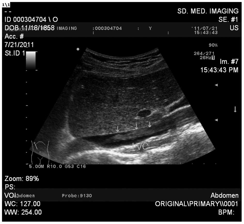
Routine pre-operative abdominal US revealed a
moderately sized echoic mass in the right internal iliac vein. The
mass extended to the common iliac vein, the inferior vena cava and
the orifice of the right atrium (Fig.
1). The common iliac vein was dilated to 20 mm in diameter on
the right side compared with 12 mm on the left side. The mass had
well-demarcated borders and was not attached to the vessel walls.
Magnetic resonance imaging (MRI) demonstrated a large mixed-signal
intramural mass on the right side of the uterus and a mass with a
similar signal extending to the right atrium via the right iliac
vein and inferior vena cava. The initial diagnosis was of ICL.
Following careful preparation, the patient underwent a one-stage
thoraco-abdominal surgical procedure with a total hysterectomy,
bilateral salpingo-oophorectomy and removal of the ICL pedicle,
which measured 630 mm in length and 5 mm in diameter, together with
several smaller pedicles. The pathology report confirmed the
diagnosis of ICL. The patient was discharged following an
uneventful post-operative course. No signs of recurrence occurred
in the following 13 months.
Discussion
IVL with intracardiac extension is an extremely rare
type of benign tumor associated with a high rate of mortality
(2). The etiology of the disease is
not yet fully understood. In total, ∼90% of reported cases have
occurred in parous females and 10% of patients have presented with
a history of previous pelvic surgery and hysterectomy (4). It has been suggested that an
incomplete hysterectomy may promote the proliferation of IVL
(5), or alternatively, that IVL may
originate in the smooth muscle cells of the vessel wall (6). The present study patient had a normal
pregnancy and delivered via cesarean section four years earlier.
This was not followed by growth of the myoma. However, growth was
initially observed following an abortion two years earlier, which
may have been stimulated by either the pregnancy or the abortion.
Hormones have been suggested to stimulate IVL (7).
The clinical course of IVL is variable and dependent
on the burden of the disease. It is most commonly observed in
middle-aged females (5) and in
conjunction with leiomyoma of the uterus or an ovarian tumor
(8). Two-thirds of patients exhibit
diffuse symptoms, including pelvic discomfort or abdominal pain;
however, IVL may also be diagnosed accidentally during abdominal
US, unrelated surgeries or at autopsy (9). As IVL extends to the larger veins and
the right atrium (ICL), symptoms of impaired venous circulation
develop, including Budd-Chiari syndrome (10), congestive heart failure and sudden
fatalities (8). Although the lesion
reached the orifice of the right atrium in the current study, the
patient presented with no symptoms of impaired circulation. This
may have been as the diameter of the tumor (5 mm) was not large
enough to significantly affect the blood circulation.
A diagnosis of ICL is usually made at the time of
surgery. Abdominal US, computed tomography, MRI and
echocardiography possess different advantages in identifying IVL
and ICL. In the present case, IVL was suspected following abdominal
US and confirmed by MRI. Detailed pre-operative information with
regard to the localization and extension of the tumor is essential
for a successful outcome (11).
Surgical treatment with extensive resection has been
demonstrated to provide the optimal mid- and long-term prognosis
(9). The procedure should include
removal of the intravenous tumor extension and total hysterectomy
and bilateral salpingo-oophorectomy, as the tumor is considered to
be estrogen-dependent (12).
Anti-estrogenic drugs have been used pre- and post-operatively to
reduce the tumor burden and control residual tumors (13).
Histologically, IVL resembles a typical leiomyoma
and the rate of mitosis is low. Perinodular hydropic degeneration
in a myoma (which was identified in our patient) may be a precursor
of IVL (14), thus physicians
should be aware of this when diagnosing IVL.
For middle-aged females with rapidly growing myomas
and a history of pelvic surgery, IVL and ICL should be considered.
Abdominal US is essential for the pre-operative assessment of
ICL.
Acknowledgements
This study was supported by the
science and technology research projects of the Population and
Family Planning Commission of Shandong (no. 9, 2012). The authors
would like to thank Dr Bertil Casslen of Skanes University Hospital
(Lund, Sweden) for assistance in reviewing the manuscript for
linguistic correctness.
References
|
1.
|
Lam PM, Lo KW, Yu MY, Wong WS, Lau JY,
Arifi AA and Cheung TH: Intravenous leiomyomatosis: two cases with
different routes of tumor extension. J Vasc Surg. 39:465–469. 2004.
View Article : Google Scholar : PubMed/NCBI
|
|
2.
|
Butler MW and Sanders A: Obstructive shock
in a 47 year old female with a deep venous thrombosis due to
intravascular leiomyomatosis: a case report. Cases J.
22:81592009.PubMed/NCBI
|
|
3.
|
Du J, Zhao X, Guo D, Li H and Sun B:
Intravenous leiomyomatosis of the uterus: a clinicopathologic study
of 18 cases, with emphasis on early diagnosis and appropriate
treatment strategies. Hum Pathol. 42:1240–1246. 2011. View Article : Google Scholar
|
|
4.
|
To WW, Ngan HY and Collins RJ: Intravenous
leiomyomatosis with intracardiac involvement. Int J Gynaecol
Obstet. 42:37–40. 1993. View Article : Google Scholar : PubMed/NCBI
|
|
5.
|
Lou YF, Shi XP and Song ZZ: Intravenous
leiomyomatosis of the uterus with extension to the right heart.
Cardiovasc Ultrasound. 9:252011. View Article : Google Scholar : PubMed/NCBI
|
|
6.
|
Nishida N, Nonoshita A, Kojiro S, Takemoto
Y and Kojiro M: Intravenous leiomyomatosis with uterine leiomyoma
and adenomyosis: a case presentation and brief comment on the
histogenesis. Kurume Med J. 50:173–175. 2003. View Article : Google Scholar : PubMed/NCBI
|
|
7.
|
Kokawa K, Yamoto M, Yata C, Mabuchi Y and
Umesaki N: Postmenopausal intravenous leiomyomatosis with high
levels of estradiol and estrogen receptor. Obstet Gynecol.
100:1124–1126. 2002. View Article : Google Scholar : PubMed/NCBI
|
|
8.
|
Lo KW and Lau TK: Intracardiac
leiomyomatosis. Case report and literature review. Arch Gynecol
Obstet. 264:209–210. 2001. View Article : Google Scholar : PubMed/NCBI
|
|
9.
|
Stolf NA, dos Santos GG and Haddad VL:
Unusual abdominal tumors with intracardiac extension. Two cases
with successful surgical resection. Rev Hosp Clin Fac Med Sao
Paulo. 54:159–164. 1999. View Article : Google Scholar : PubMed/NCBI
|
|
10.
|
Kuenen BC, Slee PH, Seldenrijk CA and
Wagenaar SS: Intravenous leiomyomatosis complicated by Budd-Chiari
syndrome. Postgrad Med J. 72:686–688. 1996. View Article : Google Scholar : PubMed/NCBI
|
|
11.
|
Cohen DT, Oliva E, Hahn PF, Fuller AF Jr
and Lee SI: Uterine smooth-muscle tumors with unusual growth
patterns: imaging with pathologic correlation. AJR Am J Roentgenol.
188:246–255. 2007. View Article : Google Scholar : PubMed/NCBI
|
|
12.
|
Ling FT, David TE, Merchant N, Yu E and
Butany JW: Intracardiac extension of intravenous leiomyomatosis in
a pregnant woman: A case report and review of the literature. Can J
Cardiol. 16:73–79. 2000.PubMed/NCBI
|
|
13.
|
Tresukosol D, Kudelka AP, Malpica A, Varma
DG, Edwards CL and Kavanagh JJ: Leuprolide acetate and
intravascular leiomyomatosis. Obstet Gynecol. 86:688–692. 1995.
View Article : Google Scholar : PubMed/NCBI
|
|
14.
|
Andrade LA, Torresan RZ, Sales JF Jr,
Vicentini R and De Souza GA: Intravenous leiomyomatosis of the
uterus. A report of three cases Pathol Oncol Res. 4:44–47. 1998.
View Article : Google Scholar
|