Spandidos Publications Logo
  • About
    • About Spandidos
    • Aims and Scopes
    • Abstracting and Indexing
    • Editorial Policies
    • Reprints and Permissions
    • Job Opportunities
    • Terms and Conditions
    • Contact
  • Journals
    • All Journals
    • Oncology Letters
      • Oncology Letters
      • Information for Authors
      • Editorial Policies
      • Editorial Board
      • Aims and Scope
      • Abstracting and Indexing
      • Bibliographic Information
      • Archive
    • International Journal of Oncology
      • International Journal of Oncology
      • Information for Authors
      • Editorial Policies
      • Editorial Board
      • Aims and Scope
      • Abstracting and Indexing
      • Bibliographic Information
      • Archive
    • Molecular and Clinical Oncology
      • Molecular and Clinical Oncology
      • Information for Authors
      • Editorial Policies
      • Editorial Board
      • Aims and Scope
      • Abstracting and Indexing
      • Bibliographic Information
      • Archive
    • Experimental and Therapeutic Medicine
      • Experimental and Therapeutic Medicine
      • Information for Authors
      • Editorial Policies
      • Editorial Board
      • Aims and Scope
      • Abstracting and Indexing
      • Bibliographic Information
      • Archive
    • International Journal of Molecular Medicine
      • International Journal of Molecular Medicine
      • Information for Authors
      • Editorial Policies
      • Editorial Board
      • Aims and Scope
      • Abstracting and Indexing
      • Bibliographic Information
      • Archive
    • Biomedical Reports
      • Biomedical Reports
      • Information for Authors
      • Editorial Policies
      • Editorial Board
      • Aims and Scope
      • Abstracting and Indexing
      • Bibliographic Information
      • Archive
    • Oncology Reports
      • Oncology Reports
      • Information for Authors
      • Editorial Policies
      • Editorial Board
      • Aims and Scope
      • Abstracting and Indexing
      • Bibliographic Information
      • Archive
    • Molecular Medicine Reports
      • Molecular Medicine Reports
      • Information for Authors
      • Editorial Policies
      • Editorial Board
      • Aims and Scope
      • Abstracting and Indexing
      • Bibliographic Information
      • Archive
    • World Academy of Sciences Journal
      • World Academy of Sciences Journal
      • Information for Authors
      • Editorial Policies
      • Editorial Board
      • Aims and Scope
      • Abstracting and Indexing
      • Bibliographic Information
      • Archive
    • International Journal of Functional Nutrition
      • International Journal of Functional Nutrition
      • Information for Authors
      • Editorial Policies
      • Editorial Board
      • Aims and Scope
      • Abstracting and Indexing
      • Bibliographic Information
      • Archive
    • International Journal of Epigenetics
      • International Journal of Epigenetics
      • Information for Authors
      • Editorial Policies
      • Editorial Board
      • Aims and Scope
      • Abstracting and Indexing
      • Bibliographic Information
      • Archive
    • Medicine International
      • Medicine International
      • Information for Authors
      • Editorial Policies
      • Editorial Board
      • Aims and Scope
      • Abstracting and Indexing
      • Bibliographic Information
      • Archive
  • Articles
  • Information
    • Information for Authors
    • Information for Reviewers
    • Information for Librarians
    • Information for Advertisers
    • Conferences
  • Language Editing
Spandidos Publications Logo
  • About
    • About Spandidos
    • Aims and Scopes
    • Abstracting and Indexing
    • Editorial Policies
    • Reprints and Permissions
    • Job Opportunities
    • Terms and Conditions
    • Contact
  • Journals
    • All Journals
    • Biomedical Reports
      • Information for Authors
      • Editorial Policies
      • Editorial Board
      • Aims and Scope
      • Abstracting and Indexing
      • Bibliographic Information
      • Archive
    • Experimental and Therapeutic Medicine
      • Information for Authors
      • Editorial Policies
      • Editorial Board
      • Aims and Scope
      • Abstracting and Indexing
      • Bibliographic Information
      • Archive
    • International Journal of Epigenetics
      • Information for Authors
      • Editorial Policies
      • Editorial Board
      • Aims and Scope
      • Abstracting and Indexing
      • Bibliographic Information
      • Archive
    • International Journal of Functional Nutrition
      • Information for Authors
      • Editorial Policies
      • Editorial Board
      • Aims and Scope
      • Abstracting and Indexing
      • Bibliographic Information
      • Archive
    • International Journal of Molecular Medicine
      • Information for Authors
      • Editorial Policies
      • Editorial Board
      • Aims and Scope
      • Abstracting and Indexing
      • Bibliographic Information
      • Archive
    • International Journal of Oncology
      • Information for Authors
      • Editorial Policies
      • Editorial Board
      • Aims and Scope
      • Abstracting and Indexing
      • Bibliographic Information
      • Archive
    • Medicine International
      • Information for Authors
      • Editorial Policies
      • Editorial Board
      • Aims and Scope
      • Abstracting and Indexing
      • Bibliographic Information
      • Archive
    • Molecular and Clinical Oncology
      • Information for Authors
      • Editorial Policies
      • Editorial Board
      • Aims and Scope
      • Abstracting and Indexing
      • Bibliographic Information
      • Archive
    • Molecular Medicine Reports
      • Information for Authors
      • Editorial Policies
      • Editorial Board
      • Aims and Scope
      • Abstracting and Indexing
      • Bibliographic Information
      • Archive
    • Oncology Letters
      • Information for Authors
      • Editorial Policies
      • Editorial Board
      • Aims and Scope
      • Abstracting and Indexing
      • Bibliographic Information
      • Archive
    • Oncology Reports
      • Information for Authors
      • Editorial Policies
      • Editorial Board
      • Aims and Scope
      • Abstracting and Indexing
      • Bibliographic Information
      • Archive
    • World Academy of Sciences Journal
      • Information for Authors
      • Editorial Policies
      • Editorial Board
      • Aims and Scope
      • Abstracting and Indexing
      • Bibliographic Information
      • Archive
  • Articles
  • Information
    • For Authors
    • For Reviewers
    • For Librarians
    • For Advertisers
    • Conferences
  • Language Editing
Login Register Submit
  • This site uses cookies
  • You can change your cookie settings at any time by following the instructions in our Cookie Policy. To find out more, you may read our Privacy Policy.

    I agree
Search articles by DOI, keyword, author or affiliation
Search
Advanced Search
presentation
Oncology Letters
Join Editorial Board Propose a Special Issue
Print ISSN: 1792-1074 Online ISSN: 1792-1082
Journal Cover
August 2013 Volume 6 Issue 2

Full Size Image

Sign up for eToc alerts
Recommend to Library

Journals

International Journal of Molecular Medicine

International Journal of Molecular Medicine

International Journal of Molecular Medicine is an international journal devoted to molecular mechanisms of human disease.

International Journal of Oncology

International Journal of Oncology

International Journal of Oncology is an international journal devoted to oncology research and cancer treatment.

Molecular Medicine Reports

Molecular Medicine Reports

Covers molecular medicine topics such as pharmacology, pathology, genetics, neuroscience, infectious diseases, molecular cardiology, and molecular surgery.

Oncology Reports

Oncology Reports

Oncology Reports is an international journal devoted to fundamental and applied research in Oncology.

Experimental and Therapeutic Medicine

Experimental and Therapeutic Medicine

Experimental and Therapeutic Medicine is an international journal devoted to laboratory and clinical medicine.

Oncology Letters

Oncology Letters

Oncology Letters is an international journal devoted to Experimental and Clinical Oncology.

Biomedical Reports

Biomedical Reports

Explores a wide range of biological and medical fields, including pharmacology, genetics, microbiology, neuroscience, and molecular cardiology.

Molecular and Clinical Oncology

Molecular and Clinical Oncology

International journal addressing all aspects of oncology research, from tumorigenesis and oncogenes to chemotherapy and metastasis.

World Academy of Sciences Journal

World Academy of Sciences Journal

Multidisciplinary open-access journal spanning biochemistry, genetics, neuroscience, environmental health, and synthetic biology.

International Journal of Functional Nutrition

International Journal of Functional Nutrition

Open-access journal combining biochemistry, pharmacology, immunology, and genetics to advance health through functional nutrition.

International Journal of Epigenetics

International Journal of Epigenetics

Publishes open-access research on using epigenetics to advance understanding and treatment of human disease.

Medicine International

Medicine International

An International Open Access Journal Devoted to General Medicine.

Journal Cover
August 2013 Volume 6 Issue 2

Full Size Image

Sign up for eToc alerts
Recommend to Library

  • Article
  • Citations
    • Cite This Article
    • Download Citation
    • Create Citation Alert
    • Remove Citation Alert
    • Cited By
  • Similar Articles
    • Related Articles (in Spandidos Publications)
    • Similar Articles (Google Scholar)
    • Similar Articles (PubMed)
  • Download PDF
  • Download XML
  • View XML
Case Report

Osteolytic myxopapillary ependymoma with marked hyaline degeneration in a 72‑year‑old male: A case report

  • Authors:
    • Hai Wang
    • Zhiyuan Zhang
    • Mohammad Shahidul Makki
    • Qunli Shi
  • View Affiliations / Copyright

    Affiliations: Department of Pathology, Jinling Hospital, Nanjing University School of Medicine, Nanjing, Jiangsu 210002, P.R. China, Department of Neurosurgery, Nanjing General Hospital of Nanjing Military Command, Nanjing, Jiangsu 210002, P.R. China, Department of Molecular and Cellular Oncology, The University of Texas MD Anderson Cancer Center, Houston, TX 77030, USA
  • Pages: 487-489
    |
    Published online on: June 14, 2013
       https://doi.org/10.3892/ol.2013.1397
  • Expand metrics +
Metrics: Total Views: 0 (Spandidos Publications: | PMC Statistics: )
Metrics: Total PDF Downloads: 0 (Spandidos Publications: | PMC Statistics: )
Cited By (CrossRef): 0 citations Loading Articles...

This article is mentioned in:


Abstract

Myxopapillary ependymomas (MPEs) are uncommon and account for ~15% of all ependymomas. The current study presents a case of rare spinal MPE with abnormal hyaline degeneration. The patient was a 72‑year‑old male with a 10‑month history of lower back pain. Magnetic resonance imaging revealed a mass involving the L4 and L5 vertebrae with local bone destruction. The tumor was completely resected. Histologically, the majority of the tumor exhibited low cellularity. A marked change in hyaline was observed in the blood vessels and stroma. In specific areas, the tumor showed reticular or tubular patterning embedded in hyaline materials. The tumor cells were cuboidal to columnar in shape with strong immunostaining for glial fibrillary acidic protein and S‑100. A fluorescence in situ hybridization analysis for amplification of the epidermal growth factor receptor gene was negative. The results of pathological and immunohistochemical studies were consistent with the ependymal nature of neoplastic cells.

Introduction

MPE is a rare benign variant of ependymoma that occurs most commonly in the cauda equina and filum terminale of the spinal cord (1,2). The majority of patients with MPEs are young adults and only a limited number of MPEs have been reported in elderly patients. MPEs are considered to represent grade I tumors characterized by slow growth. The tumor is often intradural, although local invasion and distant metastases are occasionally observed (3,4). The typical histopathological features of MPEs have been well described, whereas reports of hyaline changes and low cellular variants are rarely published. The current study presents an unusual case of MPE with marked hyaline degeneration in an elderly male. The clinicopathological observations of the case are described and analyzed. Written informed consent was obtained from the patient.

Case report

Clinical presentation and diagnosis

A 72-year-old male presented with a 10-month history of lower back pain and dysesthesia at the lower abdominal level. The pain was of sudden onset with radiation to the legs. Weakness in the legs was also reported and the clinical signs were worsened by sitting. There was no history of trauma. The patient showed 
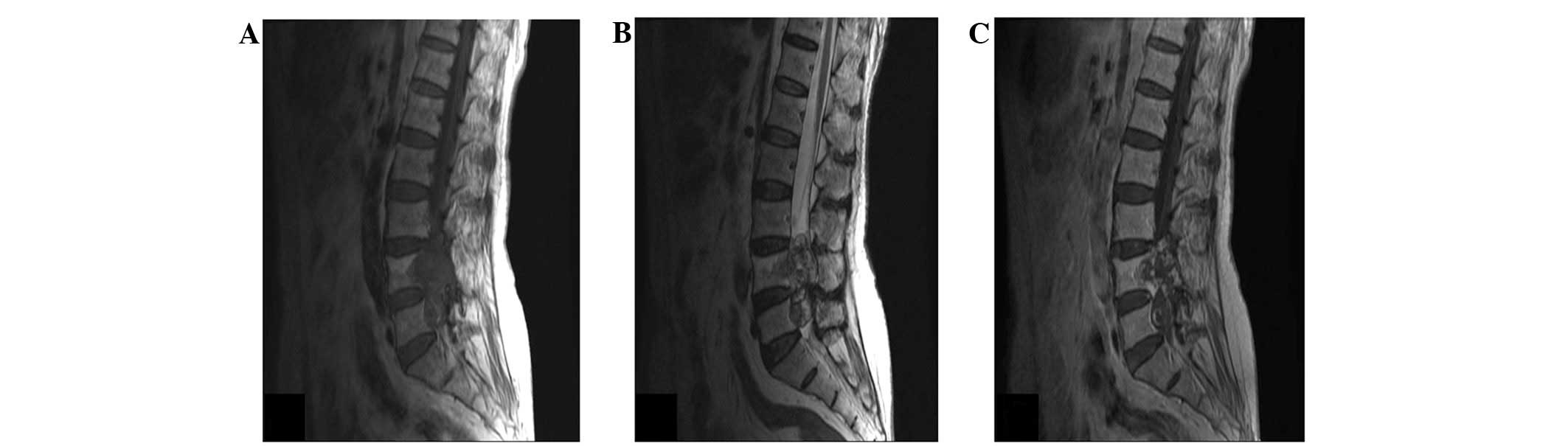
intermittent claudication after walking 200 m. Magnetic resonance imaging revealed a mass involving the L4 and L5 vertebrae with local bone destruction. The mass was hypointense on T1-weighted images, hyperintense on T2-weighted images and enhanced heterogeneously on post-contrast T1-weighted images (Fig. 1A–C). A total resection was performed and the patient’s post-operative course was uneventful. No subsequent adjuvant therapy was deemed necessary.

Figure 1.

(A) T1-weighted sagittal image showing the hypointense lesion involving the L4 and L5 vertebrae with local bone destruction. (B) T2-weighted sagittal image showing the hyperintense lesion. (C) Post-contrast T1-weighted sagittal image showing the heterogeneous enhancement of the lesion.

Pathological analysis

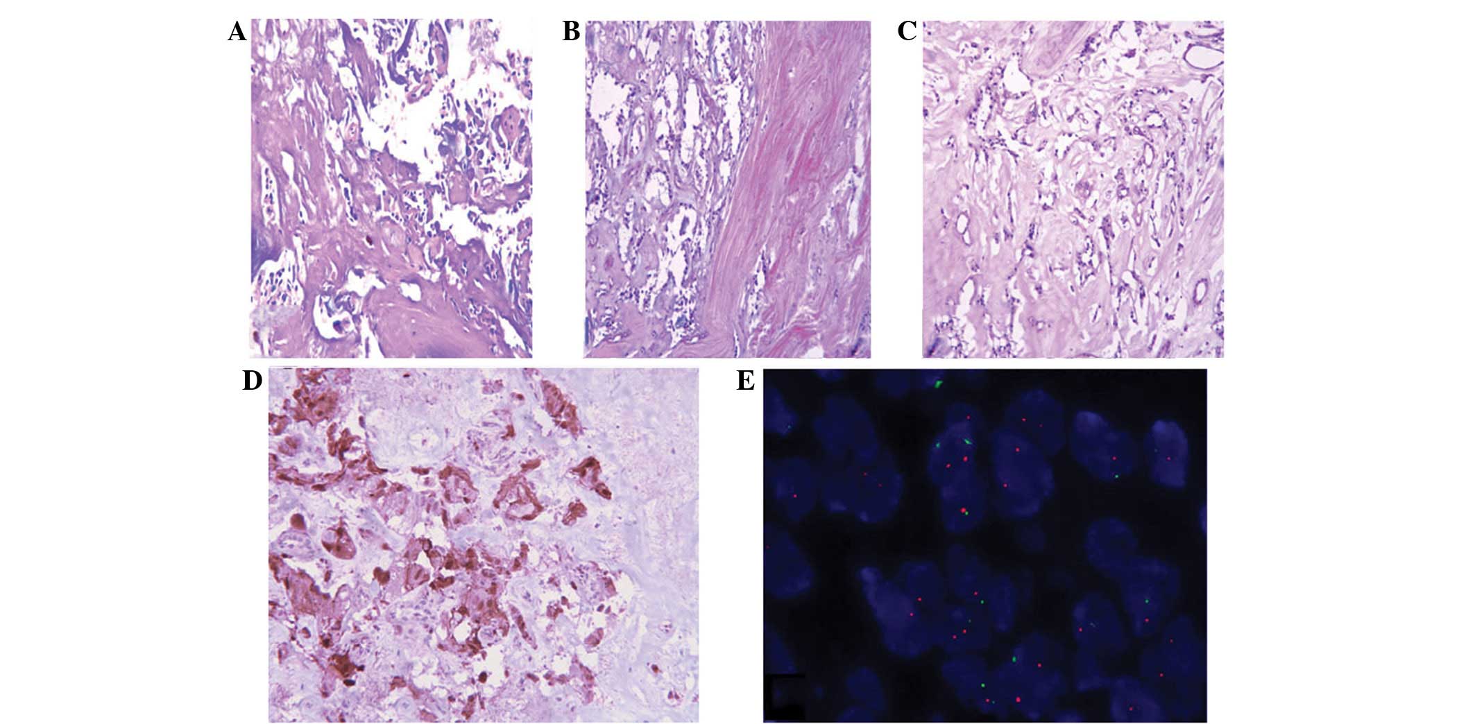
Microscopically, the tumor largely consisted of areas with low cellularity. In less cellular areas, marked hyaline changes were observed in the blood vascular walls and stroma (Fig. 2A). In other areas, the tumor showed pseudopapillary or reticular patterning formed by cuboidal cells on a hyaline background (Fig. 2B and C). Mitotic figures and necrosis were absent. The tumor cells showed marked positivity for glial fibrillary acidic protein (GFAP; Fig. 2D) and S-100 proteins, whereas the cells were negative for epithelial membrane antigen (EMA), cytokeratins (CK) and epidermal growth factor receptor (EGFR). The Ki-67 labeling index was ∼3%. Since EGFR may be a predictor of relapse in MPE (5), the EGFR gene was analyzed by fluorescence in situ hybridization. No amplification of the EGFR gene was observed (Fig. 2E). The results of pathological and immunohistochemical studies were consistent with the ependymal nature of neoplastic cells.

Figure 2.

Photomicrographs of the tumor mass. (A) Acellular tumor mass with isolated neoplastic cells and a marked hyaline change observed in the blood vascular walls and stroma (hematoxylin and eosin; magnification, ×100). (B) Cystic and pseudopapillary component (hematoxylin and eosin; magnification, ×100). (C) Irregualr reticular neoplastic cells embedded in hyaline stroma (hematoxylin and eosin; magnification, ×100). (D) GFAP-positive neoplastic cells (immunoperoxidase; magnification, ×200). (E) Fluorescence in situ hybridization displaying two centromere 7 (green) and two EGFR (orange) signals. No amplification of the EGFR gene was observed. GFAP, glial fibrillary acidic protein; EGFR, epidermal growth factor receptor.

Discussion

MPE is a benign variant of ependymoma that has a peak incidence between the third and fifth decades of life. It generally occurs in the filum terminale, but has also been identified in extra-spinal locations, including subcutaneous tissue and the brain (6,7). Clinical symptoms are directly associated with the mass location of the tumor. Histologically, it is typically characterized by papillae formed by the arrangement of cuboidal to columnar cells surrounding a central core, which contains blood vessels and myxoid change. The genesis of the stromal myxoid changes are indicative of an inundation of the plasma proteins generally located within blood vessels (8). In the current case, the pronounced deposition of hyaline materials was distributed among the tumor cells and within the blood vessel walls. Lim et al (9) previously hypothesized that the ‘proteinaceous’ deposits and hyaline vascular change were a result of increased vascular permeability that led to inundation of plasma proteins surrounding extravascular spaces. MPEs are generally slow-growing, therefore, the chronic long-standing anoxic conditions may lead to various regressive changes and necrosis. An additional explanation for the stromal change is the complicated structure of the conus medullaris and filum terminale, as this region is composed of an admixture of connective tissue, nerve fibers and neuroglia. It may be possible that these unique structural features result from the anatomical relationships of the MPE (10). Certain studies have indicated that, in specific circumstances, MPE cells produce basal lamina material, particularly in regions where ependymal cells are opposed to connective tissue (11,12). In the present case, tumor cells showed marked cytoplasmic staining for GFAP and S-100 protein, whereas immunoreactivity for CK, EMA, chromogranin, EGFR and synaptophysin were all negative. In addition, marked hyaline deposits were identified around the tumor cells, and pseudopapillary patterns or ependymal rosettes were rare. Therefore, in cases such as these, it is important to rule out the possibility of schwannomas, chordomas and metastatic mucinous adenocarcinomas. Spinal schwannomas appear on the myelin sheath of the spine and represent between one-quarter to one-third of all spinal tumors. Diagnostic features include a fibrous capsule and Antoni A and Antoni B areas. Similar to in MPEs, hyaline vessels and stroma are common in schwannomas and S100 is markedly expressed. However, GFAP is negative. It is extremely important to differentiate between a schwannoma and MPE prior to surgery, as an MPE has the potential to disseminate through the cerebrospinal fluid throughout the neuraxis and must be removed completely. The most common differential diagnosis at this location is that of a chordoma. The tumor cells are large with characteristic physaliferous cytoplasm, and the immunocytochemistry of CK is positive (13). Metastatic mucinous adenocarcinomas show epithelial cords and groups with an acinar arrangement. The cancer cells are more pleomorphic and are CK- and EMA-positive. An analysis using electron microscopy is extremely important for forming a differential diagnosis. Specific ultrastructural features, including microvilli, cilia, desmosomal attachments and cytoplasmic filaments, are indicative of a diagnosis of MPE (14,15).

In conclusion, specific regressive changes are observed in MPE and marked hyaline degeneration may lead to an acellular growth pattern, therefore, it is important to rule out the possibility of other tumors. The best curative treatment lies in a complete surgical resection, and long-term follow-up of the whole neuraxis must be performed.

Acknowledgements

The authors would like to thank Dr Wanchun Li (Department of Pathology, Nanjing Jinling Hospital) for gathering clinical information from medical records and Dr Ozgur Sahin (Department of Molecular and Cellular Oncology, The University of Texas MD Anderson Cancer Center) for linguistic assistance.

References

1. 

Bagley CA, Kothbauer KF, Wilson S, Bookland MJ, Epstein FJ and Jallo GI: Resection of myxopapillary ependymomas in children. J Neurosurg. 106(4 Suppl): 261–267. 2007.PubMed/NCBI

2. 

Nakamura M, Ishii K, Watanabe K, et al: Long-term surgical outcomes for myxopapillary ependymomas of the cauda equina. Spine. (Phila Pa 1976). 34:E756–E760. 2009. View Article : Google Scholar : PubMed/NCBI

3. 

Kulesza P, Tihan T and Ali SZ: Myxopapillary ependymoma: cytomorphologic characteristics and differential diagnosis. Diagn Cytopathol. 26:247–250. 2002. View Article : Google Scholar : PubMed/NCBI

4. 

Zacharoulis S, Ji L, Pollack IF, et al: Metastatic ependymoma: a multi-institutional retrospective analysis of prognostic factors. Pediatr Blood Cancer. 50:231–235. 2008. View Article : Google Scholar : PubMed/NCBI

5. 

Verma A, Zhou H, Chin S, Bruggers C, Kestle J and Khatua S: EGFR as a predictor of relapse in myxopapillary ependymoma. Pediatr Blood Cancer. 59:746–748. 2012. View Article : Google Scholar : PubMed/NCBI

6. 

Tseng YC, Hsu HL, Jung SM and Chen CJ: Primary intracranial myxopapillary ependymomas: report of two cases and review of the literature. Acta Radiol. 45:344–347. 2004. View Article : Google Scholar : PubMed/NCBI

7. 

Lee KJ, Min BW, Seo HJ and Cho CH: Subcutaneous sacrococcygeal myxopapillary ependymoma in asian female: a case report. J Clin Med Res. 4:61–63. 2012.PubMed/NCBI

8. 

Sato H, Ohmura K, Mizushima M, Ito J and Kuyama H: Myxopapillary ependymoma of the lateral ventricle. A study on the mechanism of its stromal myxoid change. Acta Pathol Jpn. 33:1017–1025. 1983.PubMed/NCBI

9. 

Lim SC and Jang SJ: Myxopapillary ependymoma of the fourth ventricle. Clin Neurol Neurosurg. 108:211–214. 2006. View Article : Google Scholar : PubMed/NCBI

10. 

Rawlinson DG, Herman MM and Rubinstein LJ: The fine structure of a myxopapillary ependymoma of the filum terminale. Acta Neuropathol. 25:1–13. 1973. View Article : Google Scholar : PubMed/NCBI

11. 

Miller C: The ultrastructure of the conus medullaris and filum terminale. J Comp Neurol. 132:547–566. 1968. View Article : Google Scholar : PubMed/NCBI

12. 

Matyja E, Naganska E, Zabek M and Koziara H: Myxopapillary ependymoma of the lateral ventricle with local recurrences: histopathological and ultrastructural analysis of a case. Folia Neuropathol. 41:51–57. 2003.

13. 

Estrozi B, Queiroga E, Bacchi CE, et al: Myxopapillary ependymoma of the posterior mediastinum. Ann Diagn Pathol. 10:283–287. 2006. View Article : Google Scholar : PubMed/NCBI

14. 

Stern JB and Helwig EB: Ultrastructure of subcutaneous sacrococcygeal myxopapillary ependymoma. Arch Pathol Lab Med. 105:524–526. 1981.PubMed/NCBI

15. 

Kindblom LG, Lodding P, Hagmar B and Stenman G: Metastasizing myxopapillary ependymoma of the sacrococcygeal region. A clinico-pathologic, light- and electronmicroscopic, immunohistochemical, tissue culture, and cytogenetic analysis of a case. Acta Pathol Microbiol Immunol Scand A. 94:79–90. 1986.

Related Articles

  • Abstract
  • View
  • Download
  • Twitter
Copy and paste a formatted citation
Spandidos Publications style
Wang H, Zhang Z, Makki MS and Shi Q: Osteolytic myxopapillary ependymoma with marked hyaline degeneration in a 72‑year‑old male: A case report. Oncol Lett 6: 487-489, 2013.
APA
Wang, H., Zhang, Z., Makki, M.S., & Shi, Q. (2013). Osteolytic myxopapillary ependymoma with marked hyaline degeneration in a 72‑year‑old male: A case report. Oncology Letters, 6, 487-489. https://doi.org/10.3892/ol.2013.1397
MLA
Wang, H., Zhang, Z., Makki, M. S., Shi, Q."Osteolytic myxopapillary ependymoma with marked hyaline degeneration in a 72‑year‑old male: A case report". Oncology Letters 6.2 (2013): 487-489.
Chicago
Wang, H., Zhang, Z., Makki, M. S., Shi, Q."Osteolytic myxopapillary ependymoma with marked hyaline degeneration in a 72‑year‑old male: A case report". Oncology Letters 6, no. 2 (2013): 487-489. https://doi.org/10.3892/ol.2013.1397
Copy and paste a formatted citation
x
Spandidos Publications style
Wang H, Zhang Z, Makki MS and Shi Q: Osteolytic myxopapillary ependymoma with marked hyaline degeneration in a 72‑year‑old male: A case report. Oncol Lett 6: 487-489, 2013.
APA
Wang, H., Zhang, Z., Makki, M.S., & Shi, Q. (2013). Osteolytic myxopapillary ependymoma with marked hyaline degeneration in a 72‑year‑old male: A case report. Oncology Letters, 6, 487-489. https://doi.org/10.3892/ol.2013.1397
MLA
Wang, H., Zhang, Z., Makki, M. S., Shi, Q."Osteolytic myxopapillary ependymoma with marked hyaline degeneration in a 72‑year‑old male: A case report". Oncology Letters 6.2 (2013): 487-489.
Chicago
Wang, H., Zhang, Z., Makki, M. S., Shi, Q."Osteolytic myxopapillary ependymoma with marked hyaline degeneration in a 72‑year‑old male: A case report". Oncology Letters 6, no. 2 (2013): 487-489. https://doi.org/10.3892/ol.2013.1397
Follow us
  • Twitter
  • LinkedIn
  • Facebook
About
  • Spandidos Publications
  • Careers
  • Cookie Policy
  • Privacy Policy
How can we help?
  • Help
  • Live Chat
  • Contact
  • Email to our Support Team