Introduction
Camellia sinensis is the species of plant
whose leaves and leaf buds are used to produce Chinese tea. White,
green, oolong, pu-erh and black tea are all harvested from this
species, but are processed differently to attain different levels
of oxidation. Black tea is a completely oxidized tea. Black tea
contains relatively high levels of polyphenolics with the major
phenolics being the flavan-3-ols, the flavonols (mono-, di- and
tri-glycoside conjugates of myricetin, quercetin and kaempferol),
the flavones and the quinic acid esters of gallic, coumaric and
caffeic acids (1). Black tea has a
reduced flavan-3-ol monomer content and higher levels of
polymerized derivatives, theaflavins, which account for ∼10–30% of
the converted catechins and thearubigins (2,3).
Although there is growing interest in the hypothesis that tea has a
preventive effect against cardiovascular diseases and that tea
polyphenols may mediate the observed benefits (4,5), the
intricate mechanisms of polyphenolic action require further and
comprehensive understanding.
Buccal mucosa cancer is the most common type of
cancer of the oral cavity (6). The
U14 mouse tumor is a squamous cell carcinoma, which was ectopically
induced by treating the uterine cervix with 20-methylcholanthrene
(7). After U14 cells were
transplanted into mice, buccal mucosa cancer was induced (8). The present study aimed to determine
the anti-mutagenic activities of black tea with
N-methyl-N-nitro-N-nitrosoguanidine (MNNG) and to evaluate the
cancer preventive effect of black tea using a mouse model of buccal
mucosa cancer, in order to determine whether, as a functional food,
black tea would demonstrate oral health benefits.
Materials and methods
Tea extract preparation
Qimen black tea (producer, Wuhu Huifu Tea Co., Ltd.,
Wuhu, China) was purchased from Anhui in China. To prepare the
methanol extracts, black tea was freeze-dried and powdered. A
ten-fold quantity of boiling water was added to the powdered sample
and extracted twice by agitating. The water extract was evaporated
using a rotary evaporator (N-1100; Eywla, Tokyo, Japan).
Anti-mutagenic experiment
The Salmonella typhimurium strain, TA100, a
histidine-requiring mutant bacterium, was maintained as described
by Maron and Ames (9). In brief,
0.5 ml of phosphate buffer containing the direct mutagen of MNNG
was distributed into sterilized capped tubes, and then 0.1 ml of
test bacterial suspension from an overnight culture
(1–2×10⁹ cells/ml) and 0.1 ml of test sample compound
(50 μl of mutagen and/or 50 μl of test sample) were
added. After agitating gently and pre-incubating at 37°C for 30
min, 2 ml of top agar, supplemented with L-histidine and D-biotin,
kept at 45°C, was added to each tube and agitated for 3 sec. The
entire resulting mixture was overlaid on a minimal agar plate. The
plates were incubated at 37°C for 48 h and the revertant bacterial
colonies on each plate were counted.
Animals
Seven-week-old female Kunming (KM) mice were
purchased from the Experimental Animal Center of Chongqing Medical
University (Chongqing, China). The animals were maintained in a
temperature-controlled (23±1°C; relative humidity, 50±5%) facility
with a 12-h light/dark cycle and had unlimited access to a standard
mouse chow diet and water. The protocol for these experiments was
approved by the Animal Ethics Committee of Chongqing Medical
University (Chongqing, China).
Cell preparation
U14 squamous cell carcinoma cells obtained from the
Chinese Academy of Medical Sciences (Beijing, China) were used in
this study. The cancer cells were cultured in RPMI-1640 medium
(Gibco Co., Birmingham, MI, USA) supplemented with 10% fetal bovine
serum (FBS) and 1% penicillin-streptomycin (Gibco-BRL, Grand
Island, NY, USA) at 37°C in a humidified atmosphere with 5%
CO2 (incubator model, 311 S/N29035; Forma, Waltham, MA,
USA). The medium was changed 2 or 3 times a week (10). In vitro cultured U14 cells
(5×10⁶/mouse) were injected into the abdominal cavity of
the 7-week-old female KM mice. After 1 week, the carcinoma ascites
were collected and diluted in sterile saline to a concentration of
1×10⁷/ml.
Induction of buccal mucosa cancer
To investigate the preventive effects of the black
tea against buccal mucosa cancer induced by injecting U14 cells
into the mice, the animals were divided into 4 groups with 10 mice
in each. The experimental design was as follows; the mice in the
black tea sample groups were smeared with 0.2 ml black tea solution
(200 or 100 mg/ml) onto the buccal mucosa every 12 h for 14 days.
The control and black tea sample groups were then inoculated with
0.05 ml cancer cell suspension (1×10⁷/ml) on the buccal
mucosa. The black tea samples continued to be smeared on the buccal
mucosa of the mice every 12 h. The normal group were not treated
with the cancer cell suspension. The mice were sacrificed 14 days
later and their tumor volumes and lymph node metastasis rates were
determined (8).
Histological grading of buccal mucosa
cancer
Buccal mucosa tissues were removed and embedded into
paraffin for histological analysis using hematoxylin and eosin (HE)
staining. Buccal mucosa cancer was graded as follows: i)
well-differentiated carcinoma, cells resembling the adjacent benign
squamous epithelium; ii) moderately-differentiated carcinoma, cells
forming large anastomosing areas in which keratin pearls are
formed, they are not numerous and the main component consists of
cells with pronounced cytonuclear atypia; and iii)
poorly-differentiated carcinoma, cells that have lost the majority
of their squamous epithelial characteristics and architecture
(11).
Reverse transcription polymerase chain
reaction (RT-PCR) analysis of Bcl-2-associated X protein (Bax) and
B cell lymphoma-2 (Bcl-2) mRNA expression
Total RNA was isolated using TRIzol reagent
(Invitrogen, Carlsbad, CA, USA) according to the manufacturer’s
instructions. RNA was digested with RNase-free DNase (Roche, Basel,
Switzerland) for 15 min at 37°C and purified using an RNeasy kit
(Qiagen, Hilden, Germany) according to the manufacturer’s
instructions. cDNA was synthesized from 2 μg total RNA by
incubation at 37°C for l h with avian myeloblastosis virus (AMV)
reverse transcriptase (GE Healthcare, Uppsala, Sweden) with random
hexanucleotides, according to the manufacturer’s instructions. The
sequences of the primers used to specifically amplify the genes of
interest are shown in Table I.
Amplification was performed in a thermal cycler (Eppendorf,
Hamburg, Germany) with 29 Bax cycles, 34 Bcl-2 cycles and 25 GAPDH
cycles of denaturation. The amplified PCR products were run on 1.0%
agarose gels and visualized by ethidium bromide (EtBr) staining
(12).
 | Table I.Sequences of RT-PCR primers used in
this study. |
Table I.
Sequences of RT-PCR primers used in
this study.
| Gene name | Sequence |
|---|
| Bax | Forward: 5′-AAG CTG
AGC GAG TGT CTC CGG CG-3′ |
| Reverse: 5′-CAG ATG
CCG GTT CAG GTA CTC AGT C-3′ |
| Bcl-2 | Forward: 5′-CTC GTC
GCT ACC GTC GTG ACT TGG-3′ |
| Reverse: 5′-CAG ATG
CCG GTT CAG GTA CTC AGT C-3′ |
| GAPDH | Forward: 5′-CGG AGT
CAA CGG ATT TGG TC-3′ |
| Reverse: 5′-AGC CTT
CTC CAT GGT CGT GA-3′ |
Statistical analysis
Data are presented as the mean ± standard deviation.
Differences between the mean values for the individual groups were
assessed using a one-way analysis of variance (ANOVA) with Duncan’s
multiple range tests. P<0.05 was considered to indicate a
statistically significant difference. The SAS v9.1 statistical
software package (SAS Institute Inc., Cary, NC, USA) was used for
the analysis.
Results
Antimutagenic effects of black tea
The black tea demonstrated inhibitory effects on
spontaneous mutations in the Salmonella typhimurium TA100
strain (Table II). At 1.25
mg/plate, the spontaneous mutation inhibitory rate of black tea was
57%. At 2.5 mg/plate, black tea revealed an inhibition rate of 79%.
These results indicate that the black tea exerted a decreasing
effect on the spontaneous levels of mutation.
| Table II.Effect of black tea on spontaneous
mutagenicity. |
Table II.
Effect of black tea on spontaneous
mutagenicity.
| Treatment | No. of
revertants/plate
|
|---|
| 1.25 mg/plate | 2.5 mg/plate |
|---|
| Spontaneous (No
Mutation) | 128±15a |
| Black tea | 55±9b (57) | 27±7c (79) |
Black tea showed an anti-mutagenic effect in the
Salmonella typhimurium TA100 strain when treated with MNNG
(Table III). At 1.25 mg/plate, the
mutagenic inhibition rate of black tea was 32%, demonstrating an
anti-mutagenic effect. When the black tea concentration was 2.5
mg/plate, black tea further showed significantly increased
anti-mutagenic effects, with an inhibition rate of 63%. This
suggested that black tea had a strong anti-mutagenic effect.
| Table III.Effect of black tea on the
mutagenicity induced by MNNG (0.4 μg/plate) in Salmonella
typhimurium TA100. |
Table III.
Effect of black tea on the
mutagenicity induced by MNNG (0.4 μg/plate) in Salmonella
typhimurium TA100.
| Treatment | No. of
revertants/plate
|
|---|
| 1.25 mg/plate | 2.5 mg/plate |
|---|
| Spontaneous (No
mutation) | 128±15 |
| MNNG (control) | 922±37a |
| Black tea | 668±22b (32) | 422±24c (63) |
Tumor volumes and lymph node metastasis
rates
Buccal mucosa cancer was induced by injecting U14
cells into mice. After 14 days, the mice in all groups presented
with carcinogenesis. The tumor volumes of the buccal mucosa tissues
were measured. The tumor volumes for the control, 100 mg/ml black
tea and 200 mg/ml black tea groups were 10.8, 9.7 and 5.2
mm3, respectively (Table
IV). There were 6 mice demonstrating lymph node metastasis in
the control group, 4 in the black tea (100 mg/ml) group and 1 in
the black tea (200 mg/ml) group. Consequently, the lymph node
metastasis rate was 60, 40 and 10%, respectively. These results
demonstrate that black tea is effective in impeding carcinogenesis,
proliferation and metastasis.
| Table IV.Tumor sizes and lymph node metastasis
rates of black tea sample smeared on mice. |
Table IV.
Tumor sizes and lymph node metastasis
rates of black tea sample smeared on mice.
| Normal group | Control group | Black tea groups
|
|---|
| 100 mg/ml | 200 mg/ml |
|---|
| Tumor volume
(mm3) | 0 | 10.8±0.6a | 9.7±0.5b | 5.2±0.2c |
| Lymph node
metastasisd | 0 | 6/10 (60%) | 4/10 (40%) | 1/10 (10%) |
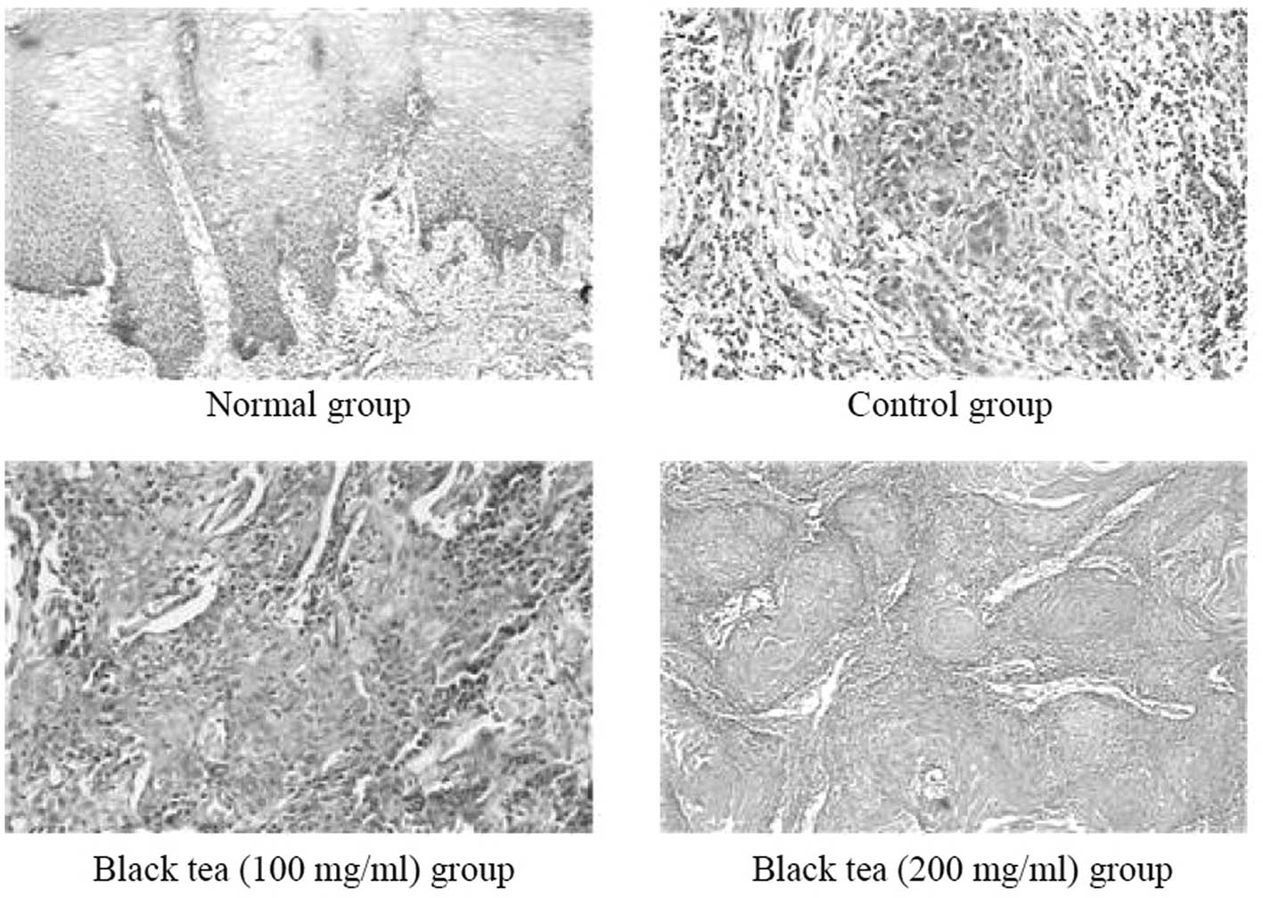
Histopathology of buccal mucosa
tissues
Histological changes in the buccal mucosa of mice
injected with U14 cells were examined by HE staining. The
histological tissue sections of the mice in the normal group
demonstrated a normal histological morphology for squamous
epithelial tissue. The histopathological evaluation revealed
indications of buccal mucosa cancer in the two groups administered
with U14 cells (Fig. 1). The
sections from the mice in the control group revealed that the
tissue had lost its squamous epithelial characteristics and
architecture (grade iii). The tissue sections of the black tea (100
mg/ml) group looked less like normal squamous epithelium (grade
ii). The tumor cells remained in nests but there were few larger,
eosinophilic, polygonal cells that were trying to layer themselves
in a squamous-like fashion. The tissue sections of the black tea
(200 mg/ml) group appeared much like the adjacent benign squamous
epithelium (grade i). From these sections, it was demonstrated that
black tea provided a preventive effect against buccal mucosa
cancer.
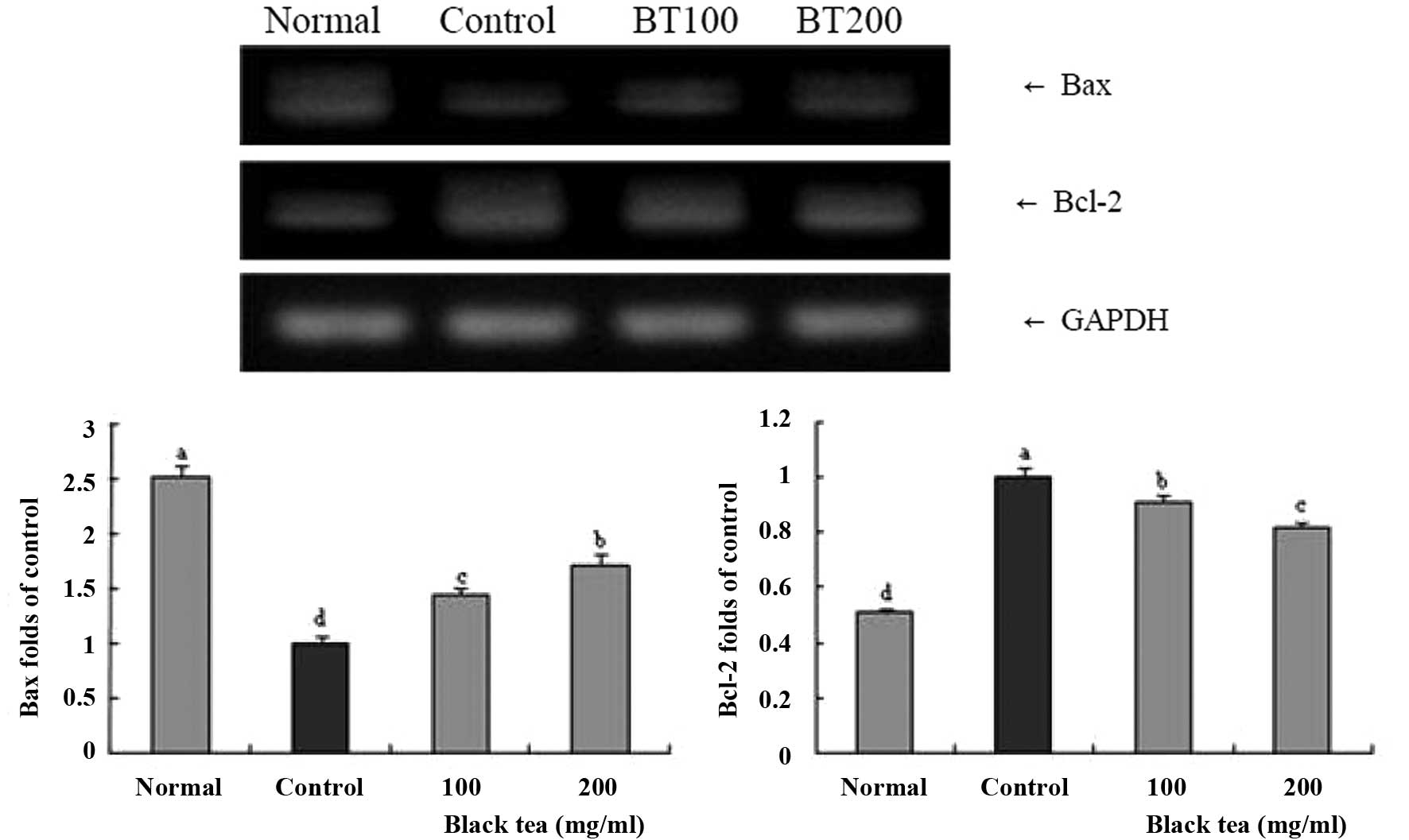
Gene expression of Bax and Bcl-2 in
buccal mucosa tissues
To determine the protective mechanisms against
buccal mucosa cancer, the expression of the Bax and Bcl-2 genes in
the buccal mucosa tissues was determined by RT-PCR. As shown in
Fig. 2, in the group treated with
black tea (100 mg/ml), significant changes were demonstrated in the
pro-apoptotic activities of Bax and the anti-apoptotic activities
of Bcl-2. Accordingly, the results suggest that the black tea
induced apoptosis in buccal mucosa tissues via a Bax- and a
Bcl-2-dependent pathway. Thus, apoptosis induction by the black tea
(20 mg/ml) group was related to an increase in Bax and a decrease
in Bcl-2 in terms of mRNA and protein expression compared to black
tea (100 mg/ml) and control groups. From the results, it was
observed that black tea showed a good buccal mucosa cancer
protective effect.
Discussion
Tea has numerous functional compounds, including
polyphenols, catechins, amino acids and vitamins (13). Studies have demonstrated that there
are a number of important compounds to aid in the prevention of
cancer (14,15). Green tea extract has the highest
amount of epigallocatechin gallate (EGCG) and EGC among the four
extracts (green, Oolong, black and Pu-erh tea). However, EGCG and
EGC have not been detected in Pu-erh tea. The features of fermented
tea (Oolong, black and Pu-erh tea) greatly differ from those of
green tea, which is non-fermented and is preferably drunk as fresh
as possible (16).
The various health benefits of black tea have made
it become known as the medicinal tea plant. Pasha et al
(17) proposed that the intake of
fermented tea is superior to black tea in terms of its nutritive
and therapeutic value; it also does not show much change in taste
and color after fermentation. This may be recommended for
consumption as a modified beverage with higher nutritive and
medicinal values.
Deleterious mutations produced by mutagens in the
DNA may result in aberrant, impaired or lost function in a
particular gene, and accumulation of such mutations may lead to
cancer (18). The Ames test is a
rapid test that is able to screen for chemical carcinogens.
Biological genetic mutation is regarded as a causative key factor
of cancer (19), and Salmonella
typhimurium is used as a testing strain in a biological assay
to assess the mutagenic potential of chemical compounds (20).
Apoptosis induction in cancer cells is initially
identified by morphological changes including cell shrinkage,
membrane blebbing, chromatin condensation and nuclear fragmentation
(21). Apoptosis is an important
defense against cancer. This process involves the elimination of
potentially harmful cells. Numerous diseases have been associated
with dysregulated apoptotic processes, ultimately leading to the
inhibition of cell death and propagation of diseases such as
cancer. Elucidating the critical events associated with
carcinogenesis provides an opportunity for preventing cancer
development via dietary intervention by inducing apoptosis,
particularly with bioactive agents or functional foods. Diet and
drink are significant environmental factor in the overall cancer
process, and may exacerbate or interfere with disease progression.
In addition to dietary effects on protein expression and function,
evidence is also accumulating that a large number of food
components may exert effects on the human genome, by either
directly or indirectly modulating gene expression (22,23).
Bax, a protein that is a member of the Bcl-2 gene family, promotes
apoptosis by competing with Bcl-2 (24). In a healthy cell, the anti-apoptotic
protein Bcl-2 is expressed on the outer mitochondrial membrane
surface (25). As Bax and Bcl-2
genes are mainly expressed during apoptosis, we determined that
these genes regulate apoptotic activity.
Histopathology is an important tool in anatomical
pathology, since the accurate diagnosis of cancer usually requires
a histopathological examination of the samples. Histopathology is a
clinical standard for the diagnosis of oral cancer (26). Histopathological assays permit the
precise determination of the anti-cancer effects of black tea.
In summary, the present study employed in
vitro and in vivo experimental methods, such as the Ames
test and histopathology and RT-PCR analyses, to evaluate the
anti-cancer effects of black tea. The results from the present
study demonstrate that black tea may decrease the spontaneous
revertants in Salmonella typhimurium TA100 and may also
decrease the mutagenic effect of MNNG. A mouse model bearing tumors
produced by squamous cell carcinoma U14 cells was used to study the
in vivo effects of black tea. The results showed that black
tea had strong anti-cancer activities against buccal mucosa cancer.
Overall, black tea demonstrated in vitro anti-mutagenic
effects and in vivo anti-cancer and anti-metastatic
activities. In conclusion, an increased concentration may be used
to increase the oral cancer preventive effect of black tea.
Acknowledgements
This study was partially supported by
the Educational Reform Project JG20132206 and JG201234 of Chongqing
University of Education.
References
|
1.
|
Bahorun T, Luximon-Ramma A, Gunness TK,
Sookar D, Bhoyroo S, Jugessur R, Reebye D, Googoolye K, Crozier A
and Aruoma OI: Black tea reduces uric acid and C-reactive protein
levels in humans susceptible to cardiovascular diseases.
Toxicology. 278:68–74. 2010. View Article : Google Scholar : PubMed/NCBI
|
|
2.
|
de Mejia EG, Ramirez-Mares MV and
Puangpraphant S: Bioactive components of tea: cancer, inflammation
and behavior. Brain Behav Immun. 23:721–731. 2009.PubMed/NCBI
|
|
3.
|
Rouanet JM, Décordé K, Del Rio D, Auger C,
Borges G, Cristol JP, Lean MEJ and Crozier A: Berry juices, teas,
anti-oxidants and the prevention of atherosclerosis in hamsters.
Food Chem. 118:266–271. 2010. View Article : Google Scholar
|
|
4.
|
Sharangi AB: Medicinal and therapeutic
potentialities of tea (Camellia sinensis L.) - A review.
Food Res Int. 42:529–535. 2009. View Article : Google Scholar
|
|
5.
|
Stangl V, Lorenz M and Stangl K: The role
of tea and tea flavonoids in cardiovascular health. Mol Nutr Food
Res. 50:218–228. 2006. View Article : Google Scholar : PubMed/NCBI
|
|
6.
|
Kolanjiappan K, Ramachandran CR and
Manoharan S: Biochemical changes in tumor tissues of oral cancer
patients. Clin Biochem. 36:61–65. 2003. View Article : Google Scholar : PubMed/NCBI
|
|
7.
|
Gu B, Feng HL, Dong JH, Zhang H, Bian XC
and Liu YQ: The establishment and characterization of a continuous
cell line of mouse cervical carcinoma. Chin J Clin Oncol. 5:44–48.
2008. View Article : Google Scholar
|
|
8.
|
Zhao X, Deng XX, Park KY, Qiu LH and Pang
L: Purple bamboo salt has anticancer activity in TCA8113 cells in
vitro and preventive effects on buccal mucosa cancer in mice in
vivo. Exp Ther Med. 5:549–554. 2013.PubMed/NCBI
|
|
9.
|
Maron DM and Ames BN: Revised methods for
the Salmonella mutagenicity test. Mutat Res. 113:173–215.
1983.
|
|
10.
|
Zhao X, Kim SY and Park KY: Bamboo salt
has in vitro anti-cancer activity in HCT-116 cells and exerts
anti-metastatic effects in vivo. J Med Food. 16:9–19. 2013.
View Article : Google Scholar : PubMed/NCBI
|
|
11.
|
Schrader M and Laberke HG: Differential
diagnosis of verrucous carcinoma in the oral cavity and larynx. J
Laryngol Otol. 102:700–703. 1988. View Article : Google Scholar : PubMed/NCBI
|
|
12.
|
Zhao X: Hawk tea (Litsea coreana
Levl. var. lanuginose) attenuates CCl4-induced
hepatic damage in Sprague-Dawley rats. Exp Ther Med. 5:555–560.
2013.PubMed/NCBI
|
|
13.
|
Fujiki H, Suganuma M, Imai K and Nakachi
K: Green tea: cancer preventive beverage and/or drug. Cancer Lett.
118:9–13. 2002. View Article : Google Scholar : PubMed/NCBI
|
|
14.
|
Leone M, Zhai D, Sareth S, Kitada S, Reed
JC and Pellecchia M: Cancer prevention by tea polyphenols is linked
to their direct inhibition of antiapoptotic Bcl-2-family proteins.
Cancer Res. 63:8118–8121. 2003.
|
|
15.
|
Kim J, Zhang X, Rieger-Christ KM,
Summerhayes IC, Wazer DE, Paulson KE and Yee AS: Suppression of Wnt
signaling by the green tea compound (−)-epigallocatechin 3-gallate
(EGCG) in invasive breast cancer cells. Requirement of the
transcriptional repressor HBP1. J Biol Chem. 281:10865–10875.
2006.PubMed/NCBI
|
|
16.
|
Duh PD, Yen GC, Yen WJ, Wang BS and Chang
LW: Effects of Pu-erh tea on oxidative damage and nitric oxide
scavenging. J Agric Food Chem. 52:8169–8176. 2004. View Article : Google Scholar : PubMed/NCBI
|
|
17.
|
Pasha C and Reddy G: Nutritional and
medicinal improvement of black tea by yeast fermentation. Food
Chem. 89:449–453. 2005. View Article : Google Scholar
|
|
18.
|
Huang L, Snyder AR and Morgan WF:
Radiation-induced genomic instability and its implications for
radiation carcinogenesis. Oncogene. 22:5848–5854. 2003. View Article : Google Scholar : PubMed/NCBI
|
|
19.
|
Hwang KM, Jung KO, Song CH and Park KY:
Increased anti-mutagenic and anticlastogenic effects of doenjang
(Korean fermented soybean paste) prepared with bamboo salt. J Med
Food. 11:717–722. 2008. View Article : Google Scholar : PubMed/NCBI
|
|
20.
|
Mortelmans K and Zeiger E: The Ames
Salmonella/microsome mutagenicity assay. Mutat Res.
455:29–60. 1999.
|
|
21.
|
Lowe SW and Lin AW: Apoptosis in cancer.
Carcinogenesis. 21:485–495. 1999. View Article : Google Scholar
|
|
22.
|
Koo JY, Kim HJ, Jung KO and Park KY:
Curcumin inhibits the growth of AGS human gastric carcinoma cells
in vitro and shows synergism with 5-fluorouracil. J Med Food.
7:117–121. 2004. View Article : Google Scholar : PubMed/NCBI
|
|
23.
|
Kwon JI, Kim GY, Park KY, Ryu CH and Choi
YH: Induction of apoptosis by linoleic acid is associated with the
modulation of Bcl-2 family and Fas/FasL system and activation of
caspases in AGS human gastric adenocarcinoma cells. J Med Food.
11:1–8. 2008. View Article : Google Scholar : PubMed/NCBI
|
|
24.
|
Oltvai ZN, Milliman CL and Korsmeyer SJ:
Bcl-2 heterodimerizes in vivo with a conserved homolog, Bax, that
accelerates programed cell death. Cell. 74:609–619. 1993.
View Article : Google Scholar : PubMed/NCBI
|
|
25.
|
Chao DT and Korsmeyer SJ: Bcl-2 family:
regulators of cell death. Annu Rev Immunol. 16:395–419. 1998.
View Article : Google Scholar
|
|
26.
|
Sankaranarayanan R, Ramadas K, Thomas G,
Muwonge R, Thara S, Mathew B and Rajan B; Trivandrum Oral Cancer
Screening Study Group: Effect of screening on oral cancer mortality
in Kerala, India: a cluster-randomised controlled trial. Lancet.
365:1927–1933. 2005. View Article : Google Scholar : PubMed/NCBI
|