Introduction
Choriocarcinoma is a rare malignant neoplasm
composed of mononucleated and multinucleated trophoblasts, mainly
arising in the uterus of pregnant females and in the ovaries. A
choriocarcinomatous component is rarely present in carcinomas of
certain sites, including the lung (1), breast (2), esophagus (3), stomach (4,5), colon
(6,7) and urinary system (8). Furthermore, uterine corpus carcinomas
with choriocarcinomatous differentiation have rarely been reported
(9–17). The present study describes a case of
endometrioid adenocarcinoma of the uterine corpus with
choriocarcinomatous differentiation and discusses the
clinicopathological features of this rare tumor. Written informed
consent was obtained from the patient.
Case report
Patient
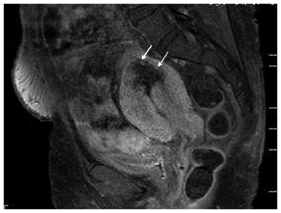
A 59-year-old post-menopausal female with
scleroderma and diabetes mellitus presented with abnormal vaginal
bleeding. A physical examination revealed the presence of pyometra
and magnetic resonance imaging demonstrated a relatively
well-circumscribed tumor, measuring 30 mm in diameter, in the
fundus of the uterus (Fig. 1).
Swelling of the internal and external iliac and paraaortic lymph
nodes was also observed. A clinical diagnosis of a malignant
uterine corpus tumor was suspected, and a total cystectomy and
bilateral salpingo-oophorectomy were performed, with dissection of
the pelvic and paraaortic lymph nodes. Serum β-human chorionic
gonadotropin (hCG) levels were not measured.
Materials and methods
Formalin-fixed, paraffin-embedded tissue blocks were
cut into 3-μm thick sections, then deparaffinized and rehydrated.
Each section was stained with hematoxylin and eosin and used for
immunostaining. Immunohistochemical analyses were performed using
an autostainer (Benchmark XT system; Ventana Medical System,
Tucson, AZ, USA) according to the manufacturer’s instructions. The
following primary antibodies were used: Mouse monoclonal antibody
against CA125 (Ov185:1; Novocastra Laboratories, Ltd., Newcastle
upon Tyne, UK), mouse monoclonal antibody against cytokeratin
(AE1/AE3; DAKO Cytomation, Glostrup, Denmark) and rabbit polyclonal
antibody against human β-hCG (Novocastra).
Histopathological findings
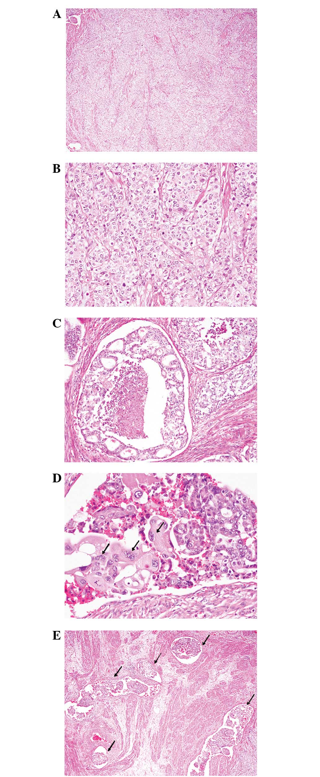
The uterine corpus tumor consisted of two distinct
histopathological components. The first component comprised ~80% of
the tumor and was composed of a poorly-differentiated
adenocarcinoma with extensive hemorrhage and necrosis. This area
involved a proliferation of sheets or variable-sized nests of
atypical epithelial cells containing large oval nuclei with coarse
chromatin and small nucleoli (Fig 2A
and B). These tumor cells contained a relatively rich,
marginally eosinophilic cytoplasm, but no intracytoplasmic mucin
was observed (Fig. 2B). Mitotic
figures were frequently observed (34/10 high-power fields). Focal
glandular differentiation showing cribriform glands with central
necrosis was present (Fig. 2C),
however, no squamous differentiation was noted. Accordingly, this
component was considered to be an endometrioid adenocarcinoma. The
remaining component consisted of mononucleated and syncytial-like
giant cells, with a rich eosinophilic cytoplasm and large
pleomorphic nuclei with coarse chromatin (Fig. 2D). There was a transition between
the endometrioid adenocarcinoma and choriocarcinomatous components
(Fig. 2D). A number of lymphatic
and vascular invasions were noted (Fig.
2E). The tumor had invaded deeply into the entire layer of the
uterine corpus wall and serosal invasion was also noted. However,
no parametrial or vaginal invasion was observed. The internal and
external iliac and paraaortic lymph nodes exhibited metastatic
carcinomas accompanying each of the components.
Immunohistochemical findings
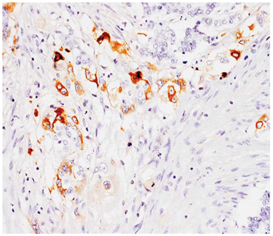
Cytokeratin (AE1/AE3) was expressed in the
endometrioid adenocarcinoma and choriocarcinomatous components.
CA125 was expressed in the endometrioid adenocarcinoma component,
but not in the choriocarcinomatous component. β-hCG was expressed
in the choriocarcinomatous component, particularly in the syncytial
giant cells (Fig. 3), but not in
the endometrial carcinoma component. Metastatic lesions of the
lymph nodes showed similar immunohistochemical features to the
primary tumor and β-hCG-positive syncytial giant cells were
scattered amongst the metastatic lesions.
According to the histopathological and
immunohistochemical features, an ultimate diagnosis of endometrioid
adenocarcinoma with choriocarcinomatous differentiation was made
[pIIIC2; International Federation of Gynecology and Obstetrics
(FIGO)].
Discussion
The present study describes a case of endometrioid
adenocarcinoma of the uterine corpus with choriocarcinomatous
differentiation. Civantos and Rywlin first reported a case of
uterine corpus carcinoma (serous papillary adenocarcinoma) with
choriocarcinomatous differentiation in 1972 (16). Subsequently, Savage et al
reported the first case of endometrioid adenocarcinoma of the
uterine corpus with choriocarcinomatous differentiation in 1987
(9). Since then, few uterine corpus
adenocarcinomas with choriocarcinomatous differentiation have been
reported (10–15). The most common histopathological
subtype of the carcinomatous component is endometrioid
adenocarcinoma (9–15), as seen in the present case. Serous
papillary adenocarcinoma (16,17),
clear cell adenocarcinoma (18) and
carcinosarcoma with choriocarcinomatous differentiation (19,20)
have also been documented. In addition, uterine cervical
adenocarcinoma with choriocarcinomatous differentiation has also
been reported (21).
Table I summarizes
the clinicopathological features of nine previously reported cases
of endometrioid adenocarcinoma of the uterine corpus with
choriocarcinomatous differentiation, in addition to the present
case. The median age of the patients was 62.3 years (range, 42–83
years) and the majority were post-menopausal females, with the
exception of the case reported by Akbulut et al, in which
the patient was a 42-year-old premenopausal female (15). Abnormal vaginal bleeding and
abdominal pain were the main presenting symptoms (14,15).
Endometrial choriocarcinoma may be considered to have a highly
aggressive clinical course, since nine of the 10 cases studied
showed metastases, with the common metastatic sites being the lung,
liver and lymph nodes, while four patients succumbed to the disease
(Table I). The histopathological
features of the metastatic sites were variable; two cases,
including the present case, shared the same features as the primary
site (adenocarcinoma with choriocarcinomatous components) and two
cases only exhibited the choriocarcinoma component (Table I).
 | Table IClinicopathological features of
endometrioid adenocarcinoma with choriocarcinomatous
differentiation. |
Table I
Clinicopathological features of
endometrioid adenocarcinoma with choriocarcinomatous
differentiation.
| Case no. | Age (years) | Histopathology of
coexisting tumor | Metastases or
invasion | Histopathology at
metastatic sites | Outcome, months | Reference |
|---|
| 1 | 70 | WD | Brain, lung, liver,
kidneys | Choriocarcinoma | DOD, 14 | 9 |
| 2 | 78 | PD | Pelvic lymph
nodes | NA | DOD, 1.5 | 10 |
| 3 | 48 | PD | Lungs | NA | AWD, 2 | 10 |
| 4 | 63 | Adenocarcinoma | Lungs, liver,
peritoneum | Same as primary
site | DOD, 14 | 10 |
| 5 | 83 | MD | Lungs | NA | AWD, 1 | 11 |
| 6 | 68 | PD, clear cell and
serous papillary | Pelvic lymph
nodes | Serous papillary
adenocarcinoma | NED, 16 | 12 |
| 7 | 54 | MD | Retroperitoneum | Adenocarcinoma | DOD, 24 | 13 |
| 8 | 58 | WD | Vaginal cuff | Choriocarcinoma | NED, 36 | 14 |
| 9 | 42 | MD | None | | NED, 6 | 15 |
| Present case | 59 | PD | Iliac and paraaortic
lymph nodes | Same as primary
site | NED, 2 | |
Although the pathogenesis of the choriocarcinomatous
component in non-gestational tumors is not well understood, studies
suggest that the choriocarcinomatous component probably represents
heterotopic or aberrant differentiation of the conventional
carcinoma components, rather than the malignant transformation of
germ cells (7,22). Zetll et al reported a case of
urothelial carcinoma with a choriocarcinomatous component and
analyzed the comparative genomic hybridization of the two
components (22). The study clearly
demonstrated that the components shared losses of chromosomes 9 and
17p, which were characteristic genetic alterations of urothelial
carcinoma, and that the choriocarcinomatous components acquired
additional chromosomal losses and gains, mostly associated with
poorly-differentiated urothelial carcinoma (22). The results suggest a close genetic
association between urothelial carcinoma and the
choriocarcinomatous component. Furthermore, Verbeek et al
reported a case of rectal adenocarcinoma with choriocarcinomatous
components and identified genetic changes that are characteristic
of colorectal adenocarcinoma (losses of chromosomes 8p and 18q and
gains of 5p and 20q) in the two components, providing evidence for
a common origin (7). According to
these results, the two histological components of endometrioid
adenocarcinoma with choriocarcinomatous differentiation may share a
common genetic origin, however, genetic analyses of this rare tumor
have not been performed.
In conclusion, the present study describes the 10th
documented case of endometrioid adenocarcinoma of the uterine
corpus with choriocarcinomatous differentiation. The
clinicopathological analyses revealed that this rare tumor has a
highly aggressive clinical course, with a high incidence of
metastases and a high mortality rate. Therefore, identifying the
choriocarcinomatous component in endometrioid adenocarcinoma is
essential for establishing an adequate therapeutic strategy.
References
|
1
|
Serno J, Zeppernick F, Jäkel J, et al:
Primary pulmonary choriocarcinoma: case report and review of the
literature. Gynecol Obstet Invest. 74:171–176. 2012. View Article : Google Scholar : PubMed/NCBI
|
|
2
|
Mohammadi A and Rosa M: Carcinoma of the
breast with choriocarcinomatous features. Arch Pathol Lab Med.
135:1097–1100. 2011. View Article : Google Scholar : PubMed/NCBI
|
|
3
|
Merimsky O, Jossiphov J, Asna N, Shmueli
E, Stabsky A and Inbar M: Choriocarcinoma arising in a squamous
cell carcinoma of the esophagus. Am J Clin Oncol. 23:203–206. 2000.
View Article : Google Scholar : PubMed/NCBI
|
|
4
|
Satake N, Chikakiyo M, Yagi T, Suzuki Y
and Hirose T: Gastric cancer with choriocarcinoma and yolk sac
tumor components: case report. Pathol Int. 61:156–160. 2011.
View Article : Google Scholar : PubMed/NCBI
|
|
5
|
Yoon JH, Kim MS, Kook EH, et al: Primary
gastric choriocarcinoma: two case reports and review of the
literatures. Cancer Res Treat. 40:145–150. 2008. View Article : Google Scholar : PubMed/NCBI
|
|
6
|
Harada M, Inoue T and Hamano K:
Choriocarcinoma of the sigmoid colon: report of a case. Surg Today.
42:93–96. 2012. View Article : Google Scholar : PubMed/NCBI
|
|
7
|
Verbeek W, Schulten HJ, Sperling M, et al:
Rectal adenocarcinoma with choriocarcinomatous differentiation:
clinical and genetic aspects. Hum Pathol. 35:1427–1430. 2004.
View Article : Google Scholar : PubMed/NCBI
|
|
8
|
Minamino K, Adachi Y, Okamura A, et al:
Autopsy case of primary choriocarcinoma of the urinary bladder.
Pathol Int. 55:216–222. 2005. View Article : Google Scholar : PubMed/NCBI
|
|
9
|
Savage J, Subby W and Okagaki T:
Adenocarcinoma of the endometrium with trophoblastic
differentiation and metastases as choriocarcinoma: a case report.
Gynecol Oncol. 26:257–262. 1987. View Article : Google Scholar : PubMed/NCBI
|
|
10
|
Pesce C, Merino MJ, Chambers JT and
Nogales F: Endometrial carcinoma with trophoblastic
differentiation. An aggressive form of uterine cancer. Cancer.
68:1799–1802. 1991. View Article : Google Scholar : PubMed/NCBI
|
|
11
|
Kalir T, Seijo L, Deligdisch L and Cohen
C: Endometrial adenocarcinoma with choriocarcinomatous
differentiation in an elderly virginal woman. Int J Gynecol Pathol.
14:266–269. 1995. View Article : Google Scholar : PubMed/NCBI
|
|
12
|
Bradley CS, Benjamin I, Wheeler JE and
Rubin SC: Endometrial adenocarcinoma with trophoblastic
differentiation. Gynecol Oncol. 69:74–77. 1998. View Article : Google Scholar : PubMed/NCBI
|
|
13
|
Tunç M, Simşek T, Trak B and Uner M:
Endometrium adenocarcinoma with choriocarcinomatous
differentiation: a case report. Eur J Gynaecol Oncol. 19:489–491.
1998.
|
|
14
|
Yamada T, Mori H, Kanemura M, Ohmichi M
and Shibayama Y: Endometrial carcinoma with choriocarcinomatous
differentiation: a case report and review of the literature.
Gynecol Oncol. 113:291–294. 2009. View Article : Google Scholar : PubMed/NCBI
|
|
15
|
Akbulut M, Tosun H, Soysal ME and Oztekin
O: Endometrioid carcinoma of the endometrium with
choriocarcinomatous differentiation: a case report and review of
the literature. Arch Gynecol Obstet. 278:79–84. 2008. View Article : Google Scholar : PubMed/NCBI
|
|
16
|
Civantos F and Rywlin AM: Carcinomas with
trophoblastic differentiation and secretion of chorionic
gonadotrophins. Cancer. 29:789–798. 1972. View Article : Google Scholar : PubMed/NCBI
|
|
17
|
Horn LC, Hänel C, Bartholdt E and Dietel
J: Serous carcinoma of the endometrium with choriocarcinomatous
differentiation: a case report and review of the literature
indicate the existence of 2 prognostically relevant tumor types.
Int J Gynecol Pathol. 25:247–251. 2006. View Article : Google Scholar
|
|
18
|
Black K, Sykes P and Ostör AG:
Trophoblastic differentiation in an endometrial carcinoma. Aus NZ J
Obstet Gynaecol. 38:472–473. 1998. View Article : Google Scholar : PubMed/NCBI
|
|
19
|
Khuu HM, Crisco CP, Kilgore L, Rodgers WH
and Conner MG: Carcinosarcoma of the uterus associated with a
nongestational choriocarcinoma. South Med J. 93:226–228. 2000.
View Article : Google Scholar : PubMed/NCBI
|
|
20
|
Nguyen CP, Levi AW, Montz FJ and Bristow
RE: Coexistent choriocarcinoma and malignant mixed mesodermal tumor
of the uterus. Gynecol Oncol. 79:499–503. 2000. View Article : Google Scholar : PubMed/NCBI
|
|
21
|
Shintaku M, Kariya M, Shime H and Ishikura
H: Adenocarcinoma of the uterine cervix with choriocarcinomatous
and hepatoid differentiation: report of a case. Int J Gynecol
Pathol. 19:174–178. 2000. View Article : Google Scholar : PubMed/NCBI
|
|
22
|
Zettl A, Konrad MA, Polzin S, et al:
Urothelial carcinoma of the renal pelvis with choriocarcinomatous
features: genetic evidence of clonal evolution. Hum Pathol.
33:1234–1237. 2002. View Article : Google Scholar : PubMed/NCBI
|