Introduction
UNC5H1-4 are the mammalian homologs of
Caenorhabditis elegans uncoordinated 5 (UNC5). These
homologs have two immunoglobulin and two thrombospondin type 1
domains in their extracellular regions, as well as a deleted in
colorectal cancer (DCC) and C terminal death domains in their
cytoplasmic regions (1,2).
The mammalian UNC5H family is composed of UNC5H1-4
(2,3) and among these, human UNC5H4 cDNA
encodes a 948 aa residue with a putative 14 aa signal peptide and
359 aa extracellular domain. The extracellular domain of human
UNC5H4 shares ~97 and 66% aa sequence identity with mouse UNC5H4
and human UNC5H3, respectively. Similar to the other UNC5H family
members, UNC5H4 contains a canonical caspase cleavage site in its
cytoplasmic region, and forced expression of UNC5H4 has been
reported to result in cell apoptosis (4,5). In
addition, Wang et al previously reported that
UNC5H4-mediated apoptosis is dependent on p53 status (6).
The hyposensitivity of non-small cell lung cancer
(NSCLC) to radiation is a major cause of failure in radiotherapy.
By increasing the radiation dose, the local control of tumor
development may be improved, however, it may also result in various
side-effects (7). Thus, the
therapeutic efficiency of X-radiation in NSCLC treatment may be
improved by increasing the sensitivity of low-sensitivity NSCLC
tissue to formulate a personalized and reasonable chemoradiotherapy
program for patients with NSCLC of varying sensitivity levels.
Radiobiology has demonstrated that cell apoptosis is an important
index of radiosensitivity since radiation induces tumor cell
apoptosis by activating p53 and additional signal pathways
(8,9). Therefore, an analysis of the
correlation between UNC5H4 and p53 expression and apoptosis in lung
cancer tissue through exposure to clinical doses of radiation under
in vitro culture conditions may be beneficial.
To date, there have been no clinicopathological
studies that have analyzed the correlation between UNC5H4
expression and human lung cancer. In the present study, the pattern
and prognostic significance of UNC5H4 expression were examined in
patients with lung cancer. The gene expression profile of UNC5H4 in
NSCLC was measured and the correlation between UNC5H4 expression
and patient pathological features and clinical outcome were
analyzed. In addition, the effects of UNC5H4 and p53 expression on
NSCLC prognosis were analyzed from follow-up data. By exposing
fresh NSCLC tissues to X-rays, the changes in UNC5H4, p53 wild type
(wt), p53 mutant type (mt) and, in particular, caspase 3
expression, were observed in order to evaluate whether UNC5H4
expression may represent an index for the radiosensitivity of NSCLC
patients.
Materials and methods
Tissue samples and tissue microarray
(TMA) development
Tumor specimens, including NSCLC and paired
non-tumor tissues (obtained >5 cm from the primary tumor edge),
were extracted from 130 patients with NSCLC between 2000 and 2005
following surgical resection at the Hunan Province People’s
Hospital (Changsha, China). This study was approved by the local
institutional review board at China Medical University (Shenyang,
China). Written informed consent was obtained from the patients.
The patients had not received radiotherapy, chemotherapy or
immunotherapy prior to the tumor excision. The patient ages at the
time of surgery ranged between 35 and 77 years old, with an average
age of 54.38 years old. According to the TNM staging revised by the
International Union Against Cancer in 2007 (10) and the classification of the World
Health Organization (11), the
patients with primary lung cancer were classified into two groups,
including 69 squamous cell carcinomas (15, 21, 30 and 3 cases at
stages I, II, III and IV, respectively) and 61 adenocarcinomas (19,
8, 33 and 1 cases at stages I, II, III and IV, respectively). The
complete follow-up records of 70/130 patients and the lymph node
metastases of 35/130 patients were available.
TMAs were constructed from formalin-fixed and
paraffin-embedded tumor tissues by selecting viable and
representative regions enriched in tumor cells. Core samples were
removed (diameter, 1.6 mm) from each tumor and rearranged on empty
paraffin-blocks using a manual TMA device (MTA-II; Beecher
Instruments, Inc., Sun Prairie, WI, USA).
Immunohistochemical staining
Immunohistochemical staining was performed using the
streptavidin-peroxidase system (Ultrasensitive; MaiXin Technology
Co., Ltd., Guangdong, China) according to the manufacturer’s
instructions. Briefly, the TMAs were deparaffinized in xylene and
rehydrated with graded alcohol solutions. Each specimen was
incubated with 3% hydrogen peroxide, followed by incubation
overnight at 4°C with primary antibodies against UNC5H4 (1:100;
sc-67978; Santa Cruz Biotechnology Inc., Santa Cruz, CA, USA), p53
mt (1:200; DO-7; Thermo Fisher Scientific Inc., Waltham, MA, USA)
and p53 wt (1:200; Ab-5; Calbiochem-Merck Co., Darmstadt, Germany).
Biotinylated immunoglobulin G (IgG) was used as a secondary
antibody (Cell Signaling Technology, Inc., Danvers, MA, USA). The
slides were washed with phosphate-buffered saline and the antibody
reaction was visualized using a fresh substrate solution containing
diaminobenzidine. The TMAs were counter-stained with hematoxylin
and then the immunostained TMAs were independently reviewed by two
pathologists. Sections were defined as negative or positive
according to the positive staining rate for p53 as follows: <5%,
negative; and >5%, positive (12). For UNC5H4, normal expression was
defined by >90% cell membrane staining of the tumor cells. The
abnormal expression of UNC5H4 was defined by <90% cell membrane
staining (reduced membranous expression) and >10% cytoplasmic
staining (ectopic cytoplasmic expression) of the tumor cells.
NSCLC tissue exposure to X-rays
Fresh tissues were isolated from 20 NSCLC patients,
including 9 squamous cell carcinomas and 11 adenocarcinomas (5
well-, 13 moderately- and 2 poorly-differentiated tissues), and
normal lung tissue was obtained from the resected lung of an NSCLC
patient. The samples were immediately sectioned into 4×4×1-mm
blocks and cultured in Dulbecco’s modified Eagle’s medium (DMEM;
Gibco-BRL, Carlsbad, CA, USA) with 10% fetal calf serum at 37°C.
Tissues were exposed to 1 Gy irradiation with 6 MeV X-rays at 37°C
using a linear accelerator (PRIMUS; Siemens, Munich, Germany), as
described previously (13). Exposed
tissues were cultured in DMEM for 5 h at 37°C in a 5%
CO2 incubator and then harvested.
Apoptosis assay
Apoptosis was detected in the tissues by terminal
deoxynucleotidyl transferase-mediated dUTP nick end labeling
(TUNEL) assays using an In Situ Cell Death Detection kit
(Roche Diagnostics GmbH, Mannheim, Germany), according to the
manufacturer’s instructions.
Sections were dewaxed in xylene, taken through
gradient ethanol (100, 95, 90, 80 and70%, each for 10 min), diluted
in double distilled water and incubated with 30 ml/l hydrogen
peroxide in methanol for 30 min. The sections were then washed with
PBS, incubated in dialysate solution for 2 min on ice and incubated
with TUNEL reaction mixture at 37°C for 30 min. Next, the samples
were washed with PBS, incubated with converter peroxidase at 37°C
for 1 h and stained with 3,3′-diaminobenzidine tetrahydrochloride.
A negative control was performed in parallel using all reagents,
with the exception of terminal transferase. The nuclei of positive
cells were stained brown and detected under light microscopy. A
total of 10 optical fields of 500–1,000 cells were counted in each
slide under high power microscopy (magnification, ×400). The
apoptosis index (AI) was calculated as the percentage of positive
cells in 1,000 cells and then divided into two groups; low AI, ≤5%
and high AI, >5%.
Western blot analysis
Tissue lysate (50 mg) was resolved by 12% sodium
dodecyl sulfate-polyacrylamide gel electrophoresis and transferred
onto polyvinylidene difluoride membranes (Sigma-Aldrich, St Louis,
MO, USA). The membranes were blocked with 3% fetal bovine serum and
incubated with antibodies against UNC5H4 (1:200; sc-14029, Santa
Cruz Biotechnology Inc.), p53 mt (1:200), p53 wt (1:200; Ab-5;
Calbiochem-Merck Co.), caspase 3 (1:400; ab7980; Abcam, Cambridge,
UK) and β-actin (1:200) overnight at 4°C. The samples were then
incubated with horseradish peroxidase-conjugated IgG secondary
antibody and bands were visualized by enhanced chemiluminescence
(Thermo Fisher Scientific Inc.).
Reverse transcription-PCR
Total RNA was extracted from cell tissue using
TRIzol reagent (Invitrogen Life Technologies, Carlsbad, CA, USA)
according to the manufacturer’s instructions. A reverse
transcription-PCR analysis was performed using a Takara RNA PCR kit
(AMV) version 3.0 (Takara Bio, Inc., Shiga, Japan) and the
following primers: UNC5H4 sense, 5′-ACCGAAAGTACATTCTTGATAAC-3′ and
antisense, 5′-TCCATACCTGAACTCTCTGC-3′; and β-actin sense,
5′-AGAGCTACGAGCTGCCTGAC-3′ and antisense, 5′-AGT
ACTTGCGCTCAGGAGGA-3′. The PCR conditions were set at 94°C for 4
min, 35 cycles of 94°C for 1 min, 52°C (UNC5H4) and 55°C (β-actin)
for 30 sec, 72°C for 30 sec and then 72°C for 10 min. The products
were resolved by 1% agarose gel and the bands were visualized by
ethidium bromide staining. Densitometric analyses of the bands were
performed using a EC3 Imaging System (UVP LLC, Upland, CA,
USA).
Statistical analysis
Data are presented as the mean ± SD. SPSS version
13.0 (SPSS, Inc., Chicago, IL, USA) was used for all the analyses.
The correlation between the percentage of the UNC5H4 and p53
mt-positive staining rates and the AI was analyzed using Spearman’s
rank correlation coefficient and the correlation between UNC5H4 and
p53 mt expression and the related clinical and pathological factors
was analyzed using the χ2 test. Survival rate analyses
were performed using the correlation between UNC5H4 and p53 mt
expression and the prognosis of patients by the Kaplan-Meier method
(log-rank test). Differences in the expression of UNC5H4 and the
activation of caspase 3 in the lung cancer tissues and cells were
determined by Student’s t-test. P<0.05 was considered to
indicate a statistically significant difference.
Results
Correlation between UNC5H4 and p53 mt
expression and the AI
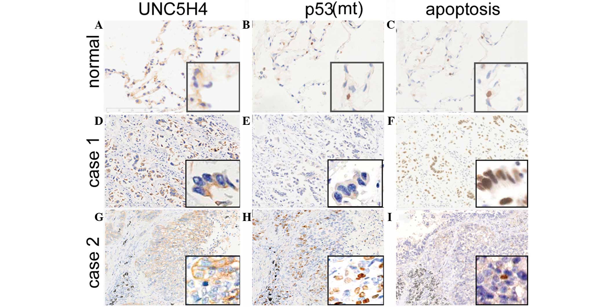
The expression of UNC5H4 occurred in the cell
membrane of the normal bronchial epithelium and particularly in the
cytoplasm of the alveolar epithelial cells (Fig. 1A). Normal or preserved membranous
UNC5H4 expression was identified in 50/130 (38.5%) patients, while
80/130 (61.5%) patients were identified with abnormal UNC5H4
expression, including absent membranous expression with ectopic
cytoplasmic expression (Fig. 1D)
and reduced membranous expression with ectopic cytoplasmic
expression (Fig. 1G). In addition,
abnormal UNC5H4 expression was shown to correlate with the degree
of differentiation (P=0.015) and TNM staging (P=0.037), as shown in
Table I. The abnormal expression
rates for the well- and moderately-differentiated cells (47.9%)
were significantly higher compared with those of the
poorly-differentiated cells (69.5%; P=0.015), and significantly
higher for TNM stages III and IV (70.1%) compared with stages I and
II (52.4%; P=0.037). In addition, the abnormal expression rate for
patients with lymph node metastasis (69.8%) was significantly
higher when compared with that of patients with no lymph node
metastasis (53.7%; P=0.059). No significant differences were
identified among patient gender, age, histological type or
lymphatic metastasis.
 | Table IClinical and histological features of
130 patients with lung cancer. |
Table I
Clinical and histological features of
130 patients with lung cancer.
| | UNC5H4 expression,
n | | p53 expression,
n | |
|---|
| |
| |
| |
|---|
| Variables | Patients, n | Abnormal | Normal | P-valuea | Positive | Negative | P valuea |
|---|
| n | 130 | 80 | 50 | | 51 | 79 | |
| Age, years |
| ≤55 | 56 | 37 | 19 | 0.355 | 25 | 31 | 0.511 |
| >55 | 74 | 43 | 31 | | 26 | 48 | |
| Gender |
| Male | 66 | 42 | 24 | 0.618 | 23 | 43 | 0.299 |
| Female | 64 | 38 | 26 | | 28 | 36 | |
| Stage |
| I/II | 63 | 33 | 30 | 0.037 | 17 | 46 | 0.006 |
| III/IV | 67 | 47 | 20 | | 34 | 33 | |
| Histology |
| Squamous cell
carcinoma | 69 | 46 | 23 | 0.201 | 24 | 45 | 0.269 |
| Adenocarcinoma | 61 | 34 | 27 | | 27 | 34 | |
| Differentiation |
| Well, moderate | 48 | 23 | 25 | 0.015 | 24 | 24 | 0.054 |
| Poor | 82 | 57 | 25 | | 27 | 55 | |
| Lymph node
metastasis |
| Yes | 63 | 44 | 19 | 0.059 | 35 | 28 | 0.000 |
| No | 67 | 36 | 31 | | 16 | 51 | |
The p53 mt-positive staining rate in the NSCLC
tissue was 39.2% (51/130 patients) and the correlation between
positive expression and the clinicopathological characteristics of
the patients with NSCLC are summarized in Table I. The expression of p53 mt was
revealed to correlate significantly with TNM staging and lymph node
metastasis (P<0.05). Cytoplasmic UNC5H4 expression was revealed
to correlate negatively with p53 mt expression (r=−0.270; P=0.002)
and positively with the AI (r=0.254; P=0.004). In addition, the
expression of p53 mt was shown to correlate negatively with the AI
(r=−0.190; P=0.09) ( Fig. 1).
Effect of abnormal UNC5H4 and p53 mt
expression on NSCLC patient prognosis
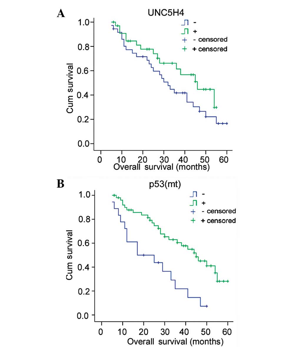
The post-surgical survival time ranged between 5 and
60 months, the average survival time was 36.6±2.4 months and the
median survival time was 38.0±5.0 months in 70 NSCLC patients. The
Kaplan-Meier method (log-rank test) was used to analyze life span.
The average and median survival times for each group were as
follows: i) Abnormal UNC5H4 expression, 33.2±3.2 and 32.0±3.6
months; ii) normal UNC5H4-positive, 39.22±3.2 and 46.0±7.2 months;
iii) p53 mt-negative, 40.9±2.7 and 45.0±5.7 months; and iv) p53
mt-positive, 24.10±3.6 and 17.0±8.8 months, respectively. The
statistical analyses indicated that the prognosis of the patients
with normal UNC5H4 expression was improved when compared with that
of patients with abnormal UNC5H4 expression, however, no
significant difference was identified (P=0.125). The prognosis of
the p53 mt-negative patients was significantly improved when
compared with that of the p53-positive patients (P=0.001) (Fig. 2).
A Cox regression analysis was used to evaluate the
significance of abnormal UNC5H4 and p53 mt expression as prognostic
factors (Table II). Age, gender,
histology, degree of differentiation, TNM stage, lymph node
metastasis, abnormal UNC5H4 expression and p53 mt-positive
expression were analyzed in 70 patients, revealing p53 mt
expression (P=0.006), lymph node metastasis (P=0.001), tumor stage
(P=0.002) and degree of differentiation (P=0.044) as independent
prognostic factors. However, abnormal UNC5H4 expression was not
identified as an independent prognostic factor (P=0.073).
| Table IICox regression model for the predicted
survival of 70 patients with lung cancer. |
Table II
Cox regression model for the predicted
survival of 70 patients with lung cancer.
| Factor | Risk | 95% CI | P-value |
|---|
| Age
(<50years) | 0.672 | 0.336–1.436 | 0.151 |
| Male gender | 0.750 | 0.424–1.653 | 0.403 |
| Histology
(adenocarcinoma) | 1.335 | 0.312–1.421 | 0.372 |
| Degree of
differentiation (Poor) | 2.847 | 1.362–3.344 | 0.044 |
| TNM stage
(III/IV) | 3.710 | 1.264–6.735 | 0.002 |
| Lymphatic
metastasis | 4.014 | 1.351–7.811 | 0.001 |
| Positive p53 mt
expression | 0.335 | 0.412–0.723 | 0.006 |
| Abnormal UNC5H4
expression | 0.302 | 0.459–1.506 | 0.073 |
Effect of X-ray exposure on UNC5H4
expression and apoptosis in NSCLC tissue
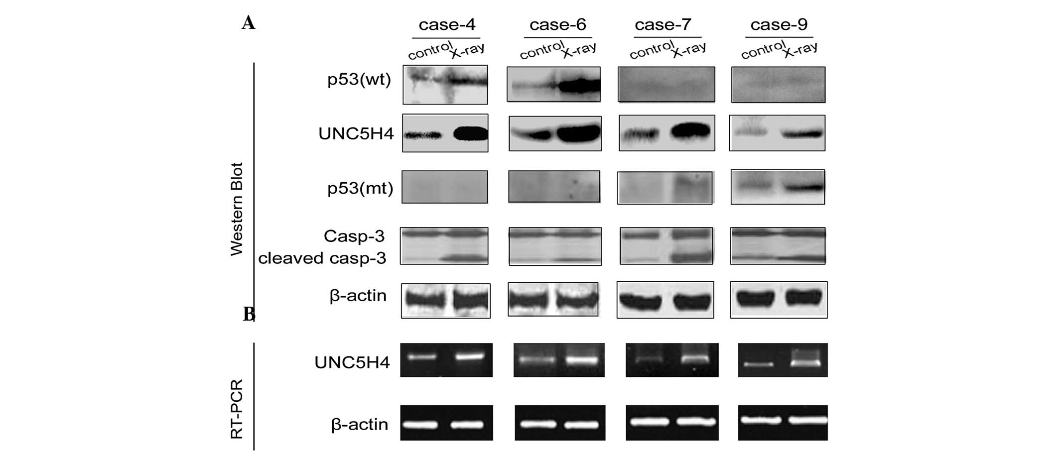
NSCLC tissue was exposed to 1 Gy of X-radiation and
cultured for 5 h. A radiation-induced increase in the UNC5H4 mRNA
and protein levels was identified in 13/20 cases, including 6
squamous cell carcinoma and 7 adenocarcinoma cases (4 well-, 8
moderately- and 1 poorly-differentiated; Fig. 3). In addition, the activation of
caspase 3 was also significantly increased in these cases,
indicating that UNC5H4 may play a role in X-ray-induced
apoptosis.
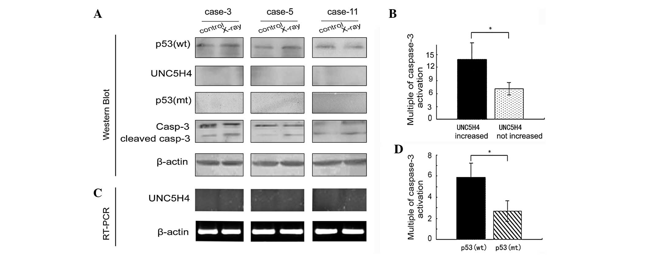
Notably, changes were not detected in the expression
of UNC5H4 in the remaining 7/20 cases, including 3 squamous cell
carcinoma and 4 adenocarcinoma cases (1 well-, 5 moderately- and 1
poorly-differentiated), however, significant increases in caspase 3
protein activation levels were observed (Fig. 4A and B), but were shown to be
significantly lower when compared with those of the 13 NSCLC cases
with increased UNC5H4 expression (P<0.05; Fig. 4C). In addition, 6/13 cases
demonstrating increased expression of UNC5H4 and p53 mt exhibited
significantly lower caspase 3 activation levels compared with the
p53 wt cells (P<0.05; Fig.
4D).
Discussion
It has been previously reported that all four
members of the UNC5H family exhibit proapoptotic activity (5,14) and
share a similar canonical caspase cleavage site in their
cytoplasmic region (13), which is
important for the induction of apoptosis. Since the
caspase-mediated cleavage of UNC5H is required for cell death
induction, in the current study, caspase 3 expression was detected
to investigate the induction of apoptosis. In contrast to UNC5H1-3,
little is known with regard to the functional significance of
UNC5H4. More recent studies have identified that the
UNC5H4-mediated induction of apoptosis is dependent on p53 status
(6). However, the correlation
between UNC5H4 expression and apoptosis via the p53 pathway remains
unclear in lung cancer.
Fresh tissues obtained from 20 NSCLC patients were
exposed to clinical doses of X-rays under in vitro culture
conditions. Notably, the exposure of NSCLC tissue to X-rays leads
to the concurrent upregulation of UNC5H4 expression and caspase 3
activation. Therefore, NSCLC tissue with high UNC5H4 expression may
be sensitive to radiation and highlight a new basis for a
radiosensitive indicator for NSCLC patients, allowing for an
improved and individualized dosing program. In addition, the
results indicated that UNC5H4 upregulation is independent of p53
status in NSCLC tissue since UNC5H4 is a direct transcriptional
target of p53 and UNC5H4-mediated apoptosis is regulated in a
p53-dependent manner.
Therefore, the present study further investigated
the correlation between UNC5H4 and p53 expression and apoptosis, as
well as the correlation between UNC5H4 expression and the
clinicopathological characteristics of NSCLC. UNC5H4 and p53
expression and apoptosis were determined in lung cancer tissue
obtained from 130 NSCLC patients at the time of treatment. UNC5H4
expression was shown to correlate significantly with the degree of
differentiation, TNM staging, lymphatic metastasis state and
prognosis of NSCLC. The prognosis was significantly improved in
patients with NSCLC tissues expressing high levels of UNC5H4
compared with patients whose NSCLC tissues expressed low levels.
Studies have also indicated that UNC5H4-mediated, radiation-induced
apoptosis correlates with p53 status and that p53 is a tumor
suppressor capable of inducing cell cycle arrest and apoptosis
(15–17). In the present study, UNC5H4
expression was demonstrated to positively correlate with
radiation-induced AI and negatively correlate with p53 mt
expression, indicating that UNC5H4-mediated apoptosis requires p53.
However, a subset of cases that responded to radiotherapy were also
shown to express p53 mt. UNC5H4 expression and caspase 3 activation
are upregulated following X-ray exposure, indicating that UNC5H4
may induce apoptosis independently of p53. In conclusion, the
results of the present study indicate that X-rays induce apoptosis
via an additional pathway when the p53 pathway is compromised.
Further analysis and identification of this pathway must be
performed in future studies.
References
|
1
|
Hong K, Hinck L, Nishiyama M, Poo MM,
Tessier-Lavigne M and Stein E: A ligand-gated association between
cytoplasmic domains of UNC5 and DCC family receptors converts
netrin-induced growth cone attraction to repulsion. Cell.
97:927–941. 1999. View Article : Google Scholar
|
|
2
|
Leonardo ED, Hinck L, Masu M, Keino-Masu
K, Ackerman SL and Tessier-Lavigne M: Vertebrate homologues of C.
elegans UNC-5 are candidate netrin receptors. Nature. 386:833–838.
1997. View
Article : Google Scholar : PubMed/NCBI
|
|
3
|
Tessier-Lavigne M and Goodman CS: The
molecular biology of axon guidance. Science. 274:1123–1133. 1996.
View Article : Google Scholar : PubMed/NCBI
|
|
4
|
Merz DC, Zheng H, Killeen MT, Krizus A and
Culotti JG: Multiple signaling mechanisms of the UNC-6/netrin
receptors UNC-5 and UNC-40/DCC in vivo. Genetics. 158:1071–1080.
2001.PubMed/NCBI
|
|
5
|
Llambi F, Causeret F, Bloch-Gallego E and
Mehlen P: Netrin-1 acts as a survival factor via its receptors
UNC5H and DCC. Embo J. 20:2715–2722. 2001. View Article : Google Scholar : PubMed/NCBI
|
|
6
|
Wang H, Ozaki T, Shamim Hossain M,
Nakamura Y, Kamijo T, Xue X and Nakagawara A: A newly identified
dependence receptor UNC5H4 is induced during DNA damage-mediated
apoptosis and transcriptional target of tumor suppressor p53.
Biochem Biophys Res Commun. 370:594–598. 2008. View Article : Google Scholar : PubMed/NCBI
|
|
7
|
Zhang X, Cheung RM, Komaki R, Fang B and
Chang JY: Radiotherapy sensitization by tumor-specific TRAIL gene
targeting improves survival of mice bearing human non-small cell
lung cancer. Clin Cancer Res. 11:6657–6668. 2005. View Article : Google Scholar : PubMed/NCBI
|
|
8
|
Masunaga S, Ono K, Takahashi A, Ohnishi T,
Kinashi Y and Takagaki M: Radiobiological characteristics of solid
tumours depending on the p53 status of the tumour cells, with
emphasis on the response of intratumour quiescent cells. Eur J
Cancer. 38:718–727. 2002. View Article : Google Scholar
|
|
9
|
Kanzawa T, Iwado E, Aoki H, Iwamaru A,
Hollingsworth EF, Sawaya R, Kondo S and Kondo Y: Ionizing radiation
induces apoptosis and inhibits neuronal differentiation in rat
neural stem cells via the c-JUN NH2-terminal kinase (JNK) pathway.
Oncogene. 25:3638–3648. 2006. View Article : Google Scholar : PubMed/NCBI
|
|
10
|
Goldstraw P, Crowley J, Chansky K, Giroux
DJ, Groome PA, Rami-Porta R, Postmus PE, Rusch V and Sobin L;
International Association for the Study of Lung Cancer
International Staging Committee; Participating Institutions. The
IASLC Lung Cancer Staging Project: proposals for the revision of
the TNM stage groupings in the forthcoming (seventh) edition of the
TNM Classification of malignant tumours. J Thorac Oncol. 2:706–714.
2007. View Article : Google Scholar
|
|
11
|
Travis WD, Brambilla E, Muller-Hermelink
HK and Harris CC: WHO and TNM classification. Pathology and
Genetics of Tumours of the Lung, Pleura, Thymus and Heart. IARC
Press; Lyon: 2004
|
|
12
|
Rusch V, Klimstra D, Venkatraman E, Oliver
J, Martini N, Gralla R, Kris M and Dmitrovsky E: Aberrant p53
expression predicts clinical resistance to cisplatin-based
chemotherapy in locally advanced non-small cell lung cancer. Cancer
Res. 55:5038–5042. 1995.PubMed/NCBI
|
|
13
|
Thornberry NA, Rano TA, Peterson EP,
Rasper DM, Timkey T, Garcia-Calvo M, Houtzager VM, Nordstrom PA,
Roy S, Vaillancourt JP, et al: A combinatorial approach defines
specificities of members of the caspase family and granzyme B.
Functional relationships established for key mediators of
apoptosis. J Biol Chem. 272:17907–17911. 1997. View Article : Google Scholar
|
|
14
|
Thiebault K, Mazelin L, Pays L, Llambi F,
Joly MO, Scoazec JY, Saurin JC, Romeo G and Mehlen P: The netrin-1
receptors UNC5H are putative tumor suppressors controlling cell
death commitment. Proc Natl Acad Sci USA. 100:4173–4178. 2003.
View Article : Google Scholar : PubMed/NCBI
|
|
15
|
Hartwell L: Defects in a cell cycle
checkpoint may be responsible for the genomic instability of cancer
cells. Cell. 71:543–546. 1992. View Article : Google Scholar : PubMed/NCBI
|
|
16
|
Ullrich SJ, Anderson CW, Mercer WE and
Appella E: The p53 tumor suppressor protein, a modulator of cell
proliferation. J Biol Chem. 267:15259–15262. 1992.PubMed/NCBI
|
|
17
|
Yin Y, Tainsky MA, Bischoff FZ, Strong LC
and Wahl GM: Wild-type p53 restores cell cycle control and inhibits
gene amplification in cells with mutant p53 alleles. Cell.
70:937–948. 1992. View Article : Google Scholar : PubMed/NCBI
|