Spandidos Publications Logo
  • About
    • About Spandidos
    • Aims and Scopes
    • Abstracting and Indexing
    • Editorial Policies
    • Reprints and Permissions
    • Job Opportunities
    • Terms and Conditions
    • Contact
  • Journals
    • All Journals
    • Oncology Letters
      • Oncology Letters
      • Information for Authors
      • Editorial Policies
      • Editorial Board
      • Aims and Scope
      • Abstracting and Indexing
      • Bibliographic Information
      • Archive
    • International Journal of Oncology
      • International Journal of Oncology
      • Information for Authors
      • Editorial Policies
      • Editorial Board
      • Aims and Scope
      • Abstracting and Indexing
      • Bibliographic Information
      • Archive
    • Molecular and Clinical Oncology
      • Molecular and Clinical Oncology
      • Information for Authors
      • Editorial Policies
      • Editorial Board
      • Aims and Scope
      • Abstracting and Indexing
      • Bibliographic Information
      • Archive
    • Experimental and Therapeutic Medicine
      • Experimental and Therapeutic Medicine
      • Information for Authors
      • Editorial Policies
      • Editorial Board
      • Aims and Scope
      • Abstracting and Indexing
      • Bibliographic Information
      • Archive
    • International Journal of Molecular Medicine
      • International Journal of Molecular Medicine
      • Information for Authors
      • Editorial Policies
      • Editorial Board
      • Aims and Scope
      • Abstracting and Indexing
      • Bibliographic Information
      • Archive
    • Biomedical Reports
      • Biomedical Reports
      • Information for Authors
      • Editorial Policies
      • Editorial Board
      • Aims and Scope
      • Abstracting and Indexing
      • Bibliographic Information
      • Archive
    • Oncology Reports
      • Oncology Reports
      • Information for Authors
      • Editorial Policies
      • Editorial Board
      • Aims and Scope
      • Abstracting and Indexing
      • Bibliographic Information
      • Archive
    • Molecular Medicine Reports
      • Molecular Medicine Reports
      • Information for Authors
      • Editorial Policies
      • Editorial Board
      • Aims and Scope
      • Abstracting and Indexing
      • Bibliographic Information
      • Archive
    • World Academy of Sciences Journal
      • World Academy of Sciences Journal
      • Information for Authors
      • Editorial Policies
      • Editorial Board
      • Aims and Scope
      • Abstracting and Indexing
      • Bibliographic Information
      • Archive
    • International Journal of Functional Nutrition
      • International Journal of Functional Nutrition
      • Information for Authors
      • Editorial Policies
      • Editorial Board
      • Aims and Scope
      • Abstracting and Indexing
      • Bibliographic Information
      • Archive
    • International Journal of Epigenetics
      • International Journal of Epigenetics
      • Information for Authors
      • Editorial Policies
      • Editorial Board
      • Aims and Scope
      • Abstracting and Indexing
      • Bibliographic Information
      • Archive
    • Medicine International
      • Medicine International
      • Information for Authors
      • Editorial Policies
      • Editorial Board
      • Aims and Scope
      • Abstracting and Indexing
      • Bibliographic Information
      • Archive
  • Articles
  • Information
    • Information for Authors
    • Information for Reviewers
    • Information for Librarians
    • Information for Advertisers
    • Conferences
  • Language Editing
Spandidos Publications Logo
  • About
    • About Spandidos
    • Aims and Scopes
    • Abstracting and Indexing
    • Editorial Policies
    • Reprints and Permissions
    • Job Opportunities
    • Terms and Conditions
    • Contact
  • Journals
    • All Journals
    • Biomedical Reports
      • Information for Authors
      • Editorial Policies
      • Editorial Board
      • Aims and Scope
      • Abstracting and Indexing
      • Bibliographic Information
      • Archive
    • Experimental and Therapeutic Medicine
      • Information for Authors
      • Editorial Policies
      • Editorial Board
      • Aims and Scope
      • Abstracting and Indexing
      • Bibliographic Information
      • Archive
    • International Journal of Epigenetics
      • Information for Authors
      • Editorial Policies
      • Editorial Board
      • Aims and Scope
      • Abstracting and Indexing
      • Bibliographic Information
      • Archive
    • International Journal of Functional Nutrition
      • Information for Authors
      • Editorial Policies
      • Editorial Board
      • Aims and Scope
      • Abstracting and Indexing
      • Bibliographic Information
      • Archive
    • International Journal of Molecular Medicine
      • Information for Authors
      • Editorial Policies
      • Editorial Board
      • Aims and Scope
      • Abstracting and Indexing
      • Bibliographic Information
      • Archive
    • International Journal of Oncology
      • Information for Authors
      • Editorial Policies
      • Editorial Board
      • Aims and Scope
      • Abstracting and Indexing
      • Bibliographic Information
      • Archive
    • Medicine International
      • Information for Authors
      • Editorial Policies
      • Editorial Board
      • Aims and Scope
      • Abstracting and Indexing
      • Bibliographic Information
      • Archive
    • Molecular and Clinical Oncology
      • Information for Authors
      • Editorial Policies
      • Editorial Board
      • Aims and Scope
      • Abstracting and Indexing
      • Bibliographic Information
      • Archive
    • Molecular Medicine Reports
      • Information for Authors
      • Editorial Policies
      • Editorial Board
      • Aims and Scope
      • Abstracting and Indexing
      • Bibliographic Information
      • Archive
    • Oncology Letters
      • Information for Authors
      • Editorial Policies
      • Editorial Board
      • Aims and Scope
      • Abstracting and Indexing
      • Bibliographic Information
      • Archive
    • Oncology Reports
      • Information for Authors
      • Editorial Policies
      • Editorial Board
      • Aims and Scope
      • Abstracting and Indexing
      • Bibliographic Information
      • Archive
    • World Academy of Sciences Journal
      • Information for Authors
      • Editorial Policies
      • Editorial Board
      • Aims and Scope
      • Abstracting and Indexing
      • Bibliographic Information
      • Archive
  • Articles
  • Information
    • For Authors
    • For Reviewers
    • For Librarians
    • For Advertisers
    • Conferences
  • Language Editing
Login Register Submit
  • This site uses cookies
  • You can change your cookie settings at any time by following the instructions in our Cookie Policy. To find out more, you may read our Privacy Policy.

    I agree
Search articles by DOI, keyword, author or affiliation
Search
Advanced Search
presentation
Oncology Letters
Join Editorial Board Propose a Special Issue
Print ISSN: 1792-1074 Online ISSN: 1792-1082
Journal Cover
December 2013 Volume 6 Issue 6

Full Size Image

Sign up for eToc alerts
Recommend to Library

Journals

International Journal of Molecular Medicine

International Journal of Molecular Medicine

International Journal of Molecular Medicine is an international journal devoted to molecular mechanisms of human disease.

International Journal of Oncology

International Journal of Oncology

International Journal of Oncology is an international journal devoted to oncology research and cancer treatment.

Molecular Medicine Reports

Molecular Medicine Reports

Covers molecular medicine topics such as pharmacology, pathology, genetics, neuroscience, infectious diseases, molecular cardiology, and molecular surgery.

Oncology Reports

Oncology Reports

Oncology Reports is an international journal devoted to fundamental and applied research in Oncology.

Experimental and Therapeutic Medicine

Experimental and Therapeutic Medicine

Experimental and Therapeutic Medicine is an international journal devoted to laboratory and clinical medicine.

Oncology Letters

Oncology Letters

Oncology Letters is an international journal devoted to Experimental and Clinical Oncology.

Biomedical Reports

Biomedical Reports

Explores a wide range of biological and medical fields, including pharmacology, genetics, microbiology, neuroscience, and molecular cardiology.

Molecular and Clinical Oncology

Molecular and Clinical Oncology

International journal addressing all aspects of oncology research, from tumorigenesis and oncogenes to chemotherapy and metastasis.

World Academy of Sciences Journal

World Academy of Sciences Journal

Multidisciplinary open-access journal spanning biochemistry, genetics, neuroscience, environmental health, and synthetic biology.

International Journal of Functional Nutrition

International Journal of Functional Nutrition

Open-access journal combining biochemistry, pharmacology, immunology, and genetics to advance health through functional nutrition.

International Journal of Epigenetics

International Journal of Epigenetics

Publishes open-access research on using epigenetics to advance understanding and treatment of human disease.

Medicine International

Medicine International

An International Open Access Journal Devoted to General Medicine.

Journal Cover
December 2013 Volume 6 Issue 6

Full Size Image

Sign up for eToc alerts
Recommend to Library

  • Article
  • Citations
    • Cite This Article
    • Download Citation
    • Create Citation Alert
    • Remove Citation Alert
    • Cited By
  • Similar Articles
    • Related Articles (in Spandidos Publications)
    • Similar Articles (Google Scholar)
    • Similar Articles (PubMed)
  • Download PDF
  • Download XML
  • View XML
Case Report

99mTcO4- scintigraphic detection of follicular thyroid cancer and multiple metastatic lesions: A case report

  • Authors:
    • Chang‑Yin Wang
    • Bang‑Ru Xiao
    • Mei‑Juan Shen
    • Ying Shen
    • Kun‑Wei Cui
  • View Affiliations / Copyright

    Affiliations: Department of Nuclear Medicine, Zhongnan Hospital of Wuhan University, Wuhan, Hubei 430071, P.R. China, Department of Nuclear Medicine, Jingzhou Center Hospital, Tongji Medical College, Huazhong University of Science and Technology, Jingzhou, Hubei 434000, P.R. China
  • Pages: 1729-1732
    |
    Published online on: October 23, 2013
       https://doi.org/10.3892/ol.2013.1639
  • Expand metrics +
Metrics: Total Views: 0 (Spandidos Publications: | PMC Statistics: )
Metrics: Total PDF Downloads: 0 (Spandidos Publications: | PMC Statistics: )
Cited By (CrossRef): 0 citations Loading Articles...

This article is mentioned in:


Abstract

99mTcO4- thyroid imaging is often used to detect thyroid diseases that are confined to the neck. However, this examination is not frequently used to detect metastatic lesions of thyroid cancer in the whole body, while 131I imaging is often used to detect the metastases of differentiated thyroid cancers. The present study performed 99mTcO4- thyroid imaging for a 69‑year‑old patient with a thyroid nodule and incidentally identified a lesion with abnormally increased 99mTcO4- uptake in the chest of the patient. Furthermore, a whole‑body scan was performed for this patient and multiple lesions with increased 99mTcO4- uptake were identified. The results confirmed that these lesions were follicular thyroid cancer and the metastatic lesions were distributed in numerous locations. The results revealed that analysis of the whole body is significant when regional lesions with abnormally increased 99mTcO4- uptake outside of thyroid tissues are identified by routine 99mTcO4- thyroid imaging.

Introduction

The incidence of follicular thyroid cancer is lower than that of papillary thyroid cancer in thyroid malignant tumors and is, therefore, the second most common thyroid malignancy (1). Follicular thyroid cancer is mainly characterized by a follicular structure. It is a differentiated thyroid cancer and demonstrates positive expression of the sodium-iodide symporter. Therefore, the tissues of follicular thyroid cancer generally uptake iodine, which is the biological basis for the detection of cancer lesions by 131I whole-body imaging and the treatment of follicular thyroid cancer by radioactive 131I (2,3). 99mTcO4− and the iodide ion have a number of similar features, so 99mTcO4− can also be absorbed by the thyorid (4). Follicular thyroid cancer may develop into regional nodal metastasis and may also progress into hematogenous metastasis (1). The accurate detection of recurrent and metastatic lesions of follicular thyroid cancer is significant in the staging of diseases and the evaluation of the therapeutic effect and prognosis (1). The present study describes a patient with follicular thyroid cancer whose metastatic lesions were detected using a 99mTcO4− whole-body scan. Informed consent was obtained from the patient prior to the study.

Case report

A 69-year-old female was admitted to Zhongnan Hospital of Wuhan University (Hubei, China) complaining of left stethalgia for three weeks. A physical examination revealed that the second rib in anterior left chest and the fourth thoracic vertebra were swollen and painful when palpated. The thyroid glands of the patient were intumescent and the left lobe to the isthmic portion was palpated as a hard and fixed nodule with an asperous surface, without pain. Laboratory examination demonstrated that the thyroid, liver and kidney functions, as well as the routine blood and urine test results, were all normal.

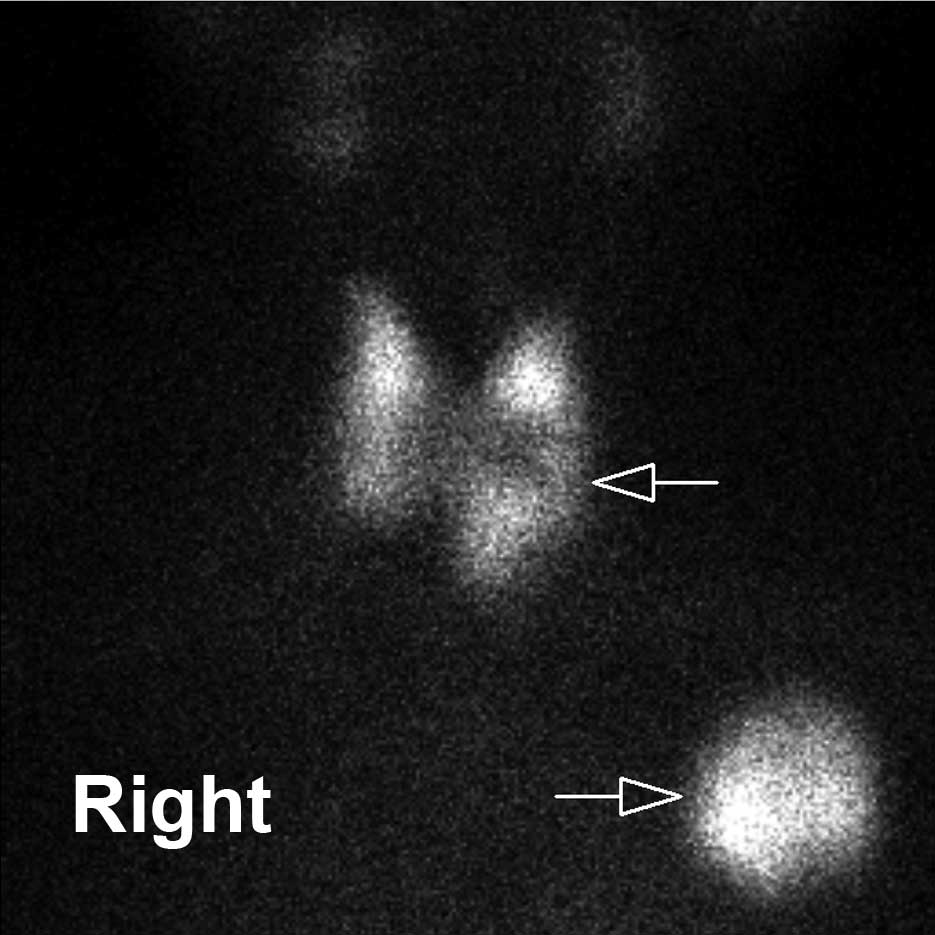
Thyroid imaging was performed at 10 min following the intravenous injection of Na99mTcO4 at 185 MBq. The result demonstrated a regional area of markedly decreased 99mTcO4− uptake with an irregular edge within the middle portion of the left lobe of the thyroid. A conglomerate area of increased 99mTcO4− uptake was identified in the left chest (Fig. 1). 
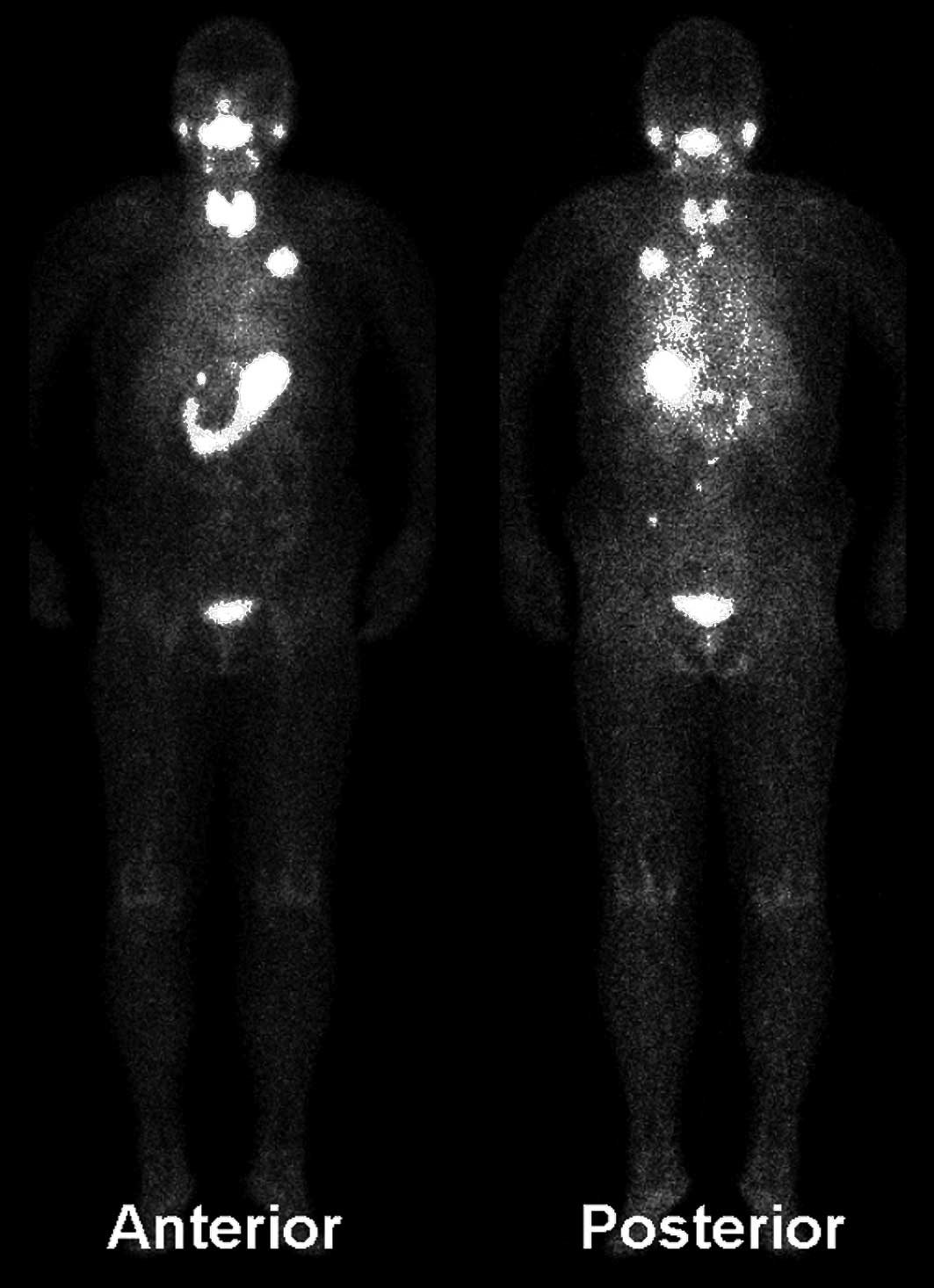
Subsequently, 99mTcO4− whole-body imaging was performed for the patient. The result revealed abnormal lesions of increased 99mTcO4− uptake in the left chest, thoracic vertebrae, lumbar vertebrae and left ilium (Fig. 2). The following day, whole-body bone imaging was performed at 3 h following an intravenous injection of 99mTc-methylene diphosphonate at 740 MBq. The imaging outcome demonstrated that the left anterior branch of the second rib, the fourth and twelfth thoracic vertebrae and the midpiece of the right thigh bone exhibited increased radioactive uptake. The third and fourth lumbar vertebrae and the left posterior inferior iliac spine were suspected of abnormal uptake, whereas the partial osseous tissue of the left anterior branch of the second rib demonstrated decreased radioactive uptake (Fig. 3).

Figure 1

Thyroid static imaging with 99mTcO4− showing a nodule in the middle portion of the left lobe of the thyroid with a decreased uptake and unclear edge, and an area of the left chest with a markedly increased uptake.

Figure 2

99mTcO4− whole-body imaging showing one site of the left chest, two sites of thoracic vertebrae, two sites of lumbar vertebrae and one site of the left ilium with an increased uptake.

Figure 3

Whole-body bone imaging showing the left anterior branch of the 2nd rib, the 4th and 12th thoracic vertebrae and the midpiece of the right thigh bone with an increased uptake of 99mTc-methylene diphosphonate. The 3rd and 4th lumbar vertebrae and the left posterior inferior iliac spine were suspected of abnormal uptake, whereas the partial osseous tissue of the 2nd rib presented with an osteolytic lesion.

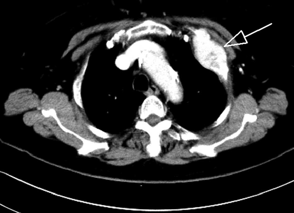
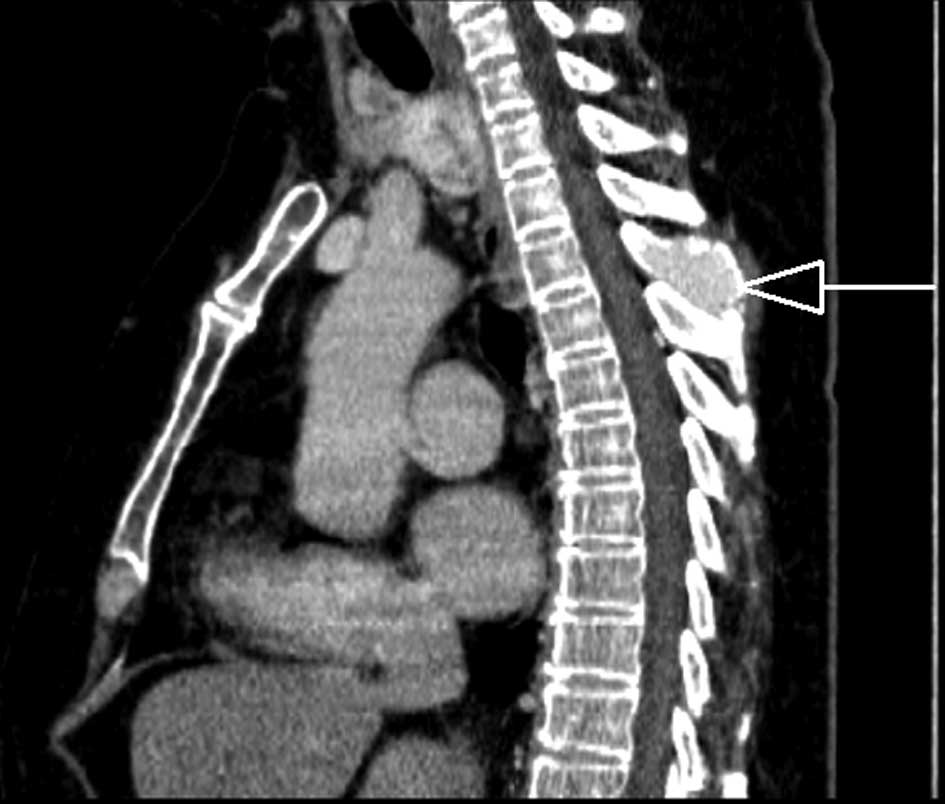
A computed tomography (CT) scan of the thoracic region revealed a mass on the left thoracic wall, bony destruction of the left anterior branch of the second rib (Fig. 4) and the destruction of the crest of the fourth thoracic vertebra (Fig. 
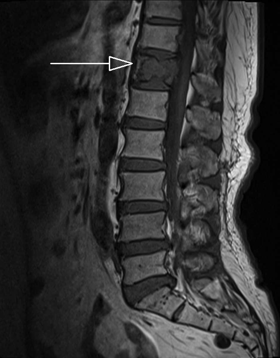
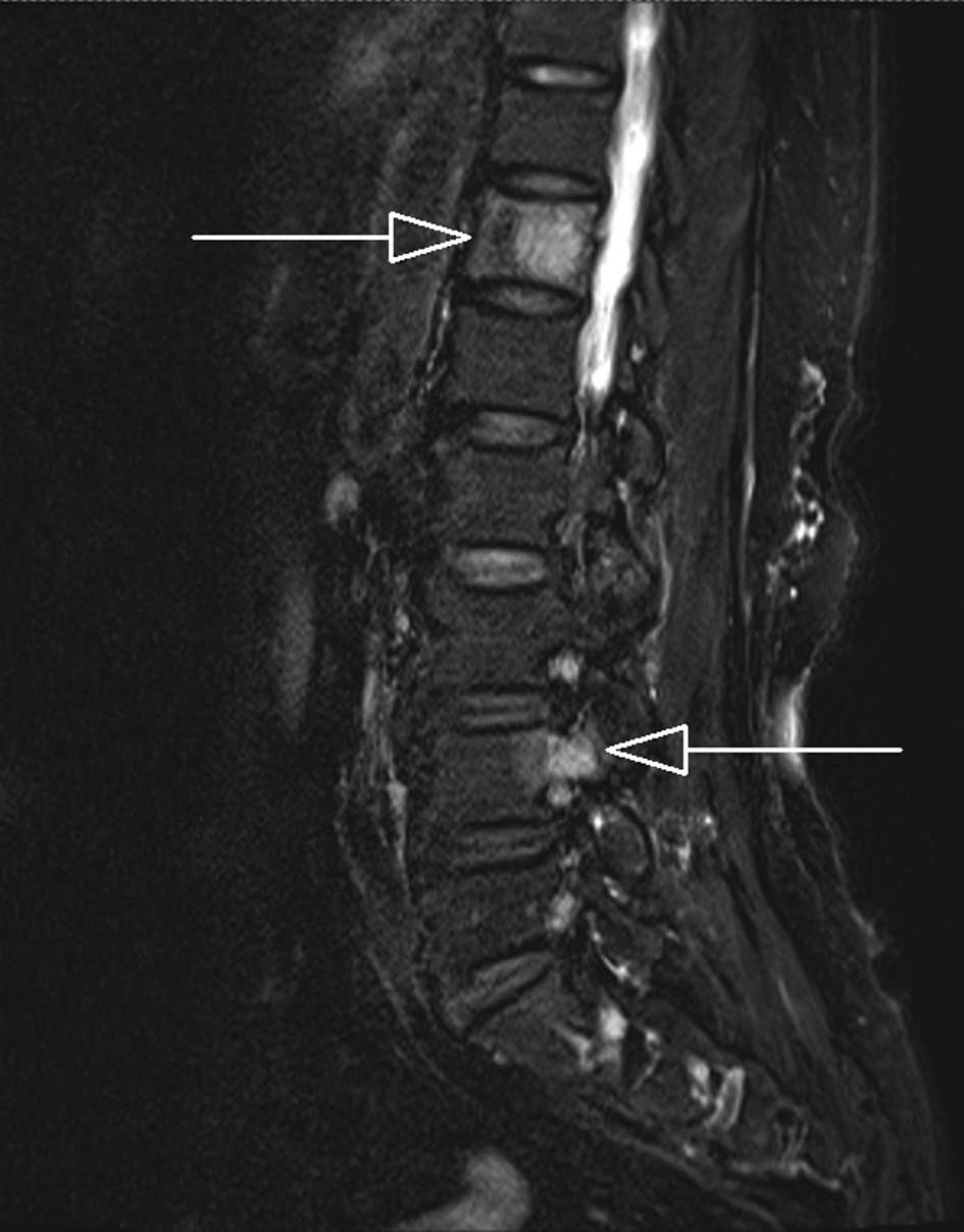
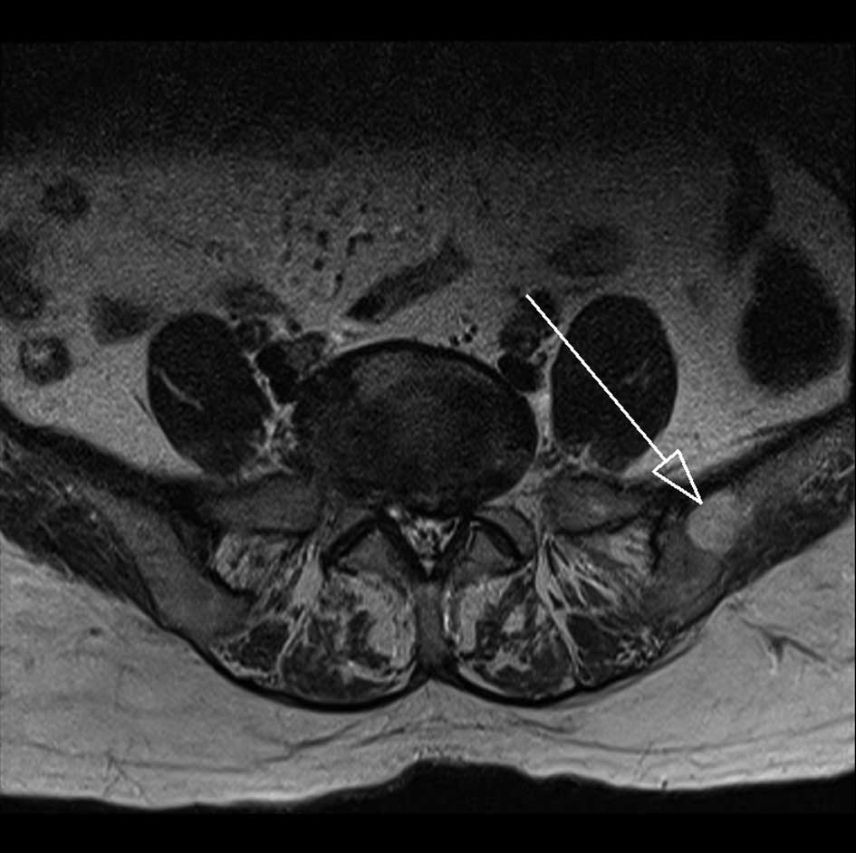
5). A magnetic resonance imaging (MRI) scan of the vertebrae and pelvic cavity demonstrated that the twelfth thoracic vertebra (Figs. 6 and 7), the pedicle of the fourth lumbar vertebral arch (Fig. 7) and the left ilium presented an abnormal signal (Fig. 8).

Figure 4

Computed tomography with contrast showing the 2nd rib with bony destruction and a markedly enhanced mass.

Figure 5

Computed tomography with contrast showing the 4th thoracic vertebra with bony destruction, and an enhanced lesion.

Figure 6

Magnetic resonance imaging showing the 12th thoracic vertebra with bony destruction.

Figure 7

Magnetic resonance imaging showing the 12th thoracic vertebra and the pedicle of the 4th lumbar vertebral arch with an abnormal signal.

Figure 8

Magnetic resonance imaging showing the left ilium with an abnormal signal.

The patient underwent a total thyroidectomy. The pathological results revealed that the left lobe nodule of thyroid was composed of follicular thyroid cancer tissue with peplos infiltration and tumor embolus formation of the small vessels.

Discussion

131I whole-body imaging is often used for the identification of metastatic lesions of differentiated thyroid cancer following total thyroidectomy (2,3). When thyroid tissues are not completely resected, 131I is largely absorbed by the existing normal thyroid tissues (5). However, metastatic lesions universally have a low uptake of 131I and, therefore, the metastatic lesions are not displayed clearly (5). As a result, 131I whole-body imaging is not generally selected to identify the metastatic lesions of thyroid cancer in the presence of normal thyroid tissues. 99mTcO4− is similar to the iodide ion to a certain extent (4); 99mTcO4− and the iodide ion are mediated by the sodium-iodide symporter and are absorbed by thyroid follicular cells (4). Therefore, the two methods are often used in thyroid imaging (6). However, 99mTcO4 imaging is not generally used to detect the metastatic lesions of thyroid cancer in the presence of thyroid tissues and also following a total thyroidectomy (6–8).

Follicular thyroid cancer tissues are mainly composed of differentiated follicular cells. The proteins of the sodium-iodide symporter are predominantly distributed in the membrane of follicular epithelial cells, and cancer tissues with follicular cells express sodium-iodide symporter proteins (9,10), which are the pacing factors for which 131I and 99mTcO4− are absorbed by the cancer tissues. The quantity of cancer tissues absorbing 99mTcO4− correlates with the level of sodium-iodide symporter protein expression. Since cancer tissues are not well-differentiated, the level of sodium-iodide symporter protein expression is low (10,11). Accordingly, the lesions of thyroid cancer frequently manifest ‘cool nodules’ or ‘cold nodules’ of decreased 99mTcO4− uptake (12). When thyroid tissues are not operated on, an increased 99mTcO4− uptake of metastatic lesions of thyroid cancer is rare. Certain studies have reported an increased uptake of neck metastases of thyroid cancer in 99mTcO4− thyroid imaging (7,8,13–15). However, to the best of our knowledge, there have been no studies with regard to the increased uptake of whole-body multiple metastatic lesions of thyroid cancer in 99mTcO4− whole-body imaging. The present study incidentally identified a mass outside of the thyroid gland in a patient with thyroid ‘cool nodules’, which exhibited increased 99mTcO4− uptake in the routine image field. Subsequently, a whole-body scan was performed. The result demonstrated that similar lesions of increased 99mTcO4− uptake existed in multiple positions of the whole body. CT, MRI and radionuclide whole-body bone imaging confirmed that the multitudinous sites of increased uptake, which were detected by 99mTcO4− imaging, contained tumor lesions. Therefore, despite the fact that 99mTcO4− imaging is not routinely used to identify metastatic lesions of thyroid cancer, when regional lesions of increased 99mTcO4− uptake are observed outside of the thyroid glands during routine field thyroid static imaging, further identification of the metastatic lesions of the whole body is significant.

Furthermore, 99mTcO4− whole-body imaging has numerous advantages. The procedure is highly sensitive, as shown by the third and fourth lumbar vertebral lesions and the ilium lesion, which were detected by 99mTcO4− imaging, but were not observed on the whole-body bone scan. MRI only identified the lesions of the fourth lumbar vertebra and the ilium, but not the third lumbar vertebral lesion. These results indicate that the sensitivity of 99mTcO4− whole-body imaging is higher than that of MRI and whole-body bone scan at this time. Furthermore, the procedure is highly specific. The fact that 99mTcO4− was able to be absorbed by thyroid cancer tissues under the mediation of sodium-iodide symporter proteins, confirms the diagnostic specificity for regional lesions. Abnormally increased 99mTcO4− uptake of the lesions outside of the thyroid glands is a characteristic of metastatic thyroid cancer tissues (7,8,13–15), suggesting that the lesions likely originated from thyroid tissues. Finally, the procedure increases the quality of the diagnosis of the thyroid nodules. For the patient of the present study, the feature of the ‘cool nodule’ was not enough to discriminate between malignant lesions and benign tumors. However, the identification of the metastatic lesions extremely supported the diagnosis of the malignant thyroid nodule. The 99mTcO4− imaging examination is able to scan the whole body of patients and, therefore, the detection area is extensive, which conduces to a complete detection of the lesions.

References

1 

Grebe SK and Hay ID: Follicular thyroid cancer. Endocrinol Metab Clin North Am. 24:761–801. 1995.

2 

Muresan MM, Olivier P, Leclère J, et al: Bone metastases from differentiated thyroid carcinoma. Endocr Relat Cancer. 15:37–49. 2008. View Article : Google Scholar : PubMed/NCBI

3 

Krishna L, Dadparvar S, Brady LW, et al: Paradoxical changes in iodine-131 scintigraphic findings in advanced follicular thyroid cancer. J Nucl Med. 34:1574–1576. 1993.PubMed/NCBI

4 

Zuckier LS, Dohan O, Li Y, Chang CJ, Carrasco N and Dadachova E: Kinetics of perrhenate uptake and comparative biodistribution of perrhenate, pertechnetate, and iodide by NaI symporter-expressing tissues in vivo. J Nucl Med. 45:500–507. 2004.PubMed/NCBI

5 

Tian R, Kuang AR, Qin WS and Zhang HM: Value of post-therapy whole-body 131I scan in the evaluation of patients with differentiated thyroid cancer. Chin J Nucl Med. 20:162–164. 2000.(In Chinese).

6 

Haugen BR and Lin EC: Isotope imaging for metastatic thyroid cancer. Endocrinol Metab Clin North Am. 30:469–492. 2001. View Article : Google Scholar : PubMed/NCBI

7 

Mathiopoulou L, Chrisoulidou A, Boudina M, Mitsakis P, Mandanas S and Pazaitou-Panayiotou K: 99mTc pertechnetate thyroid scan leads to serendipitous detection of metastatic thyroid cancer. Clin Nucl Med. 37:604–606. 2012. View Article : Google Scholar

8 

Campbell CM and Khafagi FA: Insensitivity of Tc-99m pertechnetate for detecting metastases of differentiated thyroid carcinoma. Clin Nucl Med. 15:1–4. 1990. View Article : Google Scholar : PubMed/NCBI

9 

Wang SS, Liang J, Lin YS and Yao RY: Differential expression of the Na symporter protein in thyroid cancer and adjacent normal and nodular goiter tissues. Oncol Lett. 5:368–372. 2012.PubMed/NCBI

10 

Liou MJ, Lin JD, Chan EC, Liu FH, Chao TC and Weng HF: Detection of mRNA of sodium iodide symporter in benign and malignant human thyroid tissues. Cancer Lett. 160:75–80. 2000. View Article : Google Scholar : PubMed/NCBI

11 

Peyrottes I, Navarro V, Ondo-Mendez A, et al: Immunoanalysis indicates that the sodium iodide symporter is not overexpressed in intracellular compartments in thyroid and breast cancers. Eur J Endocrinol. 60:215–225. 2009.PubMed/NCBI

12 

Summaria V, Rufini V, Mirk P, Costantini AM, Reale F and Maresca G: Diagnostic imaging of differentiated thyroid carcinoma. Rays. 25:177–190. 2000.(In English, Italian).

13 

Krausz Y and Horne T: Detection of metastatic thyroid carcinoma by 99mTc-pertechnetate in the presence of hyperfunctioning thyroid tissue. J Surg Oncol. 44:132–134. 1990. View Article : Google Scholar : PubMed/NCBI

14 

Kumaresan K and Sastry RA: Localization of Tc-99m pertechnetate in lymph node metastasis from occult thyroid carcinoma. Clin Nucl Med. 19:11121994. View Article : Google Scholar : PubMed/NCBI

15 

Khan SU, Khan AU, Khan A, Shah AS and Khan K: Extrathyroidal uptake from thyroid carcinoma on 99mTc-pertechnetate scintigraphy. J Coll Physicians Surg Pak. 21:772–774. 2011.PubMed/NCBI

Related Articles

  • Abstract
  • View
  • Download
  • Twitter
Copy and paste a formatted citation
Spandidos Publications style
Wang CY, Xiao BR, Shen MJ, Shen Y and Cui KW: 99mTcO4- scintigraphic detection of follicular thyroid cancer and multiple metastatic lesions: A case report. Oncol Lett 6: 1729-1732, 2013.
APA
Wang, C., Xiao, B., Shen, M., Shen, Y., & Cui, K. (2013). 99mTcO4- scintigraphic detection of follicular thyroid cancer and multiple metastatic lesions: A case report. Oncology Letters, 6, 1729-1732. https://doi.org/10.3892/ol.2013.1639
MLA
Wang, C., Xiao, B., Shen, M., Shen, Y., Cui, K."99mTcO4- scintigraphic detection of follicular thyroid cancer and multiple metastatic lesions: A case report". Oncology Letters 6.6 (2013): 1729-1732.
Chicago
Wang, C., Xiao, B., Shen, M., Shen, Y., Cui, K."99mTcO4- scintigraphic detection of follicular thyroid cancer and multiple metastatic lesions: A case report". Oncology Letters 6, no. 6 (2013): 1729-1732. https://doi.org/10.3892/ol.2013.1639
Copy and paste a formatted citation
x
Spandidos Publications style
Wang CY, Xiao BR, Shen MJ, Shen Y and Cui KW: 99mTcO4- scintigraphic detection of follicular thyroid cancer and multiple metastatic lesions: A case report. Oncol Lett 6: 1729-1732, 2013.
APA
Wang, C., Xiao, B., Shen, M., Shen, Y., & Cui, K. (2013). 99mTcO4- scintigraphic detection of follicular thyroid cancer and multiple metastatic lesions: A case report. Oncology Letters, 6, 1729-1732. https://doi.org/10.3892/ol.2013.1639
MLA
Wang, C., Xiao, B., Shen, M., Shen, Y., Cui, K."99mTcO4- scintigraphic detection of follicular thyroid cancer and multiple metastatic lesions: A case report". Oncology Letters 6.6 (2013): 1729-1732.
Chicago
Wang, C., Xiao, B., Shen, M., Shen, Y., Cui, K."99mTcO4- scintigraphic detection of follicular thyroid cancer and multiple metastatic lesions: A case report". Oncology Letters 6, no. 6 (2013): 1729-1732. https://doi.org/10.3892/ol.2013.1639
Follow us
  • Twitter
  • LinkedIn
  • Facebook
About
  • Spandidos Publications
  • Careers
  • Cookie Policy
  • Privacy Policy
How can we help?
  • Help
  • Live Chat
  • Contact
  • Email to our Support Team