Introduction
Sarcomatoid (spindle cell) carcinoma is an
aggressive form of carcinoma composed of malignant spindle cells,
with or without a coexisting epithelial cell component. The
carcinoma demonstrates evidence of epithelial derivation with no
specific line of mesenchymal differentiation. The current study
represents only the seventh case of sarcomatoid (spindle cell)
carcinoma of the exocrine pancreas as defined above that has
described in the English literature (1–6).
Case report
Materials and methods
Tissue from the pancreatic tumor was obtained from
the surgical pathology files of Northwestern Memorial Hospital
(Chicago, IL, USA). Light microscopic and immunohistochemical
examination were performed on hematoxylin and eosin-stained
sections prepared from formalin-fixed, paraffin-embedded tissue.
The immunomarkers used, including clones, dilutions and
manufacturers, are presented in Table
I. Cases relevant to the definition of sarcomatoid (spindle
cell) carcinoma reported in the English literature are listed in
Table II and were searched for via
PubMed.
 | Table IImmunohistochemical findings. |
Table I
Immunohistochemical findings.
| Antibody | Reactivity | Clone | Dilution | Source |
|---|
| Calponin | − | CALP | 1:400 | Dakoa |
| CD10 | F+ | 56C6 | 1:30 | Leicab |
| Chromogranin | − | LK2H10 | Predilute | Ventana Medical
Systemsc |
| CK8/18 | D+ | CAM5.2 | 1:50 |
Becton-Dickinsond |
| CK19 | − | RCK108 | 1:100 | Dakoa |
| EMA | − | E29 | 1:50 | Dakoa |
| Ki-67 | 50% | 30-9 | Predilute | Ventana Medical
Systemsc |
| MUC1 | − | Ma695 | 1:100 | Vector
Laboratoriese |
| Nuclear
β-catenin | − | 14 | 1:200 |
Becton-Dickinsond |
| p53 | 75% | Bp-53-11 | Predilute | Ventana Medical
Systemsc |
| p63 | − | 4A4 | Predilute | Ventana Medical
Systemsc |
| Pan-CK | D+ | MNF116 | 1:50 | Dakoa |
| S100 | − | 4C4.9 | Predilute | Ventana Medical
Systemsc |
| SMA | − | 1A4 | Predilute | Cell Marque
Corporationf |
| Synaptophysin | − | N/A | 1:50 | Cell Marque
Corporationf |
| Vimentin | D+ | V9 | 1:100 | Dakoa |
| Table IIClinical and pathological observations
in seven cases of sarcomatoid spindle cell (SC) carcinoma of the
pancreas reported in the English literature. |
Table II
Clinical and pathological observations
in seven cases of sarcomatoid spindle cell (SC) carcinoma of the
pancreas reported in the English literature.
| First author, year
(ref.) | Age,
years/gender | Tumor size, cm | Carcinoma | Sarcomatoid
component | Molecular | Follow-up,
months/outcome |
|---|
| Higashi et al,
1999 (1) | 74/male | 4.5×4.0×3.0 | PD adeno | SC; IHC: CK AE1,
variable CK AE3, EMA, MUC1-ARA (D+), S100, SMA (F+), desmin,
vimentin, NSE and CEA (−) | NA | 3/succumbed to
diffuse peritoneal carcinomatosis |
| Darvishian et
al, 2001 (2) | 74/male | 4.0×3.0 | MD adeno | SC; IHC:vimentin
(D+), CK (F+), CEA, SMA, desmin and CD68 (−) | NA | 4/alive and well |
| Barkatullah et
al, 2005 (3) | 67/female | 2.5×2.5×2.0 | MD adeno | SC, separate focus of
OGC; IHC (SC): CK8/18 and vimentin (D+) | NA | 8/NA |
| De la Riva et
al, 2006 (4) | 72/female | NA | Not identified, but
associated with choledochal cyst | SC; IHC: CK and
vimentin (F+) | NA | 9/succumbed to
sarcomatoid carcinoma metastatic to the liver |
| Nakano et al,
2007 (5) | 82/female | 18.0×11.0×10.0 | WD adeno | SC, foci of OGC
around hemorrhage; IHC (SC): vimentin, CD10 (D+), CK AE1/AE3 (F+),
CK7, CK20, CEA, EMA, SMA and S100 (−) | K-ras mutation at
codon 12 (and codon 34) of exon 2 in SC | 0/Succumbed to DIC on
post-operative day 13 |
| Kim et al,
2010 (6) | 48/male | 3.5×2.5×1.5 | Mucinous cyst adeno
and anaplastic carcinoma | SC, scattered OGC;
IHC (SC): vimentin (D+), pan-CK, CK, 7, CK8/18, EMA, CEA, CD34,
CD56, CD68, CD117, desmin, SMA, myogenin, S100, ER and PR (−) | K-ras mutation at
codon 12 of exon 2 in SC and epithelial components | 4/succumbed to
hepatic and peritoneal metastases |
| Current case report,
2013 | 85/male | 3.3×3.0×2.6 | PD adeno | SC; IHC: diffuse
pan-CK, CK5.2, p53 (D+), synaptophysin, chromogranin, calponin,
S100, SMA, CK19, MUC1, nuclear β-Catenin, p63, EMA and CD10
(−) | NA | 26/alive and
well |
Patient presentation and diagnosis
The need for written consent was waived by the
Institutional Review Board of Northwestern University (Chicago, IL,
USA). An 85-year-old Caucasian male presented to Northwestern
Memorial Hospital (Chicago, IL, USA) with signs and symptoms
resembling earlier episodes of pancreatitis that had been
experienced over the past 8 months. Endoscopic ultrasound
identified a well-circumscribed, hypoechoic mass adjacent to the
portal vein within the pancreatic body. A pre- and post-contrast
helical abdominal (pancreatic and portal venous phase) and pelvic
(venous phase) CT demonstrated a unilocular, non-enhancing, cystic
mass measuring 3.7×2.7 cm that obstructed the main pancreatic duct
within the body of the pancreas. The mass was homogeneously
enhanced and exhibited diffuse peripancreatic stranding. According
to these radiological observations, an initial clinical diagnosis
of an adenocarcinoma or neuroendocrine tumor was formed. A
fine-needle aspiration of the mass was performed prior to surgery
and revealed high-grade malignant epithelial cells in a
pseudopapillary pattern. A second population of more primitive
tumor cells was identified with high nuclear/cytoplasmic ratios
within a richly mucinous stromal background. In addition,
laparoscopic distal (near-total) pancreatectomy, splenectomy and
partial gastrectomy were performed. The patient was alive and well
26 months after the surgery.
Pathological observations
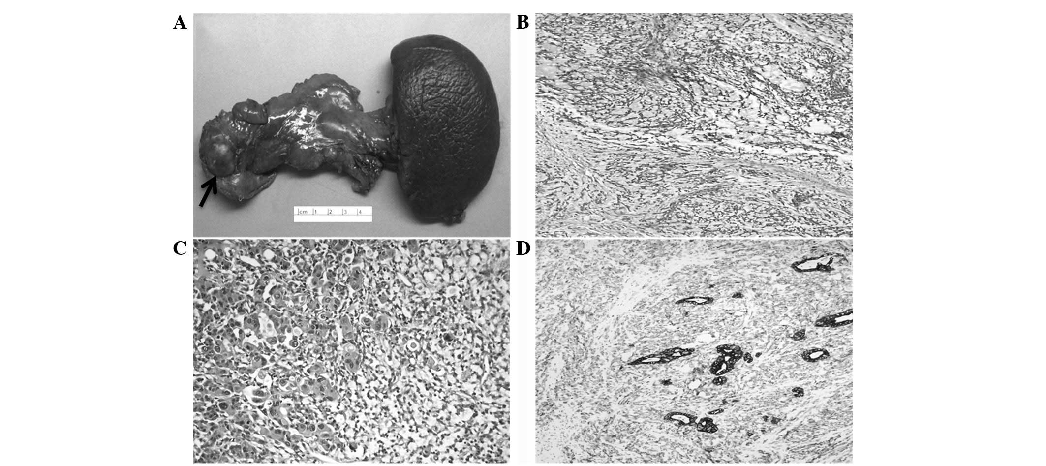
The surgical specimen consisted of the pancreatic
body and tail with the attached spleen and a portion of the stomach
(Fig. 1A). The cut surface of the
body of the pancreas revealed a poorly-circumscribed, solid, fleshy
mass of variegated yellow-tan to dark red color, measuring
3.3×3.0×2.6 cm. The tumor mass was adherent to the serosa of the
stomach, adjacent to the splenic artery and vein and externally
compressed and obstructed the main pancreatic duct (Fig. 1A).
Ill-defined, highly cellular nodules of
cytologically atypical spindle cells, intermingling with scattered
adenocarcinomatous elements within a myxoid stroma were determined
by scanning light microscopy (Fig.
1B). In addition, the lesional tissue surrounded a central area
of necrosis. At higher magnification, the minor adenocarcinomatous
component, including ill-defined ductal structures and individually
dispersed cells, blended imperceptibly with a haphazardly arranged,
cellular proliferation of cytologically atypical spindle cells. The
cells exhibited a mild to moderate degree of nuclear pleomorphism
and scanty eosinophilic cytoplasm. However, no microscopic features
indicating specific mesenchymal differentiation were identified
(Fig. 1C). Mitotic figures were
readily identified in the two components. Perineural invasion and
regional lymph node metastasis by the spindle component were
evident, but lymphovascular invasion was not identified. The
surrounding non-neoplastic parenchyma exhibited features of chronic
pancreatitis. The immunohistochemical results are presented in
Table I and indicate that the
epithelial and spindle tumor cells diffusely expressed
pan-cytokeratin MNF-116 (Fig. 1D),
cytokeratin 8/18 and nuclear p53, but not nuclear β-catenin,
calponin, CD10 or p63. Synaptophysin, chromogranin and CD56 were
expressed in scattered lesional cells and Ki-67 immunoexpression
was demonstrated in >50% of malignant cells. According to the
histological and immunohistochemical observations, a diagnosis of
primary sarcomatoid (spindle cell) carcinoma of the pancreas was
formed.
Discussion
Sarcomatoid (spindle cell) carcinoma is
characterized by malignant spindle cell proliferation that
demonstrates epithelial derivation, but no light microscopic,
ultrastructural or immunohistochemical evidence indicating a
specific line of mesenchymal differentiation. The microscopically
nondescript spindle cells, similar to the epithelial component,
typically express keratin or other epithelial-related markers,
albeit often in a focal manner, or exhibit other attributes
consistent with an epithelial pathogenesis. Carcinosarcoma is an
epithelial malignancy associated with sarcomatoid (spindle cell)
carcinoma with an equally aggressive clinical course that by
definition demonstrates biphasic epithelial and mesenchymal
differentiation (7). Therefore, for
practical diagnostic purposes, carcinosarcoma is used
interchangeably with sarcomatoid (spindle cell) carcinoma. In the
present case report, carcinosarcomas with heterologous mesenchymal
elements demonstrating light microscopic and/or immunohistochemical
evidence of specific mesenchymal (lipogenic, smooth or skeletal
muscle, peripheral nerve sheath, vascular or osteo-/cartilaginous)
differentiation were excluded. However, the World Health
Organization classification of exocrine pancreatic tumors allocates
spindle cell carcinoma, sarcomatoid carcinoma and carcinosarcoma
under the rubric of undifferentiated (anaplastic) carcinoma
(8), since the majority of these
types of tumor possess a spindle element that demonstrates an
epithelial immunohistochemical profile and/or genetic alterations
in pancreatic ductal adenocarcinomas (7).
To date, the largest study to analyze the
histological spectrum of sarcomatoid carcinoma, including examples
of malignant pancreatic neoplasms exhibiting varied sarcoma-like
features, was reported by Alguacil-Garcia et al(9). This study identified four distinctive
histological types of sarcomatoid carcinoma based on light
microscopic analysis only. The four subtypes were as follows: i)
Spindle cell carcinoma, consisting primarily of malignant spindle
cells; ii) osteoclastic giant cell tumors, demonstrating an
admixture of malignant spindle and epithelioid-appearing cells with
osteoclast-like giant cells; iii) pleomorphic giant cell carcinoma,
exhibiting highly pleomorphic mononuclear and multinucleated giant
cells; and iv) round cell anaplastic carcinoma, composed
exclusively of monotonous rounded, small- to moderately-sized tumor
cells. The reported prognosis of each of these histological
subtypes was uniformly poor (9),
consistent with the aggressive nature of this subset of malignant
tumors.
However, it is difficult to classify tumors
belonging to any of these four categories as spindle cell carcinoma
or carcinosarcoma with heterologous elements, as aforementioned, or
even to exclude pure sarcoma or carcinoma in specific cases without
the benefit of immunohistochemistry, electron microscopy or
molecular studies. In retrospect, we hypothesize that the spindle
cell variant most closely resembles the tumor identified in the
patient discussed in the present case report. The pleomorphic
epithelioid cells that typify pleomorphic giant cell carcinoma have
the potential for focal transition to a spindle morphology.
However, this neoplasm must not exhibit a distinct spindle cell
component like the sarcomatoid (spindle cell) carcinoma or
carcinosarcoma. The majority of pancreatic tumors with osteoclastic
giant cells are epithelial tumors with accompanying pleomorphic
giant cells (pleomorphic/osteoclastic or ‘mixed’ giant cell tumor)
(10) or tumors histologically
resembling giant cell tumors of the bone (osteoclastic giant cell
tumor or osteoclastoma) (11), but
whose immunohistochemical and molecular profiling are consistent
with an epithelial derivation (12). In contrast to the malignant
mononuclear cell element of the tumor presented, previous studies
have shown that the osteoclastic giant cell is a benign component
of monocytic derivation recruited into the lesional cell
environment via the production of cytokines by neoplastic cells
(13,14).
In addition to the tumor of the present case study,
six additional examples of pancreatic sarcomatoid (spindle cell)
carcinoma with confirmed epithelial derivation of the spindle
component and/or absence of specific mesenchymal differentiation
have been identified in a comprehensive review of the English
literature (PubMed; Table II)
(1–6). Of the patients with adequate
follow-up, 4/5 succumbed to their condition within 9 months of
surgery (1,4–6). In a
previous review of pancreatic tumors classified as
‘carcinosarcomas’, including two cases that are included in the
present case report as examples of sarcomatoid (spindle cell)
carcinoma (2,3), Gelos et al(15) identified that the average
post-operative survival interval was 6 months and that the longest
living patient survived for 15 months. Notably, the patient of the
present case study was alive and well 26 months after surgery and
thus, to the best of our knowledge, is the longest-living
individual with pancreatic sarcomatoid carcinoma and
carcinosarcoma.
Consistent with the results of the present case
report, an epithelial origin of the spindle cell element has been
identified by the presence of epithelial immunomarkers, including
keratin(s) and EMA, within the spindle cell component in five of
the six reported pancreatic sarcomatoid carcinomas (1–5).
Higashi et al(1) also
reported the immunoexpression of MUC1, an apomucin core protein
more specific than keratin for pancreatic ductal carcinoma, within
malignant spindle cells of pancreatic sarcomatoid (spindle cell)
carcinoma.
Molecular analysis is a useful technique for
establishing a diagnosis of sarcomatoid (spindle cell) carcinoma by
demonstrating a histogenetic link between malignant spindle cells
and pancreatic ductal adenocarcinoma. The K-ras mutation at codon
12 of exon 2 is implicated in the pathogenesis of pancreatic ductal
adenocarcinoma (16) and has been
identified in the spindle cell component of two examples of
pancreatic sarcomatoid (spindle cell) carcinoma. Nakano et
al(5) identified a mutation in
the keratin-expressing spindle component of a malignant biphasic
pancreatic tumor. Kim et al(6) identified the mutation in the
epithelial and spindle elements of a malignant biphasic pancreatic
tumor. Although the spindle component failed to express keratin, a
broad mesenchymal immunoprofile was also negative. These two cases
are consistent with the current belief that the vast majority of
malignant spindle cell neoplasms of the pancreas are sarcomatoid
carcinomas. In addition, the observation of the K-ras mutation at
codon 12 of exon 2 within a retroperitoneal-based, undifferentiated
spindle cell malignancy is likely to indicate a pancreatic
epithelial origin, particularly in cases where there is no evidence
of a coexisting or preexisting well-differentiated component
(eliminating ‘dedifferentiated’ sarcoma) and where the tumor lacks
specific mesenchymal or epithelial immunomarker expression.
The pathogenesis of sarcomatoid malignant biphasic
neoplasms remains unclear, but certain previous studies indicate a
monoclonal (stem cell) origin with divergence into carcinoma and
sarcomatous elements (17,18). Epithelial-to-mesenchymal transition
has been postulated to be an alternative mechanism explaining the
origin of a sarcomatous component from a carcinoma based on the
differential loss or gain of genes between the two components
(5,19). The identification of the
characteristic K-ras mutation in codon 12 of exon 2 in a malignant
spindle cell pancreatic tumor constitutes evidence of a monoclonal
origin of the two components of sarcomatoid carcinoma. Nakano et
al(5) postulated that an
additional K-ras mutation at codon 34 of exon 2, located within the
spindle cell element, facilitated an epithelial-to-mesenchymal
(sarcomatous) transformation. Using an alternative approach to
demonstrate monoclonality, van den Berg et al(18) identified identical genetic
aberrations in six chromosomal loci. The loci are commonly altered
in the two cellular elements of pancreatic ductal carcinoma in
cases of pancreatic mucinous neoplasm with a coexisting sarcomatous
stroma that lack keratin immunoexpression and show morphological
and/or immunohistochemical evidence of mesenchymal
differentiation.
In conclusion, the current case study documents the
seventh case of sarcomatoid (spindle cell) carcinoma of the
pancreas with data substantiating the epithelial derivation of the
nondescript malignant spindle cell element. The patient presented
here was alive and disease-free at 26 months post-surgery, and is
therefore the longest disease-free survivor reported in the English
language literature.
References
|
1
|
Higashi M, Takao S and Sato E: Sarcomatoid
carcinoma of the pancreas: a case report with immunohistochemical
study. Pathol Int. 49:453–456. 1999. View Article : Google Scholar : PubMed/NCBI
|
|
2
|
Darvishian F, Sullivan J, Teichberg S and
Basham K: Carcinosarcoma of the pancreas: a case report and review
of the literature. Arch Pathol Lab Med. 126:1114–1117.
2002.PubMed/NCBI
|
|
3
|
Barkatullah SA, Deziel DJ, Jakate SM,
Kluskens L and Komanduri S: Pancreatic carcinosarcoma with unique
triphasic histological pattern. Pancreas. 31:291–292. 2005.
View Article : Google Scholar : PubMed/NCBI
|
|
4
|
De la Riva S, Muñoz-Navas MA, Betés M,
Súbtil JC, Carretero C and Sola JJ: Sarcomatoid carcinoma of the
pancreas and congenital choledochal cyst. Gastrointest Endosc.
64:1005–1006. 2006.PubMed/NCBI
|
|
5
|
Nakano T, Sonobe H, Usui T, Yamanaka K,
Ishizuka T, Nishimura E and Hanazaki K: Immunohistochemistry and
K-ras sequence of pancreatic carcinosarcoma. Pathol Int.
58:672–677. 2008. View Article : Google Scholar : PubMed/NCBI
|
|
6
|
Kim HS, Joo SH, Yang DM, Lee SH, Choi SH
and Lim SJ: Carcinosarcoma of the pancreas: a unique case with
emphasis on metaplastic transformation and the presence of
undifferentiated pleomorphic high-grade sarcoma. J Gastrointestin
Liver Dis. 20:197–200. 2011.
|
|
7
|
Hruban RH, Pitman MB and Klimstra DS:
Adenocarcinoma variants. Tumors of the Pancreas AFIP atlas of tumor
pathology: Fourth series. American Registry of Pathology;
Washington, DC: pp. 165–190. 2007
|
|
8
|
Fukushima N, Hruban RH, Kato Y, Klimstra
DS, Kloppel G, Shimizu M and Terris B: Ductal adenocarcinoma
variants and mixed neoplasms of the pancreas. World Health
Organization Classification of Tumours of the Digestive System.
Bosman FT, Carneiro F, Hruban RH and Theise ND: 3. 4th edition.
IARC Press; Lyon: pp. 292–295. 2010
|
|
9
|
Alguacil-Garcia A and Weiland LH: The
histologic spectrum, prognosis, and histogenesis of the sarcomatoid
carcinoma of the pancreas. Cancer. 39:1181–1189. 1977. View Article : Google Scholar : PubMed/NCBI
|
|
10
|
Watanabe M, Miura H, Inoue H, Uzuki M,
Noda Y, Fujita N, Yamazaki T and Sawai T: Mixed
osteoclastic/pleomorphic-type giant cell tumor of the pancreas with
ductal adenocarcinoma: histochemical and immunohistochemical study
with review of the literature. Pancreas. 15:201–208. 1997.
View Article : Google Scholar
|
|
11
|
Rosai J: Carcinoma of pancreas simulating
giant cell tumor of bone. Electron-microscopic evidence of its
acinar cell origin. Cancer. 22:333–344. 1968. View Article : Google Scholar
|
|
12
|
Westra WH, Strum P, Drillenburg P, Choti
MA, Klimstra DS, Albores-Saavadra J, et al: K-ras oncogene
mutations in osteoclast-like giant cell tumors of the pancreas and
liver: genetic evidence to support origin from the duct epithelium.
Am J Surg Pathol. 22:1247–1254. 1998. View Article : Google Scholar : PubMed/NCBI
|
|
13
|
Goldberg RD, Michelassi F and Montag AG:
Osteoclast-like giant cell tumor of the pancreas: immunophenotypic
similarity to giant cell tumor of bone. Hum Pathol. 22:618–622.
1991. View Article : Google Scholar : PubMed/NCBI
|
|
14
|
Leighton CC and Shum DT: Osteoclastic
giant cell tumor of the pancreas: case report and literature
review. Am J Clin Oncol. 24:77–80. 2001. View Article : Google Scholar : PubMed/NCBI
|
|
15
|
Gelos M, Behringer D, Philippou S and Mann
B: Pancreatic carcinosarcoma. Case report of multimodal therapy and
review of the literature. JOP. 9:50–55. 2008.PubMed/NCBI
|
|
16
|
Almoguera C, Shibata D, Forrester K,
Martin J, Arnheim N and Perucho M: Most human carcinomas of the
exocrine pancreas contain mutant c-K-ras genes. Cell. 53:549–554.
1988. View Article : Google Scholar : PubMed/NCBI
|
|
17
|
Thompson L, Chang B and Barsky S:
Monoclonal origins of malignant mixed tumors (carcinosarcomas).
Evidence for a divergent histogenesis. Am J Surg Pathol.
20:277–285. 1996. View Article : Google Scholar : PubMed/NCBI
|
|
18
|
van den Berg W, Tascilar M, Offerhaus GJ,
et al: Pancreatic mucinous cystic neoplasms with sarcomatous
stroma: molecular evidence for monoclonal origin with subsequent
divergence of the epithelial and sarcomatous components. Mod
Pathol. 13:86–91. 2000.
|
|
19
|
Zhuang ZP, Lininger RA, Man YG,
Albuquerque A, Merino MJ and Tavassoli FA: Identical clonality of
both components of mammary carcinosarcoma with differential loss of
heterozygosity. Mod Pathol. 10:354–362. 1997.PubMed/NCBI
|