Introduction
Gastric cancer (GC) is the fourth most common
malignancy and the second leading cause of cancer mortality
worldwide (1). Helicobacter
pylori (H. pylori) is a gram-negative bacteria that
infects 50% of the global population. However, in certain regions
and countries of the world, >80% of the population is infected
with the bacteria. H. pylori has been defined by the
International Agency for Research of Cancer as a class I carcinogen
and is important for the progression from chronic superficial
gastritis to chronic atrophic gastritis, intestinal metaplasia
(IM), dysplasia (Dys) and finally GC (2).
DNA double-strand breaks (DSBs) are the most serious
type of DNA damage and are frequently caused by ionizing radiation
(IR), ultraviolet light and specific chemical agents. Recently,
H. pylori has also been shown to induce DSBs in gastric
epithelial cells in vitro(3,4).
Inappropriate repair of DSBs can result in genomic instability,
which has been shown to be a key factor in carcinogenesis (5). Phosphorylation of H2AX at Ser 139
(γH2AX) is abundant, fast, correlates well with DSBs and renders
γH2AX a sensitive marker for the detection of DSBs (6). The expression of γH2AX has been shown
to correlate with numerous types of malignant tumor and also with
prognosis in early operable non-small cell lung cancer, vulvar
squamous cell carcinoma and breast cancer (7–9).
There have been a number of studies on γH2AX
expression in GC tissues. Sentani et al(10) showed that nuclear positive staining
for GC was significantly higher than that in normal gastric
tissues. However, no previous studies have investigated γH2AX
expression in various gastric lesions or its correlation with H.
pylori infection. Therefore, the aim of the present study was
to measure the expression of γH2AX and determine its correlation
with the various stages of gastric carcinogenesis, in the presence
or absence of H. pylori infection.
Patients and methods
Patients and sample collection
Gastric tissue samples were collected from patients
who had undergone an upper gastroduodenoscopy or gastrectomy at the
First Affiliated Hospital of Nanchang University (Nanchang, China)
between January 2007 and September 2008. A total of 302 patients
ranging in age between 18 and 70 years were enrolled in the current
study. The study included 56 cases of chronic gastritis (CG), 53 of
IM, 47 of Dys and 146 of GC. None of the patients had been treated
with proton pump inhibitors or antibiotics against H.
pylori, and no GC patients had been treated with prior radio-
or chemotherapy. The clinical characteristics of these patients are
summarized in Table I. No
significant differences were identified in the age or gender
distribution among these groups. Clinicopathological
characteristics were also obtained from the pathological
reports.
 | Table IExpression of γH2AX in patients with various
histological observations. |
Table I
Expression of γH2AX in patients with various
histological observations.
| | | | Overall score of
γH2AX expression |
|---|
| | | |
|
|---|
| Group | n | Mean age (SD),
years | Gender, M/F | −, n | +, n | ++, n | +++, n | %a | P-value |
|---|
| 1 CG | 56 | 53.6 (10.7) | 30/26 | 29 | 27 | 0 | 0 | 48.2 | |
| 2 IM | 53 | 54.3 (9.6) | 29/24 | 14 | 26 | 12 | 1 | 73.5 | <0.001b |
| 3 Dys | 47 | 55.1 (10.3) | 26/21 | 2 | 9 | 27 | 9 | 95.7 | <0.001b,c |
| 4 GC | 146 | 56.8 (14.1) | 96/50 | 15 | 57 | 50 | 24 | 89.7 | <0.001b; 0.011d |
In total, 10 GC tissue samples and adjacent normal
tissues were collected from gastrectomy specimens at the First
Affiliated Hospital of Nanchang University.
The present study was approved by the Ethics
Committee of the First Affiliated Hospital of Nanchang University.
All patients provided written informed consent prior to enrollment
in the study.
Histological examination
All biopsies or surgical specimens from the patients
with gastric disease were obtained from the gastric antrum or
lesion locations. The tissues used for histological analysis were
fixed in 10% formaldehyde in Ca2+ and
Mg2+-free phosphate-buffered saline (PBS) overnight at
4°C, prior to paraffin embedding. Paraffin sections, 4 μm thick,
were sectioned with a microtome and stored at room temperature.
Pathological diagnosis and classification were performed according
to the criteria of the World Health Organization (11) and the updated Sydney system
(12).
Detection of H. pylori infection
Rapid urease test and modified Giemsa staining were
used for the detection of H. pylori infection. The modified
Giemsa staining was performed by two veteran pathologists.
Consistency in the positive or negative results of the two tests
was required.
Immunohistochemistry
Slices were deparaffinized in dimethylbenzene,
rehydrated through 100, 95 and 85% ethanol and incubated with fresh
3% H2O2 for 10 min to quench endogenous
peroxidase activity. Microwave heating was used to expose antigens
for detection. The primary antibody used for immunohistochemistry
was rabbit monoclonal anti-human γH2AX (ab81299; 1:400; Abcam,
Cambridge, UK). Slices were incubated at 4°C overnight and then
washed with PBS three times. The secondary antibody (PV-6000;
Zhongshan Golden Bridge Biotechnology Co., Ltd., Beijing, China)
was incubated at 37°C for 30 min prior to reaction with
3,3-diaminobenzidine (Zhongshan Golden Bridge Biotechnology Co.,
Ltd.). Subsequently, slices were counterstained with hematoxylin
and mounted with coverslips. Negative controls consisted of PBS
without primary antibody (13).
Review and scoring
The stained slices were reviewed and scored by two
experienced pathologists. The concordance rates were generally high
and results with any grading discrepancies were re-reviewed and
discussed to determine a final score. Epithelial cells stained
yellow or brown in the nuclei were defined as positive. Five fields
for each slice were randomly selected, reviewed and scored
(magnification, ×200). In each field, 100 immunoreactive cells were
assessed and quantified as the percentage of total cells and then
averaged from the five fields to calculate the percentage of
immunostaining, i.e. 0, ≤5.0%; 1, 5.1–25.0%; 2, 25.1–50.0%; 3,
50.1–75.0%; and 4, >75.0%. Moreover, the staining intensity was
also semi-quantitatively assessed as follows: 0, no staining; 1,
weak staining; 2, moderate staining; and 3, strong staining. The
integrals of the ‘area × intensity’ were calculated based on the
following overall scores of the expression levels of the proteins
in the sections: negative (−), 0–2; weak positive (+), 3–5;
moderate positive (++), 6–8; and strong positive (+++), 9–12
(Table I) (13).
Cell lines and culture
Five gastric mucosal cell lines were used in the
present study, including GES-1 (immortalized gastric epithelial
mucosa cell line, established by the Beijing Institute for Cancer
Research, Beijing, China) and human gastric cancer SGC7901, MKN28,
MKN45 and AGS cell lines (a gift from the Xijing Hospital of
Digestive Disease, Xi’an, China). Cell lines were cultured at 37°C
in an atmosphere of 5% CO2 in DMEM with 10% fetal bovine
serum, 100 units penicillin and 100 μg/ml streptomycin (Gibco-BRL,
Carlsbad, CA, USA) (14).
Western blotting
Tissues and cells were lysed in a buffer containing
0.5% Lubrol-PX, 50 mM KCl, 2 mM CaCl2, 20% glycerol, 5
mM Tris-HCl (pH 7.4), 0.1% protease and 1% phosphatase inhibitors
(Sigma-Aldrich, St. Louis, MO, USA). Following the addition of
sodium dodecyl sulfate-polyacrylamide gel electrophoresis
(SDS-PAGE) sample buffer, proteins were run on an SDS-PAGE gel and
transferred to nitrocellulose membranes (Whatman GmbH, Dassel,
Germany). The membranes were immunoblotted with antibodies against
γH2AX (ab81299; 1:1,000; Abcam) and actin (1:1,000; Zhongshan
Golden Bridge Biotechnology, Co., Ltd.). The reactions were
subjected to incubation with an enhanced chemiluminescence
detection system (Pierce Biotechnology, Inc., Rockford, IL, USA)
and then exposed to X-ray film for visualizing the positive
bands.
Statistical analysis
SPSS 17.0 (SPSS Inc., Chicago, IL, USA) was used to
perform the statistical analysis. Data are expressed as the mean ±
standard deviation or percentage. The χ2 test was used
to evaluate differences in categorical variables. The
Kruskal-Wallis one-way analysis of variance and Mann-Whitney U
tests were used to determine differences in numerical variables
between various groups. P<0.05 was considered to indicate a
statistically significant difference.
Results
Differential expression of γH2AX in
various gastric lesions and its association with H. pylori
infection
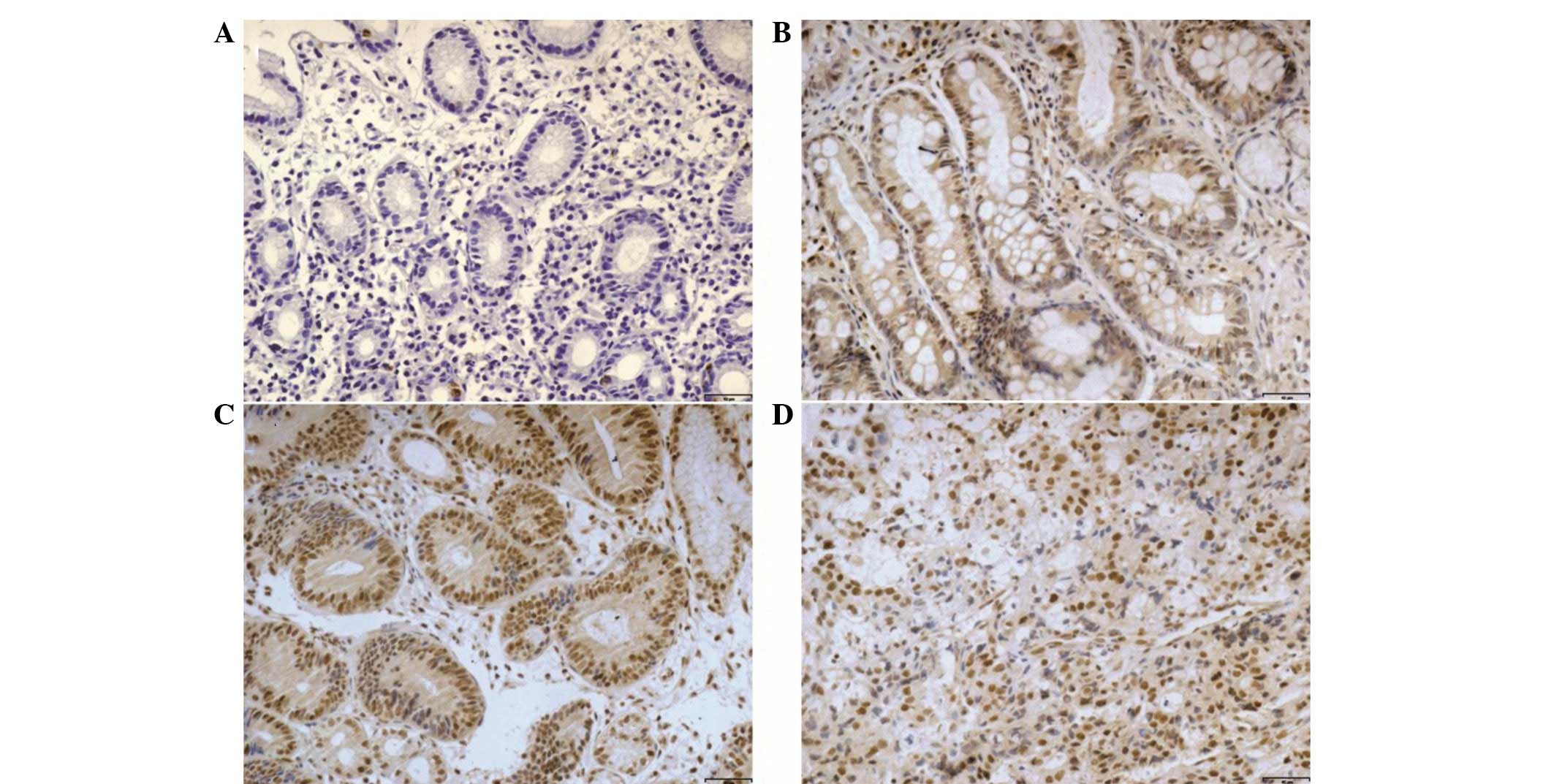
Immunohistochemical analysis showed that γH2AX was
primarily found in the nuclei of epithelial cells.
Semi-quantitative results of the expression of γH2AX are shown in
Table I. The results showed that
the expression ratio of γH2AX was 48.2% in the CG group, 73.5% in
the IM group, 95.7% in the Dys group and 89.7% in the GC group.
Consistent with the observations of Correa et al(2), the expression levels of γH2AX in the
current study were significantly increased as pathological stages
progressed from CG to Dys (P<0.001). However, expression levels
were decreased in GC (P=0.011) (Fig.
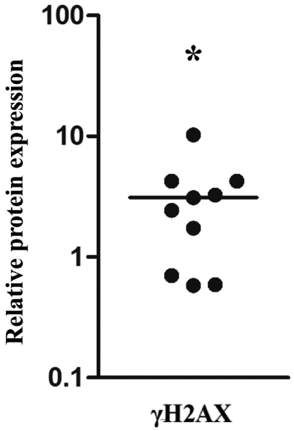
1 and Table I). The expression
of γH2AX was also measured by western blotting in GC and adjacent
normal tissues. The results showed high γH2AX expression in GC
tissues compared with adjacent normal tissues (P<0.001)
(Fig. 2), which is consistent with
the immunohistochemical results.
Moreover, in patients with IM and Dys, the
expression of γH2AX was significantly higher in the presence of
H. pylori infection (96.5 and 100%, respectively) compared
with those without the infection (45.8 and 90.4%, respectively)
(P=0.001 and P=0.008) (Table II).
However, no significant differences were detected in the CG or GC
groups.
| Table IIExpression of γH2AX in patients with various
histological observations, in relation to H. pylori
infection. |
Table II
Expression of γH2AX in patients with various
histological observations, in relation to H. pylori
infection.
| | | | | Overall score of
γH2AX expression |
|---|
| | | | |
|
|---|
| Group | H. pylori | n | Mean age (SD),
years | Gender, M/F | −, n | +, n | ++, n | +++, n | %a | P-value |
|---|
| CG | + | 26 | 54.1 (11.7) | 14/12 | 7 | 19 | 0 | 0 | 73.1 | 0.362 |
| − | 30 | 53.2 (9.1) | 16/14 | 22 | 8 | 0 | 0 | 26.7 | |
| IM | + | 29 | 54.8 (8.8) | 14/15 | 1 | 19 | 8 | 1 | 96.5 | 0.001 |
| − | 24 | 53.7 (10.3) | 15/9 | 13 | 7 | 4 | 0 | 45.8 | |
| Dys | + | 26 | 54.3 (12.2) | 13/13 | 0 | 2 | 17 | 7 | 100.0 | 0.008 |
| − | 21 | 56.1 (9.5) | 13/8 | 2 | 7 | 10 | 2 | 90.4 | |
| GC | + | 61 | 58.3 (16.3) | 39/22 | 6 | 25 | 20 | 10 | 90.2 | 0.865 |
| − | 85 | 50.8 (13.9) | 57/28 | 9 | 32 | 30 | 14 | 89.4 | |
The association of γH2AX expression with
clinicopathological parameters was analyzed in 146 GC patients
(Table III). The gastric body and
cardia cancers showed a higher γH2AX expression (97.2%) than
gastric antrum cancers (82.4%) (P<0.001). In addition, the
expression of γH2AX in Borrmann III and IV type GC (90.5%) was
significantly higher than Borrmann I and II type GC (88.9%)
(P=0.002 ). γH2AX expression in poorly- and undifferentiated GC
(93.8%) was significantly higher compared with that in well- and
moderately differentiated GC (74.1%) (P<0.001). In cancer
tissues located in the submucosa, the γH2AX expression (85.1%) was
significantly lower than that in cancer tissues that had reached
the subserosal level (91.9%) (P=0.002). Furthermore, γH2AX was
expressed more frequently in TNM III and IV stage patients (91.9%)
than in TNM I and II stage patients (86.4%) (P<0.001), and there
was higher γH2AX expression in the patients with lymph node
metastasis (91.2%) than in the patients without (84.8%)
(P=0.002).
| Table IIIClinicopathological association of
γH2AX expression in patients
with GC. |
Table III
Clinicopathological association of
γH2AX expression in patients
with GC.
| | Overall score of
γH2AX expression |
|---|
| |
|
|---|
|
Characteristics | n | −, n | +, n | ++, n | +++, n | PR, % | P-value |
|---|
| Gender |
| Male | 96 | 8 | 39 | 35 | 14 | 91.7 | 0.923 |
| Female | 50 | 7 | 18 | 15 | 10 | 86.0 | |
| Age, years |
| ≥55 | 86 | 7 | 36 | 28 | 15 | 91.8 | 0.780 |
| <55 | 60 | 8 | 21 | 22 | 9 | 86.7 | |
| Location |
| Antrum | 74 | 13 | 31 | 25 | 5 | 82.4 | <0.001 |
| Body and
cardia | 72 | 2 | 26 | 25 | 19 | 97.2 | |
| Gross type
(Borrmann) |
| I and II | 72 | 8 | 37 | 21 | 6 | 88.9 | 0.002 |
| III and IV | 74 | 7 | 20 | 29 | 18 | 90.5 | |
|
Differentiation |
| Well and
moderately | 81 | 11 | 40 | 21 | 9 | 74.1 | |
| Poorly and
undifferentiated | 65 | 4 | 17 | 29 | 15 | 93.8 | <0.001 |
| Invasive depth |
| Above
submucosa | 27 | 4 | 18 | 3 | 2 | 85.1 | |
| Muscularis
propria | 8 | 2 | 0 | 3 | 3 | 75.0 | 0.080a |
| Below
subserosa | 111 | 9 | 39 | 44 | 19 | 91.9 | 0.002a; 0.373b |
| TNM |
| I and II | 59 | 8 | 35 | 12 | 3 | 86.4 | <0.001 |
| III and IV | 87 | 7 | 22 | 38 | 20 | 91.9 | |
| Lymph node
metastasis |
| With | 113 | 10 | 37 | 45 | 21 | 91.2 | 0.002 |
| Without | 33 | 5 | 20 | 5 | 3 | 84.8 | |
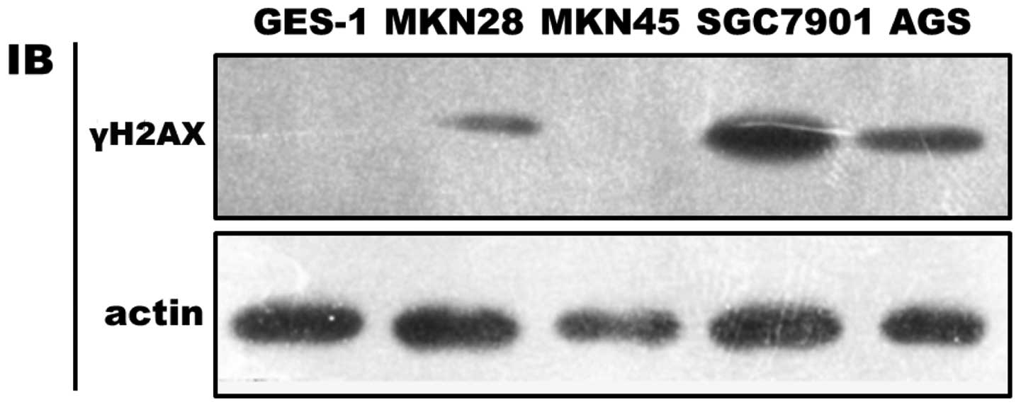
Differential expression of γH2AX in
various gastric epithelial cell lines
Expression of γH2AX was higher in GC cell lines
(i.e., SGC-7901, MKN-28 and AGS, with the exception of MKN-45)
compared with the non-cancerous cell line GES-1 (Fig. 3).
Discussion
DSBs are important threats to genome integrity
causing chromosomal aberrations, which are remarkable
characteristics of malignant tumors (15). While the response to DNA damage acts
as an anticancer barrier in early human tumorigenesis, γH2AX has
been reported to be highly overexpressed in precancerous lesions of
the urinary bladder, breast, lung and colon cancer (16). In the current study, the expression
of γH2AX was shown to progressively increase in accordance with the
pathological progression from CG to Dys. γH2AX expression in GC was
also higher than that in CG, but was slightly decreased in Dys. The
reason for lower γH2AX expression in GC compared with Dys remains
unclear; however, restoration of genomic instability during tumor
progression may explain this observation (17,18).
The western blotting results of the cancer and adjacent normal
tissues were confirmed by cell line experiments. All results showed
that the response to DNA damage was activated in gastric
precancerous lesions.
Recently, Toller et al have shown that H.
pylori triggers DSBs in gastric epithelial cells in cell
culture, primarily mediated by H. pylori blood group
antigen-binding adhesion (3). In
addition, Jang et al have shown that H.
pylori-induced DSBs may be inhibited by lycopene (4). The two investigations were based on
AGS coculture with H. pylori in cell culture, a model system
which is different from the clinical situation. The results of the
current study were based on human samples and showed that high
expression of γH2AX in IM and Dys patients appeared to correlate
with H. pylori infection, which implied that H.
pylori not only induces DSBs in cell culture, but may also be
involved in an early molecular event in gastric tumorigenesis.
However, whether the DNA damage response pathways are impaired
requires further study.
The observations of the current study have shown
that the overexpression of γH2AX in GC correlates with a number of
clinicopathological characteristics, including tumor location,
tumor gross type, differentiation, tumor invasion depth, TNM stage
and lymph node metastasis. All these correlations implied that, as
the degree of malignancy increased, the genomic instability was
also significantly increased. These results confirm previous
observations by Sentani et al(10), in which the expression of the γH2AX
protein in GC was significantly higher in non-neoplastic gastric
mucosa and its high expression was also found to correlate with
stage II–IV cases. Exposure to IR was an important factor in the
study.
In conclusion, DSBs appear to be an early molecular
event in gastric carcinogenesis, which is associated with H.
pylori infection and a number of clinical characteristics.
Although the exact mechanism by which DSBs are induced by H.
pylori infection remains unclear, the expression of γH2AX may
serve as a valuable biomarker for the diagnosis and progression of
GC.
Acknowledgements
The current study was supported in part by grants
from the National Natural Science Foundation of China (no.
81270479)and the Graduate Innovative Foundation of Jiangxi
Province, China (no. YC2013-B004).
References
|
1
|
Ferlay J, Shin HR, Bray F, Forman D,
Mathers C and Parkin DM: Estimates of worldwide burden of cancer in
2008: GLOBOCAN 2008. Int J Cancer. 127:2893–2917. 2010. View Article : Google Scholar : PubMed/NCBI
|
|
2
|
Correa P: Human gastric carcinogenesis: a
multistep and multifactorial process - First American Cancer
Society Award Lecture on Cancer Epidemiology and Prevention. Cancer
Res. 52:6735–6740. 1992.
|
|
3
|
Toller IM, Neelsen KJ, Steger M, Hartung
ML, Hottiger MO, Stucki M, et al: Carcinogenic bacterial pathogen
Helicobacter pylori triggers DNA double-strand breaks and a
DNA damage response in its host cells. Proc Natl Acad Sci USA.
108:14944–14949. 2011.PubMed/NCBI
|
|
4
|
Jang SH, Lim JW, Morio T and Kim H:
Lycopene inhibits Helicobacter pylori-induced
ATM/ATR-dependent DNA damage response in gastric epithelial AGS
cells. Free Radic Biol Med. 52:607–615. 2012.PubMed/NCBI
|
|
5
|
Smith J, Tho LM, Xu N and Gillespie DA:
The ATM-Chk2 and ATR-Chk1 pathways in DNA damage signaling and
cancer. Adv Cancer Res. 108:73–112. 2010. View Article : Google Scholar : PubMed/NCBI
|
|
6
|
Sharma A, Singh K and Almasan A: Histone
H2AX phosphorylation: a marker for DNA damage. Methods Mol Biol.
920:613–626. 2012. View Article : Google Scholar : PubMed/NCBI
|
|
7
|
Matthaios D, Foukas PG, Kefala M, Hountis
P, Trypsianis G, Panayiotides IG, et al: γ-H2AX expression detected
by immunohistochemistry correlates with prognosis in early operable
non-small cell lung cancer. Onco Targets Ther. 5:309–314. 2012.
|
|
8
|
Brustmann H, Hinterholzer S and Brunner A:
immunohistochemical expression of survivin and γ-H2AX in vulvar
intraepithelial neoplasia and low-stage squamous cell carcinoma.
Int J Gynecol Pathol. 30:583–590. 2011.
|
|
9
|
Nagelkerke A, van Kuijk SJ, Sweep FC,
Nagtegaal ID, Hoogerbrugge N, Martens JW, et al: Constitutive
expression of γ-H2AX has prognostic relevance in triple negative
breast cancer. Radiother Oncol. 101:39–45. 2011.
|
|
10
|
Sentani K, Oue N, Sakamoto N, Nishisaka T,
Fukuhara T, Matsuura H and Yasui W: Positive immunohistochemical
staining of gammaH2AX is associated with tumor progression in
gastric cancers from radiation-exposed patients. Oncol Rep.
20:1131–1136. 2008.
|
|
11
|
Hamilton SR and Aaltonen LA: Pathology and
genetics of tumours of the digestive system. World Health
Organization Classification of Tumours. IARC Press; Lyon: 2000
|
|
12
|
Dixon MF, Genta RM, Yardley JH and Correa
P: Classification and grading of gastritis. The updated Sydney
System International Workshop on the Histopathology of Gastritis,
Houston 1994. Am J Surg Pathol. 20:1161–1181. 1996.
|
|
13
|
Yang Z, Shu X, Chen L, Chen J, Xie Y and
Lu NH: Expression of p53-MDM2 feedback loop related proteins in
different gastric pathologies in relation to Helicobacter
pylori infection: implications in gastric carcinogenesis. Clin
Res Hepatol Gastroenterol. 36:235–243. 2012. View Article : Google Scholar : PubMed/NCBI
|
|
14
|
Yang Z, Yuan XG, Chen J, Luo SW, Luo ZJ
and Lu NH: Reduced expression of PTEN and increased PTEN
phosphorylation at residue Ser380 in gastric cancer tissues: a
novel mechanism of PTEN inactivation. Clin Res Hepatol
Gastroenterol. 37:72–79. 2013. View Article : Google Scholar : PubMed/NCBI
|
|
15
|
van Gent DC, Hoeijmakers JH and Kanaar R:
Chromosomal stability and the DNA double-stranded break connection.
Nat Rev Genet. 2:196–206. 2001.
|
|
16
|
Bartkova J, Horejsí Z, Koed K, Krämer A,
Tort F, Zieger K, et al: DNA damage response as a candidate
anti-cancer barrier in early human tumorigenesis. Nature.
434:864–870. 2005. View Article : Google Scholar : PubMed/NCBI
|
|
17
|
Martin RW, Orelli BJ, Yamazoe M, Minn AJ,
Takeda S and Bishop DK: RAD51 up-regulation bypasses BRCA1 function
and is a common feature of BRCA1-deficient breast tumors. Cancer
Res. 67:9658–9665. 2007. View Article : Google Scholar : PubMed/NCBI
|
|
18
|
Martin RW, Connell PP and Bishop DK: The
Yin and Yang of treating BRCA-deficient tumors. Cell. 132:919–920.
2008. View Article : Google Scholar : PubMed/NCBI
|