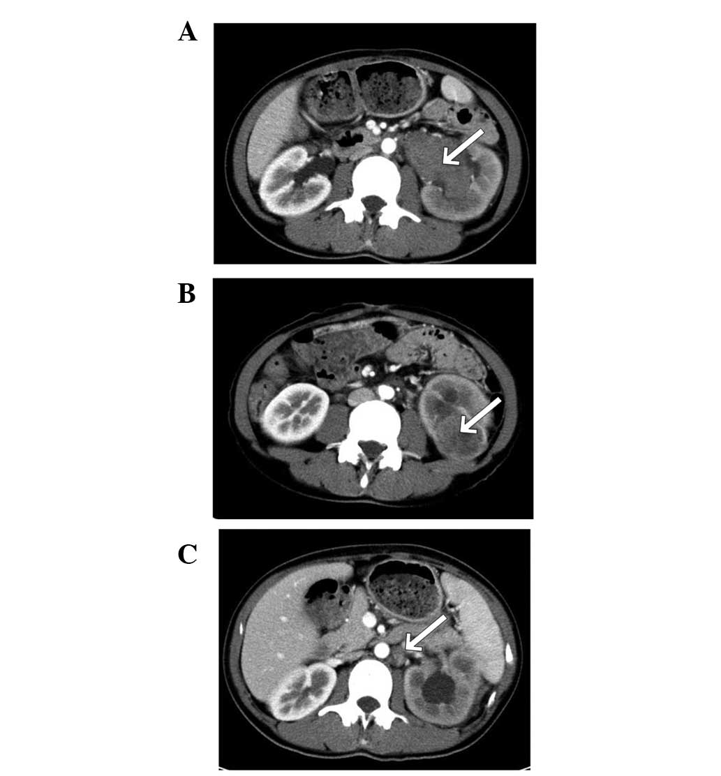
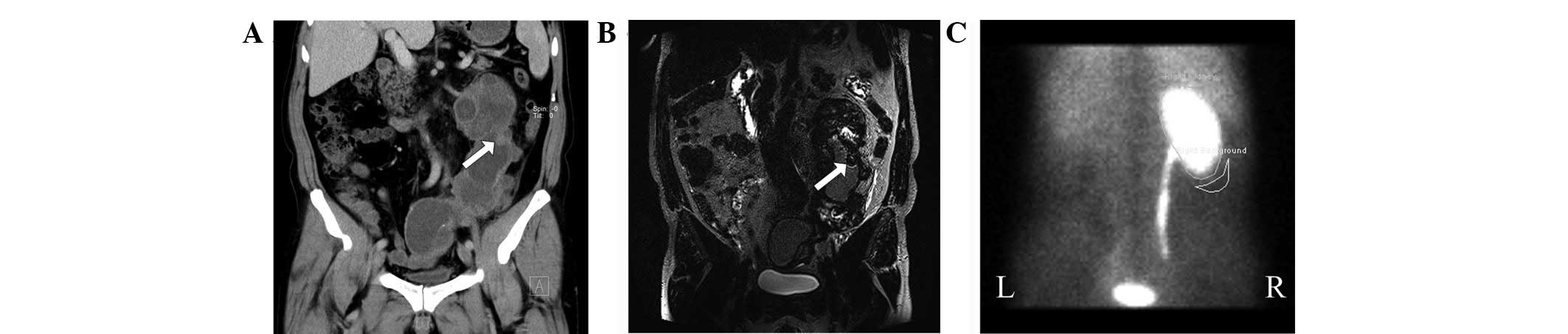
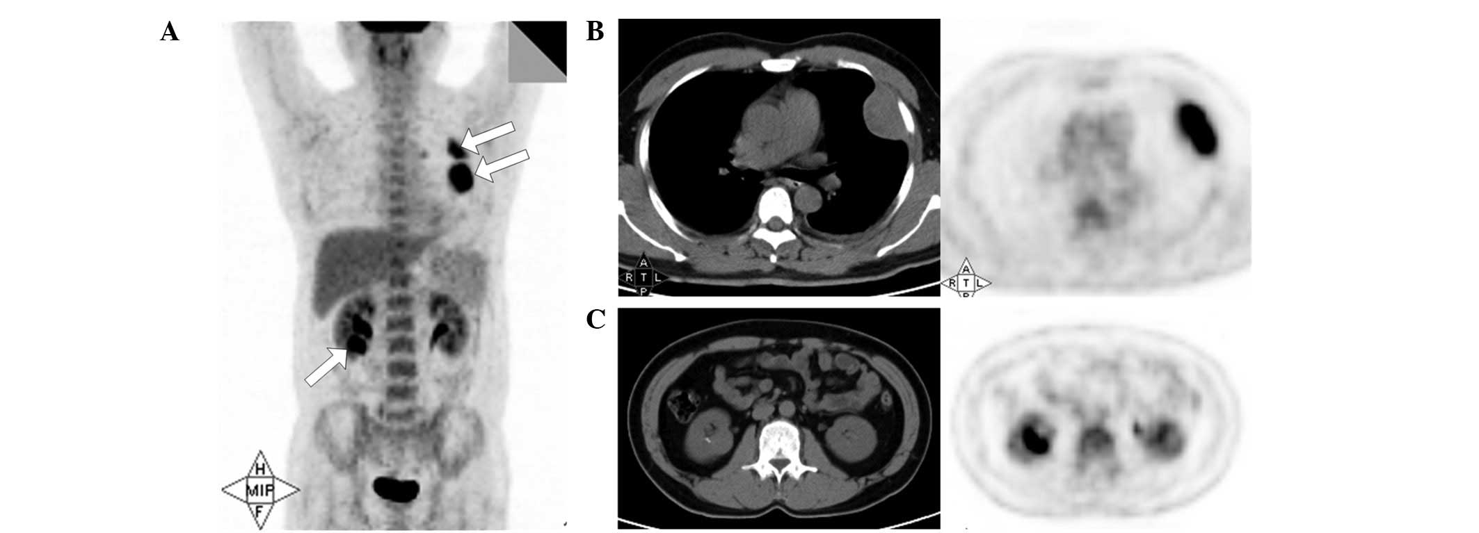
|
1
|
Yang G, Seo J and Park J: Distal ureteral
seeding metastasis of collecting duct carcinoma manifesting as deep
vein thrombosis. Clin Radiol. 67:936–939. 2012. View Article : Google Scholar
|
|
2
|
Sironi M, Delpiano C, Claren R and
Spinelli M: New cytological findings on fine-needle aspiration of
renal collecting duct carcinoma. Diagn Cytopathol. 29:239–240.
2003. View
Article : Google Scholar : PubMed/NCBI
|
|
3
|
Auguet T, Molina JC, Lorenzo A, Vila J,
Sirvent JJ and Richart C: Synchronus renal cell carcinoma and
Bellini duct carcinoma: a case report on a rare coincidence. World
J Urol. 18:449–451. 2000. View Article : Google Scholar : PubMed/NCBI
|
|
4
|
Mancilla-Jimenez R, Stanley RJ and Blath
RA: Papillary renal cell carcinoma: a clinical, radiologic, and
pathologic study of 34 cases. Cancer. 38:2469–2480. 1976.
View Article : Google Scholar : PubMed/NCBI
|
|
5
|
Cromie WJ, Davis CJ and DeTure FA:
Atypical carcinoma of kidney: possibly originating from collecting
duct epithelium. Urology. 13:315–317. 1979. View Article : Google Scholar : PubMed/NCBI
|
|
6
|
Antonelli A, Portesi E, Cozzoli A, et al:
The collecting duct carcinoma of the kidney: a cytogenetical study.
Eur Urol. 43:680–685. 2003. View Article : Google Scholar : PubMed/NCBI
|
|
7
|
Tsui K-H, Shvarts O, Smith RB, Figlin RA,
deKernion JB and Belldegrun A: Prognostic indicators for renal cell
carcinoma: a multivariate analysis of 643 patients using the
revised 1997 TNM staging criteria. J Urol. 163:1090–1095. 2000.
View Article : Google Scholar
|
|
8
|
Srigley JR and Delahunt B: Uncommon and
recently described renal carcinomas. Mod Pathol. 22(Suppl 2):
S2–S23. 2009. View Article : Google Scholar
|
|
9
|
Lopez-Beltran A, Carrasco JC, Cheng L,
Scarpelli M, Kirkali Z and Montironi R: 2009 update on the
classification of renal epithelial tumors in adults. Int J Urol.
16:432–443. 2009. View Article : Google Scholar : PubMed/NCBI
|
|
10
|
Lopez-Beltran A, Scarpelli M, Montironi R
and Kirkali Z: 2004 WHO classification of the renal tumors of the
adults. Eur Urol. 49:798–805. 2006. View Article : Google Scholar : PubMed/NCBI
|
|
11
|
Tokuda N, Naito S, Matsuzaki O, Nagashima
Y, Ozono S and Igarashi T: Collecting duct (Bellini duct) renal
cell carcinoma: a nationwide survey in Japan. J Urol. 176:40–43;
discussion 43. 2006. View Article : Google Scholar : PubMed/NCBI
|
|
12
|
Pickhardt PJ, Siegel CL and McLarney JK:
Collecting duct carcinoma of the kidney: are imaging findings
suggestive of the diagnosis? AJR Am J Roentgenol. 176:627–633.
2001. View Article : Google Scholar
|
|
13
|
Fukuya T, Honda H, Goto K, et al: Computed
tomographic findings of Bellini duct carcinoma of the kidney. J
Comput Assist Tomogr. 20:399–403. 1996. View Article : Google Scholar : PubMed/NCBI
|
|
14
|
Yoon SK, Nam KJ, Rha SH, et al: Collecting
duct carcinoma of the kidney: CT and pathologic correlation. Eur J
Radiol. 57:453–460. 2006. View Article : Google Scholar : PubMed/NCBI
|
|
15
|
Hsiao HL, Yeh HC, Chang TH, et al: Renal
collecting duct carcinoma and concomitant bladder urothelial
carcinoma: a case report. Kaohsiung J Med Sci. 24:157–162. 2008.
View Article : Google Scholar : PubMed/NCBI
|
|
16
|
Maestroni U, Ferretti S, Dinale F, et al:
A renal cancer with intermediate characteristics between collecting
(Bellini) duct carcinoma and urothelial carcinoma: case report and
review of the literature. Tumori. 92:545–548. 2006.
|
|
17
|
Ohnishi S, Dazai M, Iwasaki Y, Tsuzaka K,
Takahashi T and Miyagishima T: Undiagnosed collecting duct
carcinoma presenting as meningeal carcinomatosis and multiple bone
metastases. Intern Med. 49:1541–1544. 2010. View Article : Google Scholar : PubMed/NCBI
|
|
18
|
Nakamura H, Kuirhara Y, Matsushita K,
Sakai A, Yamaguchi T and Nakajima Y: Extrarenal multiorgan
metastases of collecting duct carcinoma of the kidney: a case
series. J Med Case Rep. 2:3042008. View Article : Google Scholar : PubMed/NCBI
|
|
19
|
Leyendecker JR and Gianini JW: Magnetic
resonance urography. Abdom Imaging. 34:527–540. 2009. View Article : Google Scholar
|
|
20
|
Leyendecker JR, Barnes CE and Zagoria RJ:
MR urography: techniques and clinical applications. Radiographics.
28:23–46; discussion 46-27. 2008. View Article : Google Scholar : PubMed/NCBI
|
|
21
|
Takahashi N, Glockner JF, Hartman RP, et
al: Gadolinium enhanced magnetic resonance urography for upper
urinary tract malignancy. J Urol. 183:1330–1365. 2010. View Article : Google Scholar
|
|
22
|
Chahal R, Taylor K, Eardley I, Lloyd S and
Spencer J: Patients at high risk for upper tract urothelial cancer:
evaluation of hydronephrosis using high resolution magnetic
resonance urography. J Urol. 174:478–482. 2005. View Article : Google Scholar
|
|
23
|
Shokeir AA, El-Diasty T, Eassa W, et al:
Diagnosis of noncalcareous hydronephrosis: role of magnetic
resonance urography and noncontrast computed tomography. Urology.
63:225–229. 2004. View Article : Google Scholar
|
|
24
|
Li Q, Zhang CL, Fu ZL, Wang RF, Ma YC and
Zuo L: Development of formulae for accurate measurement of the
glomerular filtration rate by renal dynamic imaging. Nucl Med
Commun. 28:407–413. 2007. View Article : Google Scholar : PubMed/NCBI
|
|
25
|
Xun L, Cheng W, Hua T, et al: Assessing
glomerular filtration rate (GFR) in elderly Chinese patients with
chronic kidney disease (CKD): a comparison of various predictive
equations. Arch Gerontol Geriatr. 51:13–20. 2010. View Article : Google Scholar
|
|
26
|
Ozulker F, Özülker T, Uzun AK and Özpaçacı
T: Investigation of the efficacy of 99 mTc-DTPA scintigraphic GFR
measurement with Gates method in the detection of cisplatin-induced
nephrotoxicity in comparison with plasma urea and creatinine
measurement. Med Oncol. 28:1101–1106. 2011. View Article : Google Scholar
|
|
27
|
Aide N, Cappele O, Bottet P, et al:
Efficiency of [(18)F]FDG PET in characterising renal cancer and
detecting distant metastases: a comparison with CT. Eur J Nucl Med
Mol Imaging. 30:1236–1245. 2003.
|
|
28
|
Kang DE, White R Jr, Zuger JH, Sasser HC
and Teigland CM: Clinical use of fluorodeoxyglucose F 18 positron
emission tomography for detection of renal cell carcinoma. J Urol.
171:1806–1809. 2004. View Article : Google Scholar : PubMed/NCBI
|
|
29
|
Lawrentschuk N, Davis ID, Bolton DM and
Scott AM: Positron emission tomography (PET), immuno-PET and
radioimmunotherapy in renal cell carcinoma: a developing diagnostic
and therapeutic relationship. BJU Int. 97:916–922. 2006. View Article : Google Scholar : PubMed/NCBI
|
|
30
|
Ye XH, Chen LH, Wu HB, et al: 18F-FDG
PET/CT evaluation of lymphoma with renal involvement: comparison
with renal carcinoma. South Med J. 103:642–649. 2010. View Article : Google Scholar : PubMed/NCBI
|