Open Access

Staging and monitoring of childhood rhabdomyosarcoma with flow cytometry

  • Authors:
    • Hongqiang Shen
    • Yongmin Tang
    • Ao Dong
    • Huamei Li
    • Diying Shen
    • Shilong Yang
    • Hongfeng Tang
    • Weizhong Gu
    • Qiang Shu
  • View Affiliations

  • Published online on: February 4, 2014     https://doi.org/10.3892/ol.2014.1854
  • Pages: 970-976
Metrics: Total Views: 0 (Spandidos Publications: | PMC Statistics: )
Total PDF Downloads: 0 (Spandidos Publications: | PMC Statistics: )


Abstract

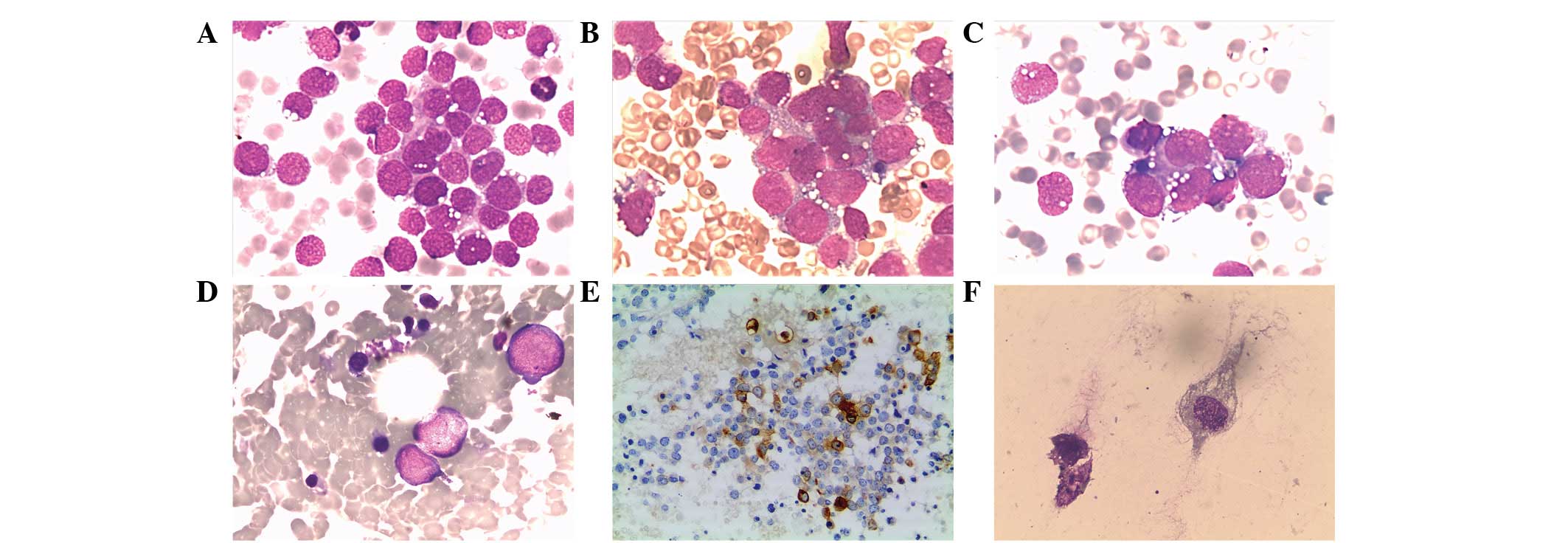
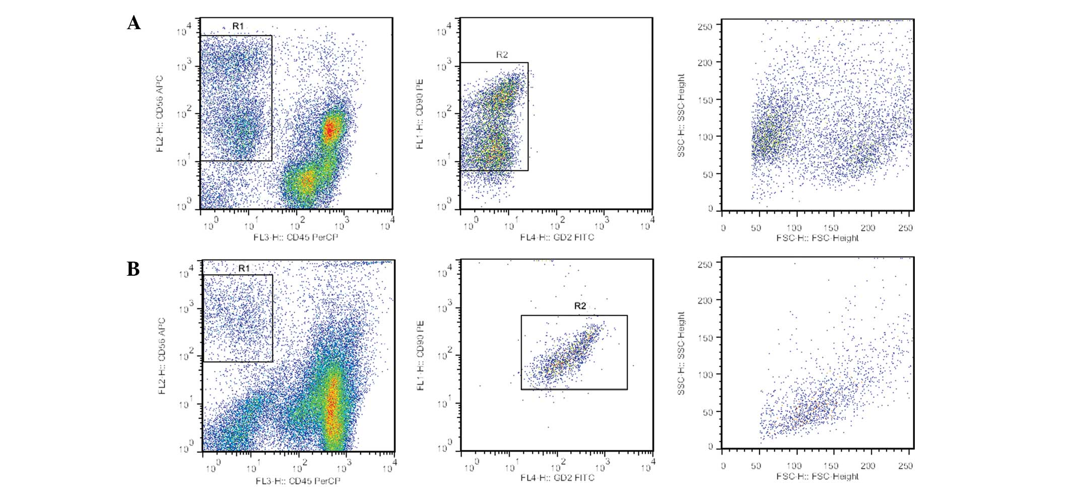
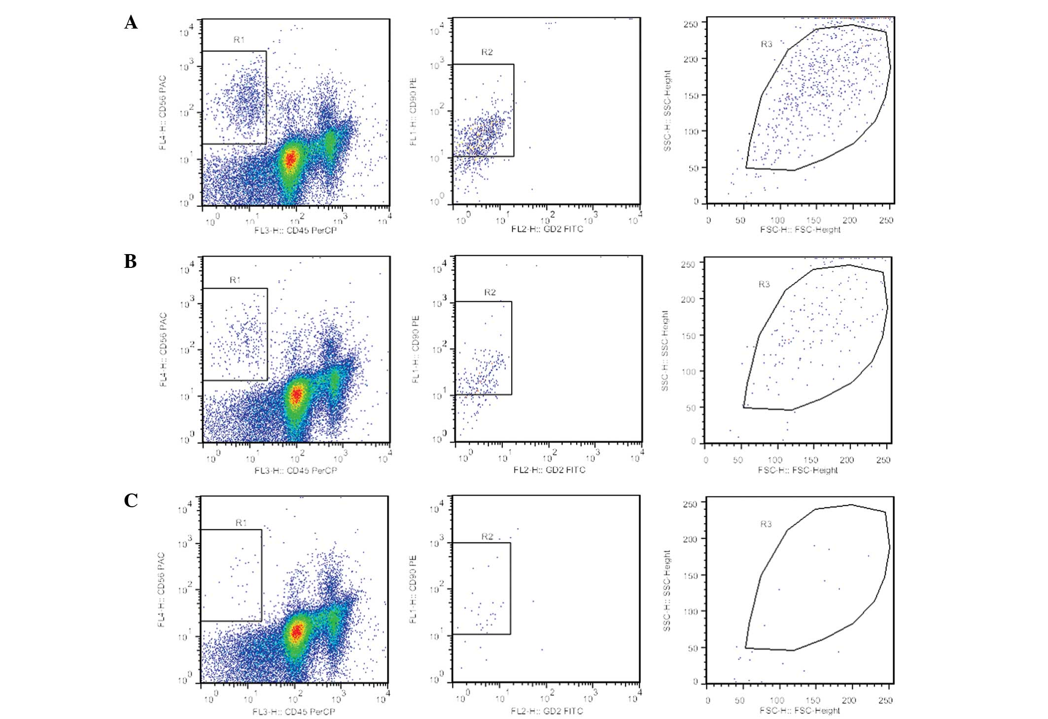
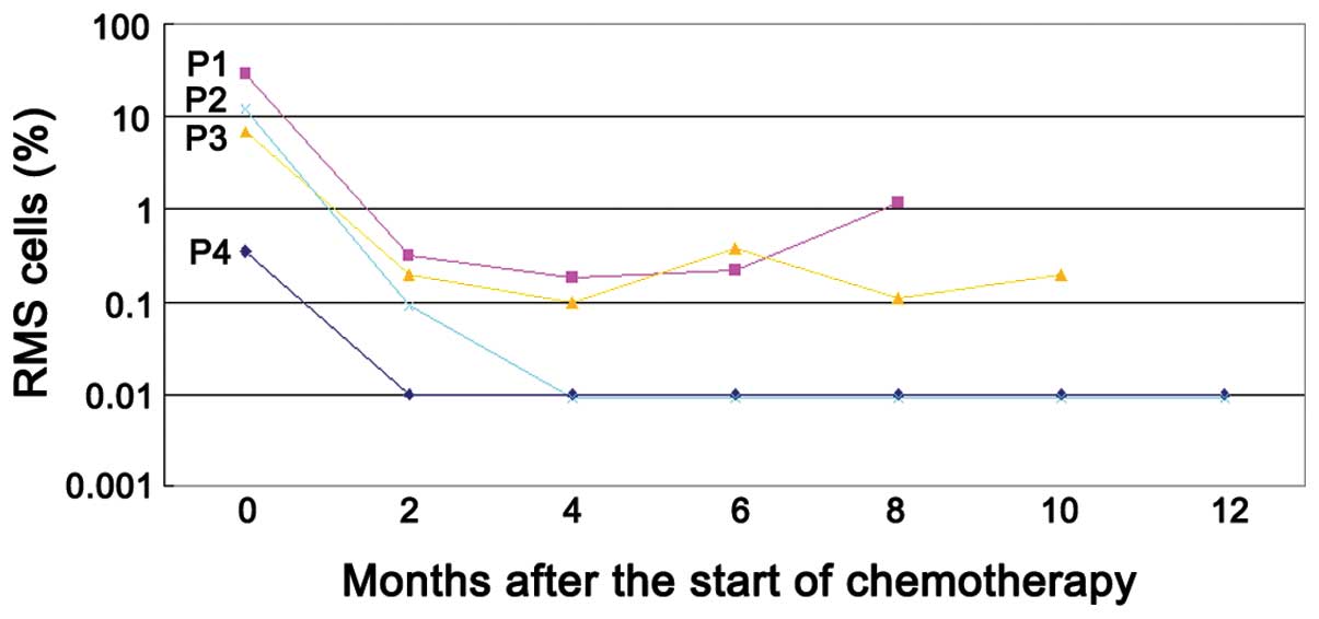
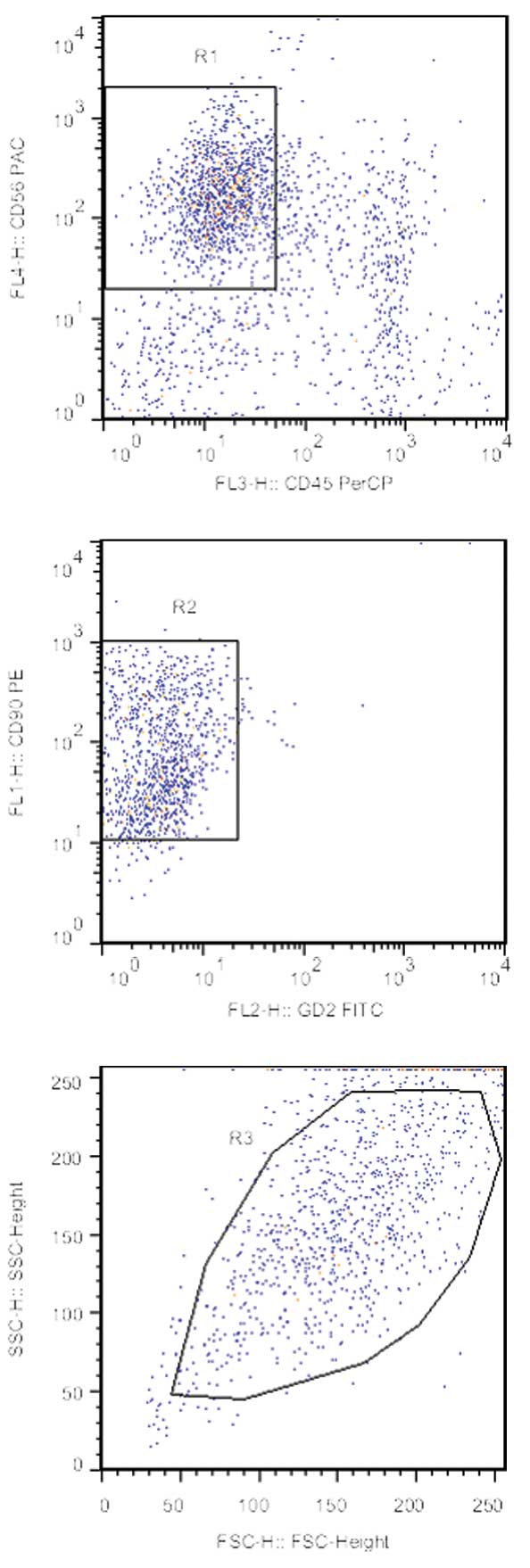
Patients with metastatic rhabdomyosarcoma (RMS) have a poor prognosis. The detection of contaminating RMS cells in the bone marrow (BM) is important in clinical staging and risk assessment. The cytological examination of the BM remains the gold standard for the diagnosis of RMS, but has a limited sensitivity. In the present study, 32 BM and two cerebrospinal fluid (CSF) samples from 11 patients with suspected metastasis were analyzed by flow cytometry (FCM) with ganglioside D2 (GD2) conjugated with fluorescein isothiocyanate, cluster of differentiation (CD)90‑phycoerythrin, CD45‑peridinin chlorophyll protein and CD56‑allophycocyanin monoclonal antibody cocktail in parallel to morphological examination at diagnosis or during treatment. Five samples (14.7%) were positive for RMS onup morphological examination. By FCM, 16 samples (47.1%) were positive for RMS. A significant difference was identified between the two methods. The four‑color FCM assay successfully detected RMS cells in BM samples to a level of 0.01% (1 per 104 cells). RMS cells demonstrated a phenotype with CD56+/CD90+/CD45-/GD2- expression, which is different from the CD56+/CD90+/CD45-/GD2+ expression phenotype in neuroblastoma cells. The follow‑up of four patients by FCM demonstrated that two patients became minimal residual disease‑negative following two and four cycles of chemotherapy, respectively, and survived. The other two cases remained FCM‑positive despite receiving four courses of chemotherapy and consequently succumbed to progressive disease. In addition, FCM analysis of the CSF samples from one patient confirmed a diagnosis of CSF metastasis with RMS. In conclusion, FCM may have a role not only in staging and monitoring the effects of therapy, but also in providing diagnostic confirmation of CSF metastasis with RMS.
View Figures
View References

Related Articles

Journal Cover

2014-April
Volume 7 Issue 4

Print ISSN: 1792-1074
Online ISSN:1792-1082

Sign up for eToc alerts

Recommend to Library

Copy and paste a formatted citation
x
Spandidos Publications style
Shen H, Tang Y, Dong A, Li H, Shen D, Yang S, Tang H, Gu W and Shu Q: Staging and monitoring of childhood rhabdomyosarcoma with flow cytometry. Oncol Lett 7: 970-976, 2014
APA
Shen, H., Tang, Y., Dong, A., Li, H., Shen, D., Yang, S. ... Shu, Q. (2014). Staging and monitoring of childhood rhabdomyosarcoma with flow cytometry. Oncology Letters, 7, 970-976. https://doi.org/10.3892/ol.2014.1854
MLA
Shen, H., Tang, Y., Dong, A., Li, H., Shen, D., Yang, S., Tang, H., Gu, W., Shu, Q."Staging and monitoring of childhood rhabdomyosarcoma with flow cytometry". Oncology Letters 7.4 (2014): 970-976.
Chicago
Shen, H., Tang, Y., Dong, A., Li, H., Shen, D., Yang, S., Tang, H., Gu, W., Shu, Q."Staging and monitoring of childhood rhabdomyosarcoma with flow cytometry". Oncology Letters 7, no. 4 (2014): 970-976. https://doi.org/10.3892/ol.2014.1854