Introduction
Salivary adenoid cystic carcinoma (SACC) is one of
the most common subtypes of malignant tumors occurring in the
salivary gland. The characteristics of SACC are the invasion of
adjacent tissue and hematogenous spread to the distant organs of
the lung, bone and liver (1).
Although aggressive surgery has been used, the rate of perineural
invasion and distant metastases after 10 years is 50 and 39%,
respectively (2). The evolution of
SACC is unpredictable in individual patients due to the lack of
strong invasion and metastasis indicators, other than advanced
stage at presentation. Therefore, the identification of
molecular-based specific markers may contribute to prognostic
evaluation, by improving our understanding of a single tumor, and
more significantly, it may aid biologically-targeted therapy in
SACC.
The process of tumor cell invasion into the stromal
tissue is closely associated with interactions between the tumor
cells and the extracellular matrix (ECM). Altered expression and
modification of ECM proteins in tumor cells plays a significant
role in their invasion into surrounding normal tissues (3). The matrix metalloproteinase (MMP)
family play critical roles in this step by degrading components of
the basement membranes and ECM (4).
Among the >20 MMPs that have been identified, MMP-2 is a
significant proteolytic enzyme (5).
MMP-2 can degrade the components of the ECM and basement membrane,
destroy the structure of local tissues and increase the ability of
vascularization induced by tumors, therefore playing a significant
role in all the different stages of the development and invasion of
tumors. Thus, the development of novel invasion inhibitors, along
with anti-proliferative therapies, may contribute to the control of
local tumor growth and tumor spread.
The reversion-inducing cysteine-rich protein with
Kazal motifs (RECK) gene was isolated as a transformation
suppressor gene; a fibroblast expression library was screened for
complementary DNA that was able to induce a non-transformed flat
morphology when expressed in v-Ki-ras-transformed NIH3T3 (a mouse
embryonic fibroblast cell line) cells (6). RECK is a 110-kDa glycoprotein that is
expressed in a number of normal tissues, however it is not
expressed in transformed and tumor-derived cells (7). RECK has been demonstrated to inhibit
MMP activity through several mechanisms, including the direct
inhibition of protease activity, the regulation of cellular release
and possible sequestration at the cell surface (8). RECK inhibits the activity of at least
three MMP members; MMP-2, MMP-9 and membrane type (MT)1-MMP
(9). An increasing amount of
evidence has now indicated that the altered expression of RECK is
involved in numerous solid tumors, including lung cancer, human
breast carcinoma, pancreatic cancer, non-small cell lung cancer and
colorectal cancer (10–12). Patients with high RECK protein
levels in tumor tissues usually have improved survival outcomes,
with tumors being less invasive, as demonstrated in clinical
investigations (13). However, our
current knowledge of the underlying molecular mechanisms mediated
by RECK in SACC is limited. Therefore, we hypothesize that RECK is
a novel suppressor gene for invasion and metastasis in SACC, and
that it is potentially a good target for SACC therapy.
The purpose of the present study was to examine the
expression of RECK and MMP-2 in vitro and in vivo,
and to analyze the correlation between RECK expression and MMP-2
expression in SACC. This study also aimed to clarify the roles of
RECK and MMP-2 in the invasion and development of SACC and to
explore their role in the prognosis of SACC.
Materials and methods
Cell lines
The human SACC cell lines, ACC-2 and ACC-M, were
kindly provided by Professor Wantao Chen (Department of Oral and
Maxillofacial Surgery, Ninth People’s Hospital, College of
stomatology, Shanghai Jiao Tong University, Shanghai, China). The
cells were cultured in RPMI 1640 medium supplemented with 10%
filtered fetal bovine serum (Hyclone, Logan, UT, USA). The cultures
were incubated at 37°C in the humid atmosphere of a 5%
CO2 incubator.
RNA isolation and quantitative polymerase
chain reaction (qPCR)
Total RNA from the cell lines was obtained using an
RNeasy Mini kit (Tiangen Biotech, Beijing, China) according to the
manufacturer’s instructions. The quantity and quality of the RNA
was assessed by measuring the absorbance at 260 nm and by agarose
gel electrophoresis. Total RNA was reverse transcribed with a High
Capacity cDNA Archive kit (Takara Bio Inc., Dalian, China), and
then an ABI Prism 7000 Sequence Detector (Applied Biosystems,
Foster City, CA, USA) was utilized to determine the expression
levels of the proteins of interest via qPCR. The thermocycler
parameters were 95°C for 15 sec, followed by 45 cycles of 95°C for
5 sec and 60°C for 30 sec. All PCR products were visualized by
electrophoresis and ethidium bromide staining in 2% agarose gels.
The RECK and MMP-2 primers are shown in Table I.
 | Table IBases of primers for qPCR. |
Table I
Bases of primers for qPCR.
| Gene | Primer |
|---|
| RECK | F:
5′-TGCAAGCAGGCATCTTCAAA-3′ |
| R:
5′-ACCGAGCCCATTTCATTTCTG-3′ |
| MMP-2 | F:
5′-AGCTCCCGGAAAAGATTGATG-3′ |
| R:
5′-CAGGGTGCTGGCTGAGTAGAT-3′ |
| GAPDH | F:
5′-GCAGGGGGGAGCCAAAAGGG-3′ |
| R:
5′-TGCCAGCCCCAGCGTCAAAG-3′ |
Western blot analysis
The cells were harvested, washed twice with cold
phosphate-buffered saline (PBS) and lysed in buffer [50 M Tris-HCl
(pH 7.4), 150 M NaCl, 2 M EDTA and 1% NP-40], containing protease
inhibitors. The protein concentration was quantified using a
bicinchoninic acid protein measurement kit (Shenneng Bocai Biology,
Co., Ltd., Shanghai, China). A total of 30 μg extract was subjected
to SDS-PAGE and then electrophoretically transferred to a
nitrocellulose membrane, which was blocked with 5% (w/v) dried
skimmed milk-tris-buffered saline and tween 20 [TBST; 10 M Tris-HCl
(pH 8.0), 150 M NaCl and 0.05% Tween 20] for 1 h at room
temperature. Subsequently, the membrane was incubated for 2 h with
antibodies against RECK (Santa Cruz Biotechnologies, Inc., Santa
Cruz, CA, USA; at 1:1000 dilution) and MMP-2 (Santa Cruz
Biotechnologies, Inc.; at 1:1000 dilution), respectively, in TBST
with 5% skimmed milk. Subsequent to being washed in TBST,
horseradish peroxidase-conjugated anti-rabbit IgG (Santa Cruz
Biotechnology, Inc.) was used as a secondary antibody (1:10,000, in
TBST with 2% bovine serum albumin incubated for 1 h). Each sample
was also probed with an anti-GAPDH antibody (Santa Cruz
Biotechnology, Inc.) as a loading control. Protein bands were
visualized by the Alpha Imager 2200 system (Alpha Innotech, San
Leandro, CA, USA).
Patients and specimens
The protocol of the study was approved by the
Institutional Ethics Committee of the Provincial Hospital
Affiliated to Shandong University (Jinan, China). In total, 83
patients with ACC of the salivary gland were recruited for the
study after providing informed consent. The patients underwent
resection of their tumors without pre-operative chemotherapy,
hormone therapy or radiotherapy at the Department of Oral and
Maxillofacial Surgery, Provincial Hospital Affiliated to Shandong
University, between May 2001 and March 2010. There were 44 male and
39 female patients aged 19–82 years old (average, 42±12.5 years
old) and the tumor sizes ranged between 0.8 and 4.5 cm (median
size, 2.1 cm). Of these patients, 36 (43.4%) exhibited stage I and
II disease and 47 (56.6%) exhibited stage III and IV disease,
according to the American Joint Committee on Cancer staging
criteria (14). A total of 27 cases
of benign salivary tumors and samples of 16 normal individuals were
collected. Pathological slides were reviewed by two pathologists
who were not provided with any information on the patients. Each
tumor was examined to determine the proportion of tubular,
cribiform or solid patterns according to the World Health
Organization’s International Histological Classification of
Salivary Gland Tumors (15). Three
histological grades were determined: Grade I, tumors with tubular
and cribriform areas, but without solid components; grade II,
cribriform tumors that were either pure or mixed with <30% solid
areas; and grade III, tumors with >30% solid patterns.
Immunohistochemistry
The rabbit polyclonal antibody against RECK (Abcam,
1:100) and the rabbit polyclonal antibody against MMP-2 (Abcam,
1:50) were used for the detection of RECK and MMP-2 in three
pathological slides, respectively. Immunohistochemistry was
performed on 4-μm thick representative sections of the
corresponding formalin-fixed and paraffin-embedded tumor specimens
by the streptavidin-peroxidase method following the manufacturer’s
instructions (ZSGB-Bio, Beijing, China). Antigen retrieval was
accomplished by 0.01 M citrate buffer solution (pH 6.0) for RECK
and MMP-2, respectively, in a 700 W microwave oven for 15 min. All
stains of RECK and MMP-2 were developed with diaminobenzidine (DAB)
for 2 and 3 min, respectively. Negative controls were performed by
replacing the primary antibody with PBS.
Evaluation of immunohistochemical
staining
The positive signals of expression of RECK and MMP-2
protein was characterized by the deposition of pale yellow, buff or
brown particles; the positive coloration was located in the
intracytoplasm. Immunohistochemical staining for RECK and MMP-2 was
evaluated in compliance with intensity and proportion. The
intensities were scored as follows: 0, no staining; 1, weak
staining; 2, moderate staining; and 3, strong staining. The
percentage of staining area was classified as: 0, <5% of tumor
cells; 1, 6–25% of tumor cells; 2, 26–50% of tumor cells; 3, 51–70%
of tumor cells; and 4, >70% of tumor cells. Finally, the two
scores were multiplied, providing the final scores: 0–1, negative
(−); 2–3, secondary positive (+); and ≥4, positive (++).
Statistical analysis
All analyses were carried out using the statistical
software, SPSS 13.0 (SPSS, Inc., Chicago, IL, USA). The association
between RECK and MMP-2 expression and several clinicopathological
variables was evaluated according to Pearson’s χ2 test
or Fisher’s exact test. Survival curves were obtained using the
Kaplan-Meier method, and the significance was analyzed by the
log-rank test. The effect of variables on survival was assessed
using Cox univariate and multivariate regression analyses. The risk
ratio and its 95% confidence interval were recorded for each
marker. P<0.05 were considered to indicate a statistically
significant difference in all of the analyses. Ooverall survival
was defined as from the date of surgery to the date a patient
succumbed due to SACC.
Results
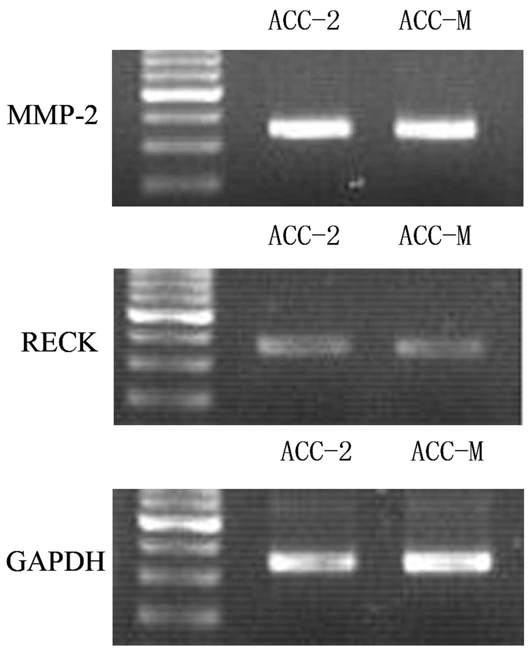
Expression of RECK and MMP-2 mRNA in ACC
cell lines
To verify whether the mRNA of RECK and MMP-2 was
expressed in ACC, the mRNA levels of these two genes were
determined in the ACC-2 and ACC-M cell lines by qPCR using specific
primers and probes for RECK and MMP-2, with GAPDH as a control. The
results revealed RECK and MMP-2 mRNA expression in the two cell
lines (Fig. 1).
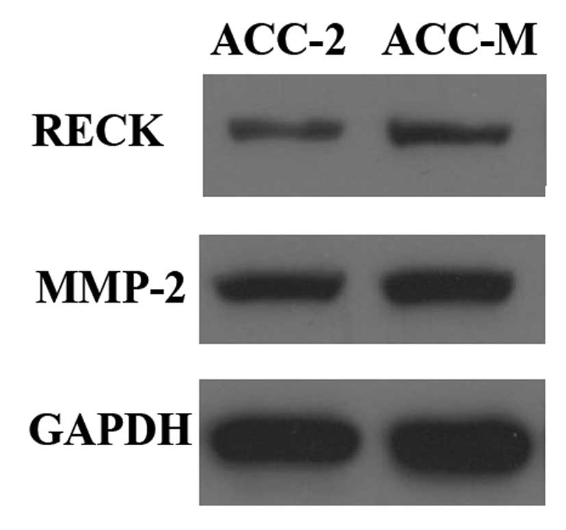
Expression of RECK and MMP-2 protein in
ACC cell lines
The protein expression levels of RECK and MMP-2 were
determined by western blot analysis in the cell lines, and the
GAPDH protein levels were also determined in the same blot to serve
as a loading control. These results revealed that RECK and MMP-2
protein was expressed in the ACC-2 and ACC-M cell lines (Fig. 2).
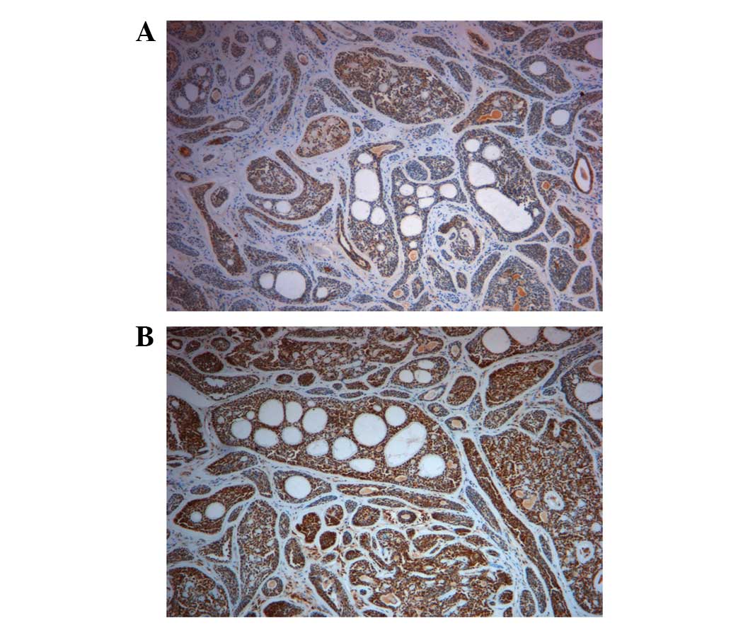
Expression of RECK and MMP-2 in
patients
The positive expression of RECK protein was mainly
located in the cytoplasm of carcinoma cells, demonstrated by a pale
yellow or buff color (Fig. 3A). The
percentage of RECK expression increased from the SACC, to the
benign salivary tumors, to the normal cases; 25.3% in the SACC,
48.1% in the benign salivary tumors and 87.5% in the normal
salivary tissues. The difference in expression rates was considered
statistically significant (P<0.05) (Table II). The positive expression of
MMP-2 protein was mainly located in the cytoplasm, demonstrated by
a buff or brown color (Fig. 3B).
The percentage of expression decreased from the SACC, to the benign
salivary tumors, to the normal salivary tissues; 83.1% in the ACC,
74.1% in the benign salivary tumors and 25.0% in the normal
salivary tissues. The difference of expression rates was considered
to be statistically significant (P<0.05) (Table II).
| Table IIExpression of RECK and MMP-2 in SACC,
benign salivary tumors and normal cases. |
Table II
Expression of RECK and MMP-2 in SACC,
benign salivary tumors and normal cases.
| Group | Total (n) | RECK | χ2 | P-value | MMP-2 | χ2 | P-value |
|---|
|
|
|---|
| − (n) | + (n) | ++ (n) | PP (%) | − (n) | + (n) | ++ (n) | PP (%) |
|---|
| SACC | 83 | 62 | 9 | 12 | 25.3 | 25.007 | 0.000 | 14 | 29 | 40 | 83.1 | 25.405 | <0.0001 |
| BST | 27 | 14 | 7 | 6 | 48.1 | | | 7 | 9 | 11 | 74.1 | | |
| NC | 16 | 2 | 5 | 9 | 87.5 | | | 12 | 4 | 0 | 25.0 | | |
Correlation between RECK and MMP-2
expression and clinicopathological parameters in SACC patients
Correlations between RECK and MMP-2 expression with
various clinicopathological features are summarized in Table III. No significant correlations
were observed between RECK and MMP-2 expression, and age, tumor
size and gender. RECK expression was significantly associated with
tumor-node-metastasis (TNM) stage (P=0.047), perineural invasion
(P=0.019) and histological grade (P=0.006). However, no significant
correlations were revealed between MMP-2 expression and
histological grade (P=0.064).
| Table IIIClinicopathological features of the
SACC patients and their primary tumors and their association with
RECK and MMP-2 expression. |
Table III
Clinicopathological features of the
SACC patients and their primary tumors and their association with
RECK and MMP-2 expression.
| | RECK | | | MMP-2 | | |
|---|
| |
| | |
| | |
|---|
| Factor | No. of cases | − (n) | + (n) | PP (%) | χ2 | P-value | − (n) | + (n) | PP (%) | χ2 | P-value |
|---|
| Gender |
| Female | 34 | 25 | 9 | 26.5 | 0.042 | 0.838 | 5 | 29 | 85.3 | 0.192 | 0.661 |
| Male | 49 | 37 | 12 | 24.5 | | | 9 | 40 | 81.6 | | |
| Age, years |
| ≥52 | 43 | 32 | 11 | 25.6 | 0.004 | 0.951 | 8 | 35 | 81.4 | 0.192 | 0.661 |
| <52 | 40 | 30 | 10 | 25.0 | | | 6 | 34 | 85.0 | | |
| Tumor size, cm |
| 3 | 51 | 39 | 12 | 23.5 | 0.220 | 0.639 | 10 | 41 | 80.4 | 0.708 | 0.400 |
| ≥3 | 32 | 23 | 9 | 28.1 | | | 4 | 28 | 87.5 | | |
| TNM stage |
| I, II | 36 | 23 | 13 | 36.1 | 3.931 | 0.047a | 10 | 26 | 72.2 | 5.397 | 0.020a |
| III, IV | 47 | 39 | 8 | 17.0 | | | 4 | 43 | 91.5 | | |
| Histological
grade |
| I | 36 | 21 | 15 | 41.7 | 10.17 | 0.006a | 10 | 26 | 72.2 | 5.489 | 0.064 |
| II | 19 | 15 | 4 | 21.1 | | | 2 | 17 | 89.5 | | |
| III | 28 | 26 | 2 | 7.1 | | | 2 | 26 | 92.9 | | |
| Perineural
invasion |
| No | 45 | 29 | 16 | 35.6 | 5.469 | 0.019a | 11 | 34 | 75.6 | 4.024 | 0.045a |
| Yes | 38 | 33 | 5 | 13.2 | | | 3 | 35 | 92.1 | | |
Correlation of RECK and MMP-2 in
SACC
As revealed in Table
IV, the MMP-2 expression rate was 45.5% (10/22) in tissues with
positive (+ and ++) RECK expression. In tissues negative (−) for
RECK expression, the MMP-2 expression rate was 95.2% (59/62).
Pearson’s χ2 test revealed that there was a significant
negative correlation between the expression of RECK and MMP-2
(χ2, 38.202; P<0.0001).
| Table IVAnalysis of the correlation between
the expression of RECK and MMP-2 in SACC. |
Table IV
Analysis of the correlation between
the expression of RECK and MMP-2 in SACC.
| RECK | Total, n | MMP-2 (n) | χ2 | P-value |
|---|
|
|---|
| − | + | ++ |
|---|
| − | 62 | 3 | 22 | 37 | 38.202 | <0.0001a |
| + | 9 | 3 | 4 | 2 | | |
| ++ | 12 | 9 | 3 | 0 | | |
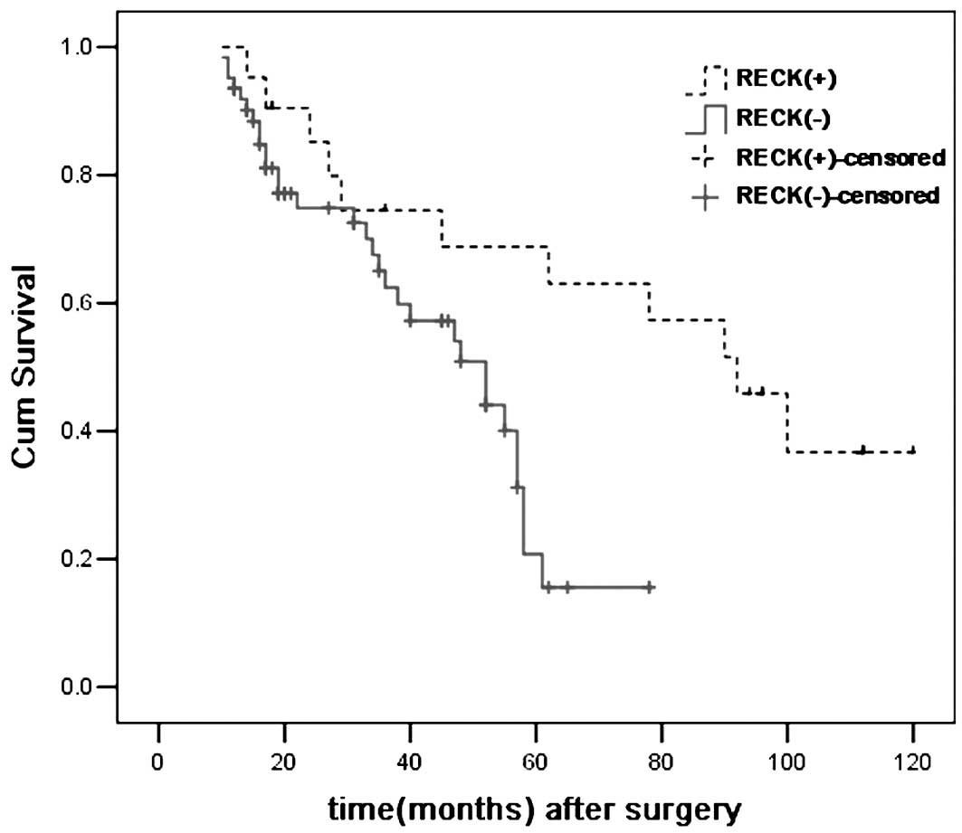
Overall survival analysis
The average follow-up period was 54 months (range,
10–120 months). In total, 42 patients (50.6%)succumbed to SACC, 5
patients (6.0%) succumbed during the follow-up period, 4 patients
(4.8%) succumbed to an unrelated cause and 32 patients (38.6%)
remained alive on the day of the study. The survival curves
demonstrated that the overall survival in the RECK-positive
expression group was improved in comparison to the RECK-negative
expression group (P=0.009), based on the use of the Kaplan-Meier
method and log-rank test (Fig. 4).
As summarized in Table V, the
univariate analysis revealed that the following variables were
significantly associated with the prognosis, including RECK and
MMP-2 expression, histological grade, TNM stage and perineural
invasion, while the multivariate analysis revealed that RECK
expression and histological grade also had an independent
prognostic effect on the overall survival of the SACC patients.
| Table VPrognostic factors in the Cox
proportional hazards model. |
Table V
Prognostic factors in the Cox
proportional hazards model.
| Univariate
analysis | Multivariate
analysis |
|---|
|
|
|
|---|
| Variables | HR (95% CI) | P-value | HR (95% CI) | P-value |
|---|
| Gender
(female/male) | 0.85
(0.28–1.47) | 0.561 | nd | nd |
| Age (<52
years/≥52 years) | 1.69
(0.38–2.79) | 0.476 | nd | nd |
| TNM stage (I,
II/III, IV) | 3.26
(1.96–6.12) | 0.038a | 2.01
(1.26–3.58) | 0.296 |
| Tumor size (≤3
cm/>3 cm) | 2.98
(1.26–5.13) | 0.233 | nd | nd |
| Histological grade
(I, II/III) | 3.65
(1.49–5.23) | 0.007a | 3.16
(1.27–5.79) | 0.038b |
| RECK expression
(positive/negative) | 2.45
(0.69–4.56) | 0.009a | 4.57
(0.85–24.55) | 0.014b |
| MMP-2 expression
(positive/negative) | 4.26
(2.13–6.39) | 0.021a | 1.69
(0.48–4.57) | 0.079 |
| Perineural invasion
(positive/negative) | 3.19
(1.26–4.81) | 0.067 | nd | nd |
Discussion
In the present study, the expression of RECK and
MMP-2 mRNA and protein was examined first in vitro using two
human ACC cell lines. The results revealed the positive expression
of RECK in the ACC-2 and ACC-M cell lines. Therefore, the in
vivo expression of RECK and MMP-2 in SACC was investigated
using an immunohistochemistry assay. First, the positive expression
of RECK was observed in 21/83 (25.3%) of SACC cases, and RECK
expression was significantly associated with the TNM stage,
histological pattern and perineural invasion of patients with SACC
(P<0.05). Furthermore, there was a significant inverse
correlation between RECK-positive expression and MMP-2-positive
expression (P=0.046), and the RECK expression was significantly
associated with overall survival.
Recurrence and metastasis is responsible for patient
mortality in most solid tumors (16–18).
Degradation and remodeling of the ECM are necessary steps in tumor
development. All ECM components can be degraded by MMPs, and each
ECM element is cleaved by a specific MMP or MMP group. In
particular, MMP-2 is upregulated in SACC and a number of other
malignant tumors, and contributes to the invasion of tumor cells by
degrading the ECM (19–21). RECK, initially identified as a
transformation suppressor gene, is normally expressed in adult
human tissues and is downregulated in numerous solid tumors,
including pancreatic cancer, non-small cell lung cancer and
colorectal cancer (11,12,22).
Similarly, the present results revealed the mRNA and protein
expression of RECK and MMP-2 in human ACC cell lines, and
identified that RECK expression was significantly lower in SACC
than in normal tissues.
Previous studies (23–25)
have reported that RECK function is correlated with the inhibition
of MMP-2, MMP-9 and MT1-MMP. Masui et al (26) reported a significant inverse
correlation between RECK and MMP-2 expression in pancreatic cancer.
To explore the correlation between MMP-2 and RECK in SACC in the
present study, 83 specimens of SACC were collected to examine the
expression of MMP-2 and RECK by immunohistochemistry assay. There
was a significant negative correlation between the expression of
RECK and MMP-2 (P<0.0001), and the result was consistent with
the aforementioned study. Furthermore, the correlation between RECK
expression and various clinicopathological features was evaluated,
revealing that RECK expression was significantly associated with
histological grade, TNM stage and the perineural invasion of
patients with SACC (P<0.05), but that MMP-2 expression had no
significant association with histological grade (P=0.064); RECK was
possibly functioning as more than an MMP inhibitor, and further
studies are therefore required in this area.
Furumoto et al (27) reported that high RECK expression
correlates with less invasive tumors and an improved prognosis in
patients with hepatocellular carcinoma, and that downregulated RECK
expression could induce tumor angiogenesis and promote tumor
regression. Masui et al (26) examined RECK expression in pancreatic
cancer and reported that tumors with positive RECK staining were
significantly less invasive in comparison to RECK-negative tumors,
indicating the potential value of RECK as a prognostic molecular
marker for pancreatic cancer. In the present study, the
multivariate survival analysis revealed that RECK expression
(P=0.014) was significantly associated with an improved survival
rate and could be an independent prognostic indicator in SACC. In
addition, histological grade (I, II/III; P=0.038) appeared to be
the significantly independent clinicopathological factor involved
with overall survival, which has been revealed to indicate a poor
prognosis for SACC in a previous study (28). Taken together, these findings
indicate that RECK may serve as a favorable prognostic predictor in
SACC, and increase the ability to predict patient prognosis when
used in combination with histological grade (I, II/III).
An increase in RECK expression appears to be a
reasonable treatment strategy to enhance the overall survival rate
of SACC patients, and several ways to upregulate RECK expression
have been suggested. Yan et al (29) reported that tomatidine inhibits the
invasion of human lung adenocarcinoma A549 cells by reducing MMP-2
expression and enhancing RECK expression. Hsu et al
(7) reported that erbB2
transcriptionally represses RECK expression through HDAC1 and Sp1,
and demonstrated that the histone deacetylase inhibitor,
trichostatin A, upregulates RECK expression in vitro.
Studies enhancing RECK expression by drug or gene knock out in
vitro and in vivo, in order to examine the effect on the
migration and invasion of SACC, are required.
In conclusion, the present data indicates that RECK
was discovered as a novel molecular biomarker and may actively be
involved in the progression, invasion and metastasis of SACC. RECK
may reduce tumor progression through functionally downregulating
MMP-2 expression. Measurements of the expression of RECK and MMP-2
are valuable, not only to assess patient prognosis, but to also
develop new strategies for cancer prevention and therapeutic
intervention.
Acknowledgements
This study was supported by grants from the Natural
Science Foundation of Shandong Province (no. ZR2012HQ022).
References
|
1
|
van der Wal JE, Becking AG, Snow GB and
van der Waal I: Distant metastases of adenoid cystic carcinoma of
the salivary glands and the value of diagnostic examinations during
follow-up. Head Neck. 24:779–783. 2002.
|
|
2
|
Terhaard CH, Lubsen H, Van der Tweel I, et
al: Salivary gland carcinoma: independent prognostic factors for
locoregional control, distant metastases and overall survival:
results of the Dutch head and neck oncology cooperative group. Head
Neck. 26:681–693. 2004. View Article : Google Scholar
|
|
3
|
Honma K, Miyata T and Ochiya T: Type I
collagen gene suppresses tumor growth and invasion of malignant
human glioma cells. Cancer Cell Int. 7:122007. View Article : Google Scholar : PubMed/NCBI
|
|
4
|
López-Otín C and Matrisian LM: Emerging
roles of proteases in tumour suppression. Nat Rev Cancer.
7:800–808. 2007.PubMed/NCBI
|
|
5
|
Visse R and Nagase H: Matrix
metalloproteinases and tissue inhibitors of metalloproteinases:
structure, function, and biochemistry. Circ Res. 92:827–839. 2003.
View Article : Google Scholar
|
|
6
|
Takahashi C, Sheng Z, Horan TP, et al:
Regulation of matrix metalloproteinase-9 and inhibition of tumor
invasion by the membrane-anchored glycoprotein RECK. Proc Natl Acad
Sci USA. 95:13221–13226. 1998. View Article : Google Scholar : PubMed/NCBI
|
|
7
|
Hsu MC, Chang HC and Hung WC: HER-2/neu
represses the metastasis suppressor RECK via ERK and Sp
transcription factors to promote cell invasion. J Biol Chem.
281:4718–4725. 2006. View Article : Google Scholar : PubMed/NCBI
|
|
8
|
Welm B, Mott J and Werb Z: Developmental
biology: vasculogenesis is a wreck without RECK. Curr Biol.
12:R209–R211. 2002. View Article : Google Scholar : PubMed/NCBI
|
|
9
|
Sasahara RM, Brochado SM, Takahashi C, et
al: Transcriptional control of the RECK metastasis/angiogenesis
suppressor gene. Cancer Detect Prev. 26:435–443. 2002. View Article : Google Scholar : PubMed/NCBI
|
|
10
|
Takagi S, Simizu S and Osada H: RECK
negatively regulates matrix metalloproteinase-9 transcription.
Cancer Res. 69:1502–1508. 2009. View Article : Google Scholar : PubMed/NCBI
|
|
11
|
Takenaka K, Ishikawa S, Yanagihara K, et
al: Prognostic significance of reversion-inducing cysteine-rich
protein with Kazal motifs expression in resected pathologic stage
IIIA N2 non-small-cell lung cancer. Ann Surg Oncol. 12:817–824.
2005. View Article : Google Scholar
|
|
12
|
Span PN, Sweep CG, Manders P, Beex LV,
Leppert D and Lindberg RL: Matrix metalloproteinase inhibitor
reversion-inducing cysteine-rich protein with Kazal motifs: a
prognostic marker for good clinical outcome in human breast
carcinoma. Cancer. 97:2710–2715. 2003. View Article : Google Scholar
|
|
13
|
Kang HG, Kim HS, Kim KJ, Oh JH, Lee MR,
Seol SM and Han I: RECK expression in osteosarcoma: correlation
with matrix metalloproteinases activation and tumor invasiveness. J
Orthop Res. 25:696–702. 2007. View Article : Google Scholar : PubMed/NCBI
|
|
14
|
Ko YH, Roh SY, Won HS, et al: Prognostic
significance of nuclear survivin expression in resected adenoid
cystic carcinoma of the head and neck. Head Neck Oncol. 2:302010.
View Article : Google Scholar : PubMed/NCBI
|
|
15
|
Seifert G and Sobin LH: The World Health
Organization’s Histological Classification of Salivary Gland
Tumors. A commentary on the second edition. Cancer. 70:379–385.
1992.
|
|
16
|
Lee JH and Welch DR: Suppression of
metastasis in human breast carcinoma MDA-MB-435 cells after
transfection with the metastasis suppressor gene, KiSS-1. Cancer
Res. 57:2384–2387. 1997.
|
|
17
|
Welch DR, Steeg PS and Rinker-Schaeffer
CW: Molecular biology of breast cancer metastasis. Genetic
regulation of human breast carcinoma metastasis. Breast Cancer Res.
2:408–416. 2000. View
Article : Google Scholar : PubMed/NCBI
|
|
18
|
Yang J, Mani SA, Donaher JL, et al: Twist,
a master regulator of morphogenesis, plays an essential role in
tumor metastasis. Cell. 117:927–939. 2004. View Article : Google Scholar
|
|
19
|
Shirasuna K, Saka M, Hayashido Y, Yoshioka
H, Sugiura T and Matsuya T: Extracellular matrix production and
degradation by adenoid cystic carcinoma cells: participation of
plasminogen activator and its inhibitor in matrix degradation.
Cancer Res. 53:147–152. 1993.
|
|
20
|
Vihinen P and Kähäri VM: Matrix
metalloproteinases in cancer: prognostic markers and therapeutic
targets. Int J Cancer. 99:157–166. 2002. View Article : Google Scholar : PubMed/NCBI
|
|
21
|
Freitas VM, Vilas-Boas VF, Pimenta DC, et
al: SIKVAV, a laminin alpha1-derived peptide, interacts with
integrins and increases protease activity of a human salivary gland
adenoid cystic carcinoma cell line through the ERK 1/2 signaling
pathway. Am J Pathol. 171:124–138. 2007. View Article : Google Scholar
|
|
22
|
Takeuchi T, Hisanaga M, Nagao M, et al:
The membrane-anchored matrix metalloproteinase (MMP) regulator RECK
in combination with MMP-9 serves as an informative prognostic
indicator for colorectal cancer. Clin Cancer Res. 10:5572–5579.
2004. View Article : Google Scholar
|
|
23
|
Oh J, Takahashi R, Kondo S, et al: The
membrane-anchored MMP inhibitor RECK is a key regulator of
extracellular matrix integrity and angiogenesis. Cell. 107:789–800.
2001. View Article : Google Scholar
|
|
24
|
Sternlicht MD and Werb Z: How matrix
metalloproteinases regulate cell behavior. Annu Rev Cell Dev Biol.
17:463–516. 2001. View Article : Google Scholar : PubMed/NCBI
|
|
25
|
Sasahara RM, Takahashi C and Noda M:
Involvement of the Sp1 site in ras-mediated downregulation of the
RECK metastasis suppressor gene. Biochem Biophys Res Commun.
264:668–675. 1999. View Article : Google Scholar
|
|
26
|
Masui T, Doi R, Koshiba T, et al: RECK
expression in pancreatic cancer: its correlation with lower
invasiveness and better prognosis. Clin Cancer Res. 9:1779–1784.
2003.PubMed/NCBI
|
|
27
|
Furumoto K, Arii S, Mori A, et al: RECK
gene expression in hepatocellular carcinoma: correlation with
invasion-related clinicopathological factors and its clinical
significance. Reverse-inducing-cysteine-rich protein with Kazal
motifs. Hepatology. 33:189–195. 2001. View Article : Google Scholar
|
|
28
|
Yang X, Dai J, Li T, et al: Expression of
EMMPRIN in adenoid cystic carcinoma of salivary glands: correlation
with tumor progression and patients’ prognosis. Oral Oncol.
46:755–760. 2010.PubMed/NCBI
|
|
29
|
Yan KH, Lee LM, Yan SH, Huang HC, Li CC,
Lin HT and Chen PS: Tomatidine inhibits invasion of human lung
adenocarcinoma cell A549 by reducing matrix metalloproteinases
expression. Chem Biol Interact. 203:580–587. 2013. View Article : Google Scholar : PubMed/NCBI
|