Introduction
Primary thyroid lymphoma accounts for only 2–5% of
all thyroid tumors (1,2) and 2.5–7% of extranodal lymphomas
(3), which predominantly originate
from B lymphocytes. The majority of B-cell lymphomas are diffuse
large B-cell lymphomas (DLBCL), extranodal marginal zone B-cell
mucosa-associated lymphoid tissue lymphomas (MALToma) and
follicular lymphomas (FL) (4).
Primary Burkitt’s lymphoma (BL) of the thyroid is even less common
than the other types of B-cell lymphoma and is a highly aggressive
non-Hodgkin’s lymphoma. Burkitt first identified BL as a sarcoma
involving the mandible in African children in 1958 (5). However, due to its rarity, little is
known concerning the origin, natural history and effective
treatment of primary BL of the thyroid. The current study presents
a case of BL and a systematic literature review on the clinical
presentation and treatment of this rare tumor. To the best of our
knowledge, this is only the fourth case of a primary BL of the
thyroid to be reported in the English literature (6–8).
Patient provided written informed consent.
Case report
An eight-year-old male presented with a mass in the
right anterior neck that had been apparent for one week. Upon
physical examination, blood pressure was recorded as 100/65 mmHg,
heart rate was 80 beats per min, respiratory rate was 20 breaths
per min and temperature was 36.1°C. A mass measuring ~4.0 cm in
size, which caused difficulty in swallowing, was identified in the
right anterior neck. The laboratory test results demonstrated a
normal blood count and serum biochemistry, as well as normal levels
of electrolytes and carcinoembryonic antigen. In addition, the test
results for Epstein-Barr virus (EBV) viral capsid antigens
immunoglobulin (Ig)M and IgG, human immunodeficiency virus (HIV)
and hepatitis C virus antibodies, hepatitis B antigen and syphilis
were negative. Furthermore, the thyroid hormone test results were
as follows: Free thyroxine (FT) 4 levels of 11.8 pmol/l (normal
range, 9–25 pmol/l); FT3 levels of 4.2 pmol/l (normal range, 3–9
pmol/l); thyroid-stimulating hormone levels of 0.720 μIU/ml (normal
range, 0.34–5.60 μIU/ml); anti-thyroglobulin levels of 20 IU/ml
(normal range, <115 IU/ml); and anti-thyroid peroxidase levels
of 25 IU/ml (normal range, <34 IU/ml). The patient had no
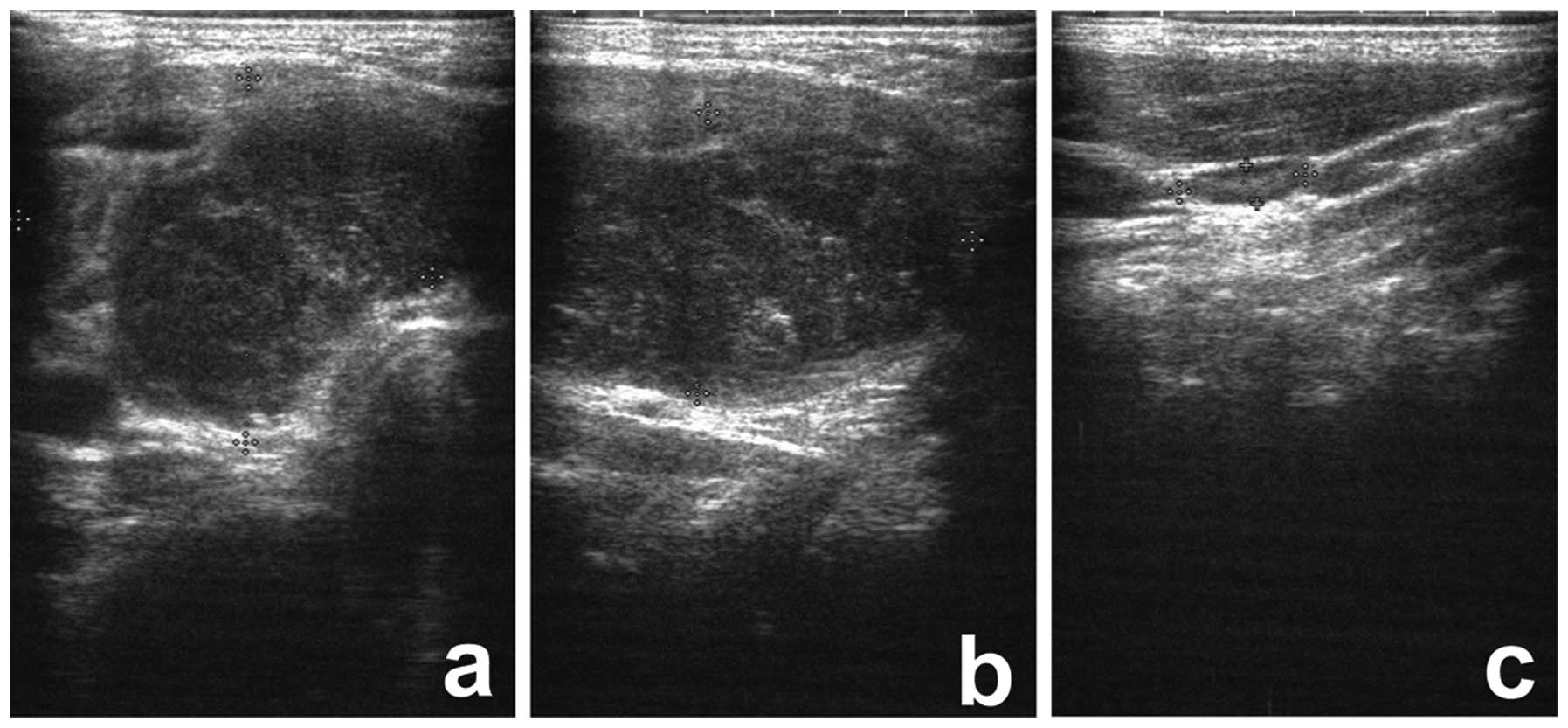
significant past medical or family history of disease. A B-mode
ultrasound examination revealed a mass measuring 4.0×3.0×2.5 cm in
the right lobe of the thyroid (Fig. 1A
and B), however, the lymph nodes surrounding the mass were
normal (Fig. 1C). The patient
underwent a right lobe and isthmus thyroidectomy whereby two lymph
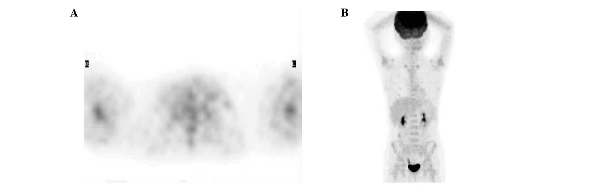
nodes were excised simultaneously. Following the surgery, positron
emission tomography-computed tomography scans showed normal
metabolism in the left lobe of the thyroid and other parts of the
body (Fig. 2). The patient’s bone
marrow cytology was also normal, however, histological examination
revealed diffuse infiltration of atypical lymphocytes and the
observation of residual thyroid follicles and necrosis (Fig. 3A). In addition, under low
magnification, the ‘starry sky’ histology was observed in certain
areas (Fig. 3B). The atypical
lymphocytes were medium-sized and consistent, with centrally
located nuclei of irregular shape, displaying dispersed and deep
basophilic chromatin and scanty cytoplasm. Additionally, certain
neoplastic cells were visible, while varying numbers of nucleoli
and apoptosis and mitosis were observed. Benign tissue cells
engulfing apoptotic bodies were also observed under high
magnification (Fig. 3C), however,
the isthmus of the thyroid was not infiltrated by the neoplastic
cells. No reactive lymphocyte infiltration or fibrosis was
identified in the stroma of the thyroid, and no oxyphilic change or
squamous metaplasia was observed in the epithelial cells of the
background thyroid tissues (Fig.
3D). The only change in the two lymph nodes that were
simultaneously excised, was the presence of reactive hyperplasia of
the lymphoid follicles (Fig. 3E and
F). Immunohistochemical staining was then performed with the
primary antibodies shown in Table I
(Zymed Corporation, Inc., San Francisco, CA, USA; Santa Cruz
Biotechnology, Inc., Santa Cruz, CA, USA). The results showed that
the neoplastic cells were diffusely positive for cluster of
differentiation (CD)20 (Fig. 3G)
and CD10 (Fig. 3H), marginally
positive for CD38, CD43 and B-cell lymphoma (Bcl)-6, but negative
for Bcl-2 and terminal deoxynucleotidyl transferase (TDT). In
addition, CD3 and CD5 stained the background T cells, and the Ki-67
proliferation index was >95% (Fig.
3I). Analysis using an EBV-encoded small RNA (EBER)
digoxin-labeled probe (PanPath B.V., Budel, Netherlands) was
performed and revealed a negative result (Fig. 3J), however, positive nuclei were
observed in the nasopharyngeal carcinoma tissue, which was used as
the positive control (Fig. 3K).
Analysis using the C-MYC break-apart detection probe (Guangzhou LBP
Medical Science Technology Co., Ltd., Guangzhou, China) was also
performed and the results revealed that ~90% of the neoplastic
cells exhibited red and green signal separation, which indicated
that chromosome breakage and translocation of the MYC gene had
occurred in the neoplastic cells (Fig.
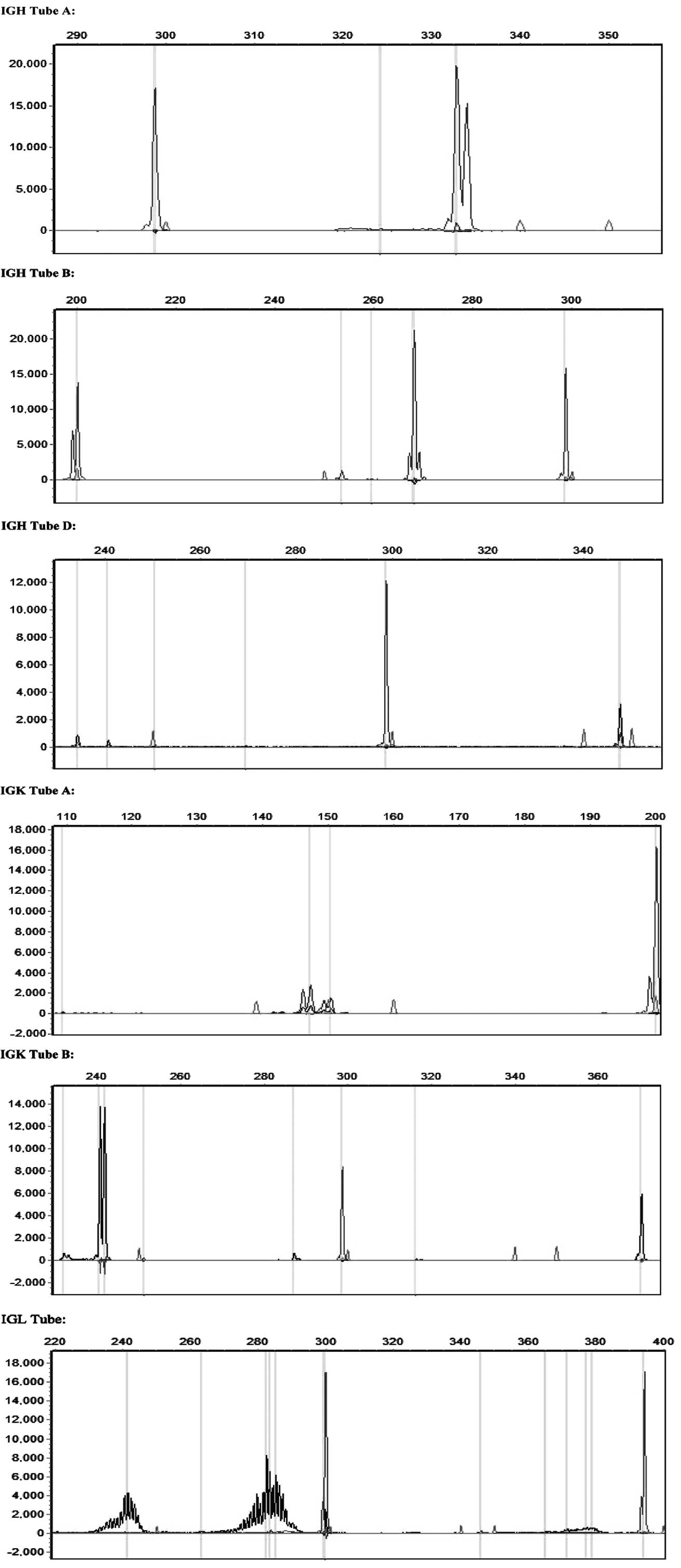
3L). Immunoglobulin gene rearrangement assays were performed
according to instructions of the Biomed-2 Polymerase Chain Reaction
kit (Invivoscribe technologies, Inc., San Diego, CA, USA), followed
by capillary electrophoresis, which was analyzed using
Genemarker® v1.5. software (SoftGenetics, LLC, State
College, PA, USA). Positive gene arrangements of IgH and IgK were
observed in the tumor tissues, however, no positive gene
rearrangements were observed for IgL (Fig. 4). Consequently, the patient was
diagnosed with primary BL of the thyroid and underwent alternate
R-B-NHL-BFM-90-A and R-B-NHL-BFM-90-B treatment, for four cycles
each. The two regimens, including the dose and duration of
chemotherapy, are described in Table
II. After almost four years of follow-up, the patient appears
well and remains free of disease.
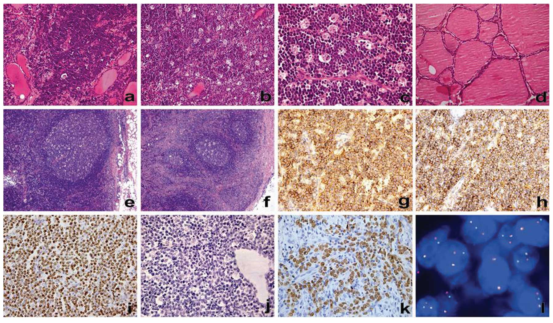 | Figure 3Histopathological observations with
hematoxylin and eosin staining. (A) Low magnification showing a
diffuse infiltration of atypical lymphoid cells in the thyroid
gland (magnification, ×200) and (B) the ‘starry sky’ histology
pattern within the tumor tissues (magnification, ×200). (C) Higher
magnification showing benign tissue cells engulfing apoptotic
bodies (magnification, ×400). (D) Lack of morphological features of
Hashimoto’s thyroiditis in the background thyroid gland, including
infiltrating lymphocytes, lymphoid follicle formation, stromal
fibrosis, eosinophilic change of the epithelial cells or squamous
metaplasia (magnification, ×200). (E and F) Reactive lymphoid
tissue hyperplasia in the two lymph nodes surrounding the tumor,
but not in the neoplastic lesions (magnification, ×100). (G–I)
Immunohistochemical staining for the expression of CD20, CD10 and
Ki-67 in the neoplastic cells, observed using anti-CD20, anti-CD10
and Ki-67 antibodies with slight hematoxylin counterstain. The
positive immunohistochemical signals are brown-yellow, and strong
and diffuse (G) anti-CD20 and (H) anti-CD10 antibody
immunoreactivity and (I) a high Ki-67 proliferation index (>95%)
are apparent in the neoplastic cells (magnification, ×400). (J and
K) EBER in situ hybridization is (J) negative in the
neoplastic cells and (K) positive in the nasopharyngeal carcinoma
tissue, which was used as a positive control (EBER digoxin-labeled
probe; magnification, ×400). (L) Fluorescence in situ
hybridization of the MYC (8q24) gene identifying chromosomal
translocation in the neoplastic cells (DAPI; magnification,
×1,000). In total, ~90% of the neoplastic cells exhibited visible
red and green signal separation, with one yellow fusion signal and
two separated red and green signals observed in the majority of the
neoplastic cells. (C-MYC break-apart probe). CD, cluster of
differentiation; EBER, Epstein-Barr virus-encoded small RNA. |
 | Table IPrimary antibodies used for
immunohistochemical staining. |
Table I
Primary antibodies used for
immunohistochemical staining.
| Antibody | Clone | Dilution | Corporation purchased
from |
|---|
| CD20 | L-26 | 1:100 | Zymed Laboratories,
Inc. |
| CD10 | 56C6 | 1:100 | Zymed Laboratories,
Inc. |
| CD3 | PS1 | 1:100 | Zymed Laboratories,
Inc. |
| CD5 | SP19 | 1:100 | Zymed Laboratories,
Inc. |
| CD43 | MT1 | 1:100 | Zymed Laboratories,
Inc. |
| CD38 | SPC32 | 1:100 | Zymed Laboratories,
Inc. |
| TDT | SEN28 | 1:100 | Zymed Laboratories,
Inc. |
| Ki-67 | K-2 | 1:100 | Zymed Laboratories,
Inc. |
| Bcl-2 | C-2 | 1:100 | Santa Cruz
Biotechnology, Inc. |
| Bcl-6 | N-3 | 1:100 | Santa Cruz
Biotechnology, Inc. |
| Table IIDetails of the R-B-NHL-BFM-90 A and
R-B-NHL-BFM-90 B regimens. |
Table II
Details of the R-B-NHL-BFM-90 A and
R-B-NHL-BFM-90 B regimens.
| Regimen | Dosage,
mg/m2 | Administration | Date |
|---|
| R-B-NHL-BFM-90 A |
| Rituximab | 375 | i.v. | Prior to D1 |
| Dexamethasone | 10 | po/i.v. | D1–5 |
| Isophosphamide | 800 | i.v. | D1–5 |
| Methotrexate | 500 | 24 h i.v. | D1 |
| Adriamycin | 150 | i.v. (every 12
h) | D4 and 5 |
| Etoposide | 100 | 1 h i.v. | D4 and 5 |
| R-B-NHL-BFM-90 B |
| Dexamethasone | 10 | po/i.v. | D1–5 |
|
Cyclophosphamide | 200 | i.v. | D1–5 |
| Methotrexate | 500 | 24 h i.v. | D1 |
| Adriamycin | 25 | i.v. | D4 and 5 |
Discussion
Primary lymphoma of the thyroid accounts for only
2–5% of thyroid malignancies and ~5% of extranodal lymphomas.
Primary thyroid lymphoma usually occurs in older individuals (mean
age, 65 years) and is significantly more common in females than in
males. Pediatric head and neck tumors commonly occur in the thyroid
between the ages of seven and 13 years (9). Lymphomas of the head and neck in
children are predominantly non-Hodgkin’s lymphomas (10) and the majority of these are B-cell
lymphomas, which include aggressive DLBCL, MALToma, FL and BL
(2,6). BL is significantly more aggressive
than the other types of non-Hodgkin’s lymphoma and was first
identified in 1958 in African children. According to the World
Health Organization (11), BL can
be classified into the following three subtypes: Endemic, sporadic
and immunodeficiency-associated BL. Endemic BL occurs in equatorial
Africa and is one of the most common types of childhood malignancy.
Sporadic BL occurs worldwide predominantly in children and young
individuals, and immunodeficiency-associated BL usually exhibits a
correlation with HIV infection. BL primarily involves extranodal
sites, the most common of which are the jaw and facial bones
(orbital), the terminal ileum, jejunum, omentum, ovaries, kidneys
and breasts. At present, only three cases of primary BL of the
thyroid have been reported in the English literature (6–8).
Pediatric lymphoma of the head and neck is likely to
present a significant diagnostic problem, particularly in cases
where the histological analysis indicates a non-Hodgkin’s lymphoma
and the initial site of involvement is extranodal (10). Pediatric lymphoma accounts for ~8.1%
of chronic cervical lymphadenopathy in children (12). Furthermore, lymphoma of the thyroid
is likely to be derived from persistent low-grade MALToma and to
frequently coexist with autoimmune thyroiditis in which the
majority of infiltrating cells are of T helper 1 cell origin. This
indicates a morphological progression from chronic lymphocytic
thyroiditis to low-grade MALToma and ultimately, to high-grade
large-cell lymphoma (13).
Hashimoto’s thyroiditis (HT), which was first identified by
Hashimoto in 1912, is an autoimmune inflammation of the thyroid
that commonly affects middle-aged females (14). The histological features of HT
include the diffuse infiltration of lymphoid cells (usually with
the formation of lymphoid follicles) and varying degrees of
fibrosis, oxyphilic change or squamous metaplasia in the epithelial
cells. When the presence of focal lymphocytic infiltration is
assumed to be an adequate criterion for the diagnosis of autoimmune
thyroiditis, the incidence appears to be as high as 16–23% in
elderly females. Evidence also exists that indicates a correlation
between primary B-cell lymphoma of the thyroid and HT, Sjögren
syndrome and rheumatoid arthritis (15,16).
The present case of BL, presenting in the thyroid of an
eight-year-old male, contradicts the typical age and gender of
onset for HT. Morphologically, no lymphocyte infiltration or
fibrosis were identified in the stroma of the residual and
surrounding thyroid tissues, and no oxyphilic change or squamous
metaplasia were observed in the epithelial cells of the background
thyroid tissues. In addition, the structures of the two lymph nodes
surrounding the mass were normal. Therefore, it may be hypothesized
that the pathogenesis of this case of BL did not originate from
chronic thyroiditis or the surrounding lymph nodes.
The histological characteristics of BL include the
following: Medium-sized and consistent atypical cells with
basophilic cytoplasm and small nucleoli; observation of apoptosis
and mitosis; the ‘starry sky’ phenomenon; a unique immunophenotype
of atypical cells with the expression of IgM, CD10, CD38, CD43 and
Bcl-6; the expression of the B-cell-associated antigens (CD20, CD19
and CD22); a Ki-67 proliferation index of ~100% (17); and usually no expression of Bcl-2
and TDT. In the present case, the immunophenotype of the atypical
lymphoid cells was consistent with these features.
The MYC gene, a proto-oncogene located at 8q24, is
an important member of the MYC gene family, which is
involved in transcriptional regulation, cell proliferation, growth,
differentiation, apoptosis and angiogenesis, and has been
associated with the development of a variety of tumors (18). The vast majority of BL cases exhibit
the MYC gene translocation, which predominantly occurs in the t(8;
14)(q24; q32) chromosomal region and less frequently in the t(8;
22) chromosomal region (17). BL
often exhibits rearrangement of the immunoglobulin heavy chain and
light chains, and a translocation between the MYC gene and the
immunoglobulin gene loci. The most common translocation is between
the MYC gene and the high-activity region of Ig, resulting in the
formation of a high-activity transcription rearrangement zone. This
subsequently initiates MYC gene transcription and the consequent
expression of MYC, which enhances the expression of malignant
cells, ultimately resulting in tumorigenesis. Dave et al
(19) determined that abnormal MYC
gene expression is a sign of BL, which indicates that abnormalities
in the MYC gene have become a genetic trait of BL and significant
for its clinical diagnosis. In the present case, the Ig heavy chain
and κ light chains exhibited rearrangement, which confirmed that
this case of BL was monoclonal. Additionally, a MYC gene
translocation was detected in the majority of the neoplastic cells,
which further confirmed the diagnosis of BL.
EBV infection and an inherited immunodeficiency
state are considered to be involved in the pathogenesis of lymphoma
(10). In addition, it has been
confirmed that the occurrence of BL is associated with viral
infections, particularly EBV infection. However, the EBV detection
rates in different subtypes of BL also vary (20). EBV infection is detected in the vast
majority of endemic BL and ~30% of sporadic BL. In addition, the
EBV detection rate may marginally differ between BL patients of
different regions; for example, a group of Brazilian studies have
shown that the EBV detection rates vary between the different
regions of Brazil, with the lowest rates observed in the southern
regions and the highest rates observed in the northern regions. In
addition to EBV infection, BL has also been associated with
Kaposi’s sarcoma-associated herpes virus and HIV. In the present
case of sporadic BL, the EBER fluorescence in situ
hybridization detection results were negative, the serological
results exhibited no evidence of HIV infection and the patient had
no organ transplantation history. Therefore, the probable cause of
this case of BL was not associated with EBV or an inherited
immunodeficiency state.
BL must be morphologically distinguished from DLBCL,
MALToma and B-cell lymphoblastic lymphoma/leukemia. Under normal
circumstances, the majority of DLBCL cases are easy to distinguish
from BL, although DLBCL may appear morphologically similar to BL,
with the appearance of medium-sized cells, the ‘starry sky’
phenomenon, adhesion growth and a high proliferation index.
However, as the treatment for these two diseases is different, the
identification of DLBCL and BL has important clinical significance
(17,21). The detection of a MYC gene
translocation is an important method used to identify them,
however, the MYC gene rearrangement is not unique to BL and may
also occur in certain cases of DLBCL (17). At present, the application of
immunohistochemistry is considered to be the most effective and
economical method of detection, as the positive results for CD20,
CD10 and Bcl-6, and the negative result for Blc-2, together with
the high Ki-67 proliferation index (usually >95%), are able to
determine a definitive diagnosis for the majority of BL cases.
Extranodal marginal zone B-cell MALToma is the most common type of
primary thyroid lymphoma, and can be morphologically differentiated
from BL. MALT is composed of heterogeous small B cells, which
include centrocyte-like cells, monocyte-like B cells, small B
lymphocytes and immunoblastic-like cells. In addition, plasma cell
differentiation is usually observed, often with the formation of
lymphoid follicles and lymphoepithelial lesions. In addition,
MALToma does not express CD10 or Bcl-6 and the Ki-67 proliferation
index is much lower than that observed in BL. B-cell lymphoblastic
lymphoma/leukemia predominantly occurs in children and mitosis is
easily observed. Occasionally, it is difficult to differentiate
B-cell lymphoblastic lymphoma/leukemia from BL according to
morphology, however, immunohistochemistry is useful for
distinguishing between them, as the former expresses TDT whereas BL
does not.
In conclusion, the current study presents a case of
sporadic primary BL of the thyroid occurring in an eight-year-old
male, which exhibited the typical morphological features and
immunophenotype of BL. This case was also found to exhibit a MYC
gene translocation, but was not associated with EBV infection. The
follow-up examinations subsequent to the surgical treatment and
systemic chemotherapy have since shown that the patient is well and
remains free of disease recurrence. Therefore, the immediate
diagnosis and timely initiation of chemotherapy may provide a
complete response, with prolonged progression-free survival, in
patients with BL.
Acknowledgements
The authors would like to thank Dr Kexin Bai, Dr
Yanan Wang and Dr Dongdong Qi (Beijing Xinji Yongkang Gene Science
and Technology Co., Ltd., Beijing, China) for their assistance with
the immunoglobulin arrangement assays.
References
|
1
|
Staunton MD and Greening WP: Clinical
diagnosis of thyroid cancer. Br Med J. 4:532–535. 1973. View Article : Google Scholar : PubMed/NCBI
|
|
2
|
Sarinah B and Hisham AN: Primary lymphoma
of the thyroid: diagnostic and therapeutic considerations. Asian J
Surg. 33:20–24. 2010. View Article : Google Scholar
|
|
3
|
Pedersen RK and Pedersen NT: Primary
non-Hodgkin’s lymphoma of the thyroid gland: a population based
study. Histopathology. 28:25–32. 1996.
|
|
4
|
Bacon CM, Diss TC, Ye H, et al: Follicular
lymphoma of the thyroid gland. Am J Surg Pathol. 33:22–34. 2009.
View Article : Google Scholar : PubMed/NCBI
|
|
5
|
Burkitt D: A sarcoma involving the jaws in
African children. Br J Surg. 46:218–223. 1958. View Article : Google Scholar
|
|
6
|
Thieblemont C, Mayer A, Dumontet C, et al:
Primary thyroid lymphoma is a heterogeneous disease. J Clin
Endocrinol Metab. 87:105–111. 2002. View Article : Google Scholar : PubMed/NCBI
|
|
7
|
Kalinyak JE, Kong CS and McDougall IR:
Burkitt’s lymphoma presenting as a rapidly growing thyroid mass.
Thyroid. 16:1053–1057. 2006.
|
|
8
|
Yildiz I, Sen F, Toz B, Kilic L, Agan M
and Basaran M: Primary Burkitt’s Lymphoma presenting as a rapidly
growing thyroid mass. Case Rep Oncol. 5:388–393. 2012.
|
|
9
|
Jaffe BF: Pediatric head and neck tumors:
a study of 178 cases. Laryngoscope. 83:1644–1651. 1973. View Article : Google Scholar : PubMed/NCBI
|
|
10
|
Weisberger EC and Davidson DD: Unusual
presentations of lymphoma of the head and neck in childhood.
Laryngoscope. 100:337–342. 1990. View Article : Google Scholar : PubMed/NCBI
|
|
11
|
Swerdlow SH, Campo E, Harris NL, Jaffe ES,
Pileri SA, Stein H, Thiele J and Vardiman JW: Burkitt lymphoma. WHO
Classification of Tumours of Haematopoietic and Lymphoid Tissue.
IARC Press; Lyon: pp. 262–264. 2011
|
|
12
|
Moore SW, Schneider JW and Schaaf HS:
Diagnostic aspects of cervical lymphadenopathy in children in the
developing world: a study of 1,877 surgical specimens. Pediatr Surg
Int. 19:240–244. 2003. View Article : Google Scholar : PubMed/NCBI
|
|
13
|
Isaacson PG and Du MQ: MALT lymphoma: from
morphology to molecules. Nat Rev Cancer. 4:644–653. 2004.
View Article : Google Scholar : PubMed/NCBI
|
|
14
|
Hashimoto H: Notes on lymphomatic thyroid
changes (struma lymphomatosa). Arch Klin Chirur. 97:219–248.
1912.(In German).
|
|
15
|
Gabryś K, Kaczmarek P, Jeleń M and Preś K:
A case of primary thyroid lymphoma with Hashimoto thyroiditis. Pol
Arch Med Wewn. 102:1101–1104. 1999.(In Polish).
|
|
16
|
Aozasa K: Hashimoto’s thyroiditis as a
risk factor of thyroid lymphoma. Acta Pathol Jpn. 40:459–468.
1990.
|
|
17
|
Chuang SS, Ye H, Du MQ, Lu CL, Dogan A,
Hsieh PP, Huang WT and Jung YC: Histopathology and
immunohistochemistry in distinguishing Burkitt lymphoma from
diffuse large B-cell lymphoma with very high proliferation index
and with or without a starry-sky pattern: a comparative study with
EBER and FISH. Am J Clin Pathol. 128:558–564. 2007. View Article : Google Scholar
|
|
18
|
Kikuchi A, Nakamura N, Kuze T, Sasaki Y,
Abe M, Ohno H, Akasaka T, Nakamura S, Ohshima K and Ando K:
Characterization of de novo diffuse large B-cell lymphoma with a
translocation of c-myc and immunoglobulin genes. Leuk Res.
32:1176–1182. 2008. View Article : Google Scholar : PubMed/NCBI
|
|
19
|
Dave SS, Fu K, Wright GW, Lam LT, Kluin P,
et al; Lymphoma/Leukemia Molecular Profiling Project. Molecular
diagnosis of Burkitt’s lymphoma. N Engl J Med. 354:2431–2442.
2006.
|
|
20
|
Queiroga EM, Gualco G, Chioato L,
Harrington WJ, Araujo I, Weiss LM and Bacchi CE: Viral studies in
burkitt lymphoma: association with Epstein-Barr virus but not
HHV-8. Am J Clin Pathol. 130:186–192. 2008. View Article : Google Scholar
|
|
21
|
Haralambieva E, Boerma EJ, van Imhoff GW,
Rosati S, Schuuring E, Müller-Hermelink HK, Kluin PM and Ott G:
Clinical, immunophenotypic, and genetic analysis of adult lymphomas
with morphologic features of Burkitt lymphoma. Am J Surg Pathol.
29:1086–1094. 2005.PubMed/NCBI
|