Spandidos Publications Logo
  • About
    • About Spandidos
    • Aims and Scopes
    • Abstracting and Indexing
    • Editorial Policies
    • Reprints and Permissions
    • Job Opportunities
    • Terms and Conditions
    • Contact
  • Journals
    • All Journals
    • Oncology Letters
      • Oncology Letters
      • Information for Authors
      • Editorial Policies
      • Editorial Board
      • Aims and Scope
      • Abstracting and Indexing
      • Bibliographic Information
      • Archive
    • International Journal of Oncology
      • International Journal of Oncology
      • Information for Authors
      • Editorial Policies
      • Editorial Board
      • Aims and Scope
      • Abstracting and Indexing
      • Bibliographic Information
      • Archive
    • Molecular and Clinical Oncology
      • Molecular and Clinical Oncology
      • Information for Authors
      • Editorial Policies
      • Editorial Board
      • Aims and Scope
      • Abstracting and Indexing
      • Bibliographic Information
      • Archive
    • Experimental and Therapeutic Medicine
      • Experimental and Therapeutic Medicine
      • Information for Authors
      • Editorial Policies
      • Editorial Board
      • Aims and Scope
      • Abstracting and Indexing
      • Bibliographic Information
      • Archive
    • International Journal of Molecular Medicine
      • International Journal of Molecular Medicine
      • Information for Authors
      • Editorial Policies
      • Editorial Board
      • Aims and Scope
      • Abstracting and Indexing
      • Bibliographic Information
      • Archive
    • Biomedical Reports
      • Biomedical Reports
      • Information for Authors
      • Editorial Policies
      • Editorial Board
      • Aims and Scope
      • Abstracting and Indexing
      • Bibliographic Information
      • Archive
    • Oncology Reports
      • Oncology Reports
      • Information for Authors
      • Editorial Policies
      • Editorial Board
      • Aims and Scope
      • Abstracting and Indexing
      • Bibliographic Information
      • Archive
    • Molecular Medicine Reports
      • Molecular Medicine Reports
      • Information for Authors
      • Editorial Policies
      • Editorial Board
      • Aims and Scope
      • Abstracting and Indexing
      • Bibliographic Information
      • Archive
    • World Academy of Sciences Journal
      • World Academy of Sciences Journal
      • Information for Authors
      • Editorial Policies
      • Editorial Board
      • Aims and Scope
      • Abstracting and Indexing
      • Bibliographic Information
      • Archive
    • International Journal of Functional Nutrition
      • International Journal of Functional Nutrition
      • Information for Authors
      • Editorial Policies
      • Editorial Board
      • Aims and Scope
      • Abstracting and Indexing
      • Bibliographic Information
      • Archive
    • International Journal of Epigenetics
      • International Journal of Epigenetics
      • Information for Authors
      • Editorial Policies
      • Editorial Board
      • Aims and Scope
      • Abstracting and Indexing
      • Bibliographic Information
      • Archive
    • Medicine International
      • Medicine International
      • Information for Authors
      • Editorial Policies
      • Editorial Board
      • Aims and Scope
      • Abstracting and Indexing
      • Bibliographic Information
      • Archive
  • Articles
  • Information
    • Information for Authors
    • Information for Reviewers
    • Information for Librarians
    • Information for Advertisers
    • Conferences
  • Language Editing
Spandidos Publications Logo
  • About
    • About Spandidos
    • Aims and Scopes
    • Abstracting and Indexing
    • Editorial Policies
    • Reprints and Permissions
    • Job Opportunities
    • Terms and Conditions
    • Contact
  • Journals
    • All Journals
    • Biomedical Reports
      • Information for Authors
      • Editorial Policies
      • Editorial Board
      • Aims and Scope
      • Abstracting and Indexing
      • Bibliographic Information
      • Archive
    • Experimental and Therapeutic Medicine
      • Information for Authors
      • Editorial Policies
      • Editorial Board
      • Aims and Scope
      • Abstracting and Indexing
      • Bibliographic Information
      • Archive
    • International Journal of Epigenetics
      • Information for Authors
      • Editorial Policies
      • Editorial Board
      • Aims and Scope
      • Abstracting and Indexing
      • Bibliographic Information
      • Archive
    • International Journal of Functional Nutrition
      • Information for Authors
      • Editorial Policies
      • Editorial Board
      • Aims and Scope
      • Abstracting and Indexing
      • Bibliographic Information
      • Archive
    • International Journal of Molecular Medicine
      • Information for Authors
      • Editorial Policies
      • Editorial Board
      • Aims and Scope
      • Abstracting and Indexing
      • Bibliographic Information
      • Archive
    • International Journal of Oncology
      • Information for Authors
      • Editorial Policies
      • Editorial Board
      • Aims and Scope
      • Abstracting and Indexing
      • Bibliographic Information
      • Archive
    • Medicine International
      • Information for Authors
      • Editorial Policies
      • Editorial Board
      • Aims and Scope
      • Abstracting and Indexing
      • Bibliographic Information
      • Archive
    • Molecular and Clinical Oncology
      • Information for Authors
      • Editorial Policies
      • Editorial Board
      • Aims and Scope
      • Abstracting and Indexing
      • Bibliographic Information
      • Archive
    • Molecular Medicine Reports
      • Information for Authors
      • Editorial Policies
      • Editorial Board
      • Aims and Scope
      • Abstracting and Indexing
      • Bibliographic Information
      • Archive
    • Oncology Letters
      • Information for Authors
      • Editorial Policies
      • Editorial Board
      • Aims and Scope
      • Abstracting and Indexing
      • Bibliographic Information
      • Archive
    • Oncology Reports
      • Information for Authors
      • Editorial Policies
      • Editorial Board
      • Aims and Scope
      • Abstracting and Indexing
      • Bibliographic Information
      • Archive
    • World Academy of Sciences Journal
      • Information for Authors
      • Editorial Policies
      • Editorial Board
      • Aims and Scope
      • Abstracting and Indexing
      • Bibliographic Information
      • Archive
  • Articles
  • Information
    • For Authors
    • For Reviewers
    • For Librarians
    • For Advertisers
    • Conferences
  • Language Editing
Login Register Submit
  • This site uses cookies
  • You can change your cookie settings at any time by following the instructions in our Cookie Policy. To find out more, you may read our Privacy Policy.

    I agree
Search articles by DOI, keyword, author or affiliation
Search
Advanced Search
presentation
Oncology Letters
Join Editorial Board Propose a Special Issue
Print ISSN: 1792-1074 Online ISSN: 1792-1082
Journal Cover
July-2014 Volume 8 Issue 1

Full Size Image

Sign up for eToc alerts
Recommend to Library

Journals

International Journal of Molecular Medicine

International Journal of Molecular Medicine

International Journal of Molecular Medicine is an international journal devoted to molecular mechanisms of human disease.

International Journal of Oncology

International Journal of Oncology

International Journal of Oncology is an international journal devoted to oncology research and cancer treatment.

Molecular Medicine Reports

Molecular Medicine Reports

Covers molecular medicine topics such as pharmacology, pathology, genetics, neuroscience, infectious diseases, molecular cardiology, and molecular surgery.

Oncology Reports

Oncology Reports

Oncology Reports is an international journal devoted to fundamental and applied research in Oncology.

Experimental and Therapeutic Medicine

Experimental and Therapeutic Medicine

Experimental and Therapeutic Medicine is an international journal devoted to laboratory and clinical medicine.

Oncology Letters

Oncology Letters

Oncology Letters is an international journal devoted to Experimental and Clinical Oncology.

Biomedical Reports

Biomedical Reports

Explores a wide range of biological and medical fields, including pharmacology, genetics, microbiology, neuroscience, and molecular cardiology.

Molecular and Clinical Oncology

Molecular and Clinical Oncology

International journal addressing all aspects of oncology research, from tumorigenesis and oncogenes to chemotherapy and metastasis.

World Academy of Sciences Journal

World Academy of Sciences Journal

Multidisciplinary open-access journal spanning biochemistry, genetics, neuroscience, environmental health, and synthetic biology.

International Journal of Functional Nutrition

International Journal of Functional Nutrition

Open-access journal combining biochemistry, pharmacology, immunology, and genetics to advance health through functional nutrition.

International Journal of Epigenetics

International Journal of Epigenetics

Publishes open-access research on using epigenetics to advance understanding and treatment of human disease.

Medicine International

Medicine International

An International Open Access Journal Devoted to General Medicine.

Journal Cover
July-2014 Volume 8 Issue 1

Full Size Image

Sign up for eToc alerts
Recommend to Library

  • Article
  • Citations
    • Cite This Article
    • Download Citation
    • Create Citation Alert
    • Remove Citation Alert
    • Cited By
  • Similar Articles
    • Related Articles (in Spandidos Publications)
    • Similar Articles (Google Scholar)
    • Similar Articles (PubMed)
  • Download PDF
  • Download XML
  • View XML
Article

Intraparietal esophageal leiomyomas diagnosed by endoscopic ultrasound‑guided fine‑needle aspiration cytology: Cytological and immunocytochemical features in two cases

  • Authors:
    • P. Todaro
    • S. F. Crinò
    • A. Ieni
    • S. Pallio
    • P. Consolo
    • G. Tuccari
  • View Affiliations / Copyright

    Affiliations: Department of Human Pathology ‘Gaetano Barresi’, Hospital Health Network ‘Polyclinic G. Martino’, University of Messina, Messina, I‑98125 Italy, Digestive Endoscopy Unit, Hospital Health Network ‘Polyclinic G. Martino’, University of Messina, Messina, I‑98125 Italy
  • Pages: 123-126
    |
    Published online on: April 16, 2014
       https://doi.org/10.3892/ol.2014.2077
  • Expand metrics +
Metrics: Total Views: 0 (Spandidos Publications: | PMC Statistics: )
Metrics: Total PDF Downloads: 0 (Spandidos Publications: | PMC Statistics: )
Cited By (CrossRef): 0 citations Loading Articles...

This article is mentioned in:


Abstract

Endoscopic ultrasound‑guided fine‑needle aspiration cytology (EUS‑FNAC) has proven to be of significant value as a diagnostic method for the evaluation of esophageal mesenchymal tumors, such as true leiomyomas. Utilizing the cell block procedure, the present study reports the diagnostic approach of EUS‑FNAC in two patients affected by this lesion, describing the cytological and immunocytochemical findings. Spindle‑shaped elements with elongated nuclei were appreciable; moreover, the cytoplasmatic immunohistochemical positivity for smooth muscle actin and desmin strongly supported the diagnosis of leiomyoma when also taking into account the constant negativity for CD34, CD117 and S100. The differential diagnosis between spindle cell mesenchymal tumors and leiomyomas, and the clinico‑therapeutic management of the latter are also discussed in the study.

Introduction

Mesenchymal tumors of the gastrointestinal tract are uncommon, representing only a small percentage of gastrointestinal neoplasms (1–7). The tumors are generally localized within the submucosa as intramural nodules that can to lead to obstruction, ulceration and bleeding (3,8,9). The majority of these tumors, including leiomyomas, schwannomas and neurofibromas, show benign behavior, even if their malignant counterparts have been reported (2,7,8). Nevertheless, among spindle cell tumors, gastrointestinal stromal tumors (GISTs) represent the most commonly occurring event, but they are characterized by a different prognosis and clinical management, and therefore require a diagnostic distinction from the other entities, mainly from leiomyomas (3,7,10–14). The differential diagnosis between GISTs and gastrointestinal leiomyomas offers certain difficulties, not only due to their overlapping clinical and ultrasound presentations, but also due to their cytological appearance, largely represented by spindle cells.

Endoscopic ultrasound-guided fine-needle aspiration cytology (EUS-FNAC) has proven itself to be a reliable method for the diagnosis of GISTs (15) and other gastrointestinal mesenchymal tumors, including true leiomyomas (15). The present study reports two cases of true intramural leiomyomas of the esophagus, in which EUS-FNAC allowed the sampling of the submucosal lesions, which are otherwise difficult to biopsy by traditional methods; moreover, the immunophenotypic profile readily obtained from cell blocks aided in the definition of these lesions, distinguishing them from other gastrointestinal stromal or mesenchymal tumors. Patients provided written informed consent.

Case reports

Case 1

A 43-year-old male presented with dysphagia that had been apparent for 2 months. A physical examination revealed no abnormalities, and the standard serum laboratory tests were in the 
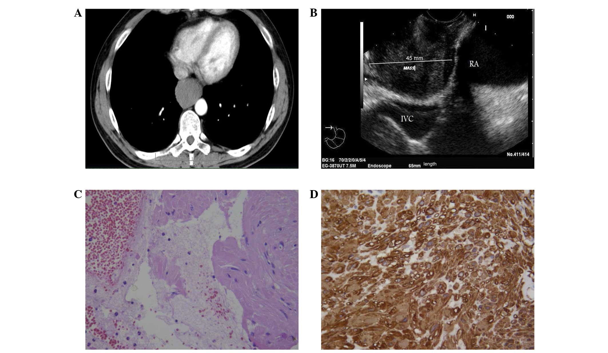
normal range. Computed tomography (CT) scans of the chest revealed a hypodense mass developing in the distal esophagus and causing substenosis of the lumen, which was extended to 65 mm in length, with non-homogeneous contrast enhancement (Fig. 1A). EUS examination revealed a 45-mm hypoechoic, round lesion with well demarcated margins, originating from the muscle layer of the distal esophagus in contact with the inferior caval vein and right atrium (Fig. 1B). EUS-FNAC was performed by using a convex array echoendoscope (EG-3870 UTK; Pentax, Co., Ltd., Tokyo, Japan) and by making two passes with a 22G needle. The specimens were processed by an in-room cytopathologist and immediately examined for adequate cellularity following staining by hematoxylin and eosin. A second slide was immediately fixed in 98% ethanol and stained with Papanicolaou. Any excess materials, including the needle and syringe utilized in the procedure, were rinsed in 10 ml 50% ethanol in a specimen container. All content was centrifuged in a 10-ml disposable centrifuge tube at 5,017 × g for 6 min to create 1 or 2 pellets; the supernatant fluid was decanted and the pelleted material was immediately fixed in a freshly prepared solution of 4% neutral buffered formalin for 45 min. The cell pellets were then placed in a cassette and stored at 80% ethanol until ready for processing in an automatic tissue processor (Leica TP1020; Leica, Buckinghamshire, UK). The cell blocks obtained were embedded in paraffin at 56°C, and 3-μm thick successive sections were cut and routinely stained by hematoxylin and eosin; parallel serial sections of the same thickness were mounted on silane-coated glasses and submitted to immunohistochemical procedures, as described previously (16,17).

Figure 1

Case 1: (A) CT scan showing a hypodense mass with non-homogeneous contrast enhancement developing from the distal esophagus causing sub-stenosis of the lumen. (B) EUS scanning results revealing a 45-mm, hypoechoic round mass, originating from the muscle layer in contact with the inferior vena cava (IVC) and the right atrium (RA). (C) Cytological smear results exhibiting aggregates of spindle cell elements with elongated nuclei (hematoxylin and eosin staining; magnification, ×160). (D) The same elements were intensely immunoreactive for SMA (immunoperoxidase and Mayer’s hemalum counterstain; magnification, ×200). CT, computed tomography; EUS, endoscopic ultrasound; SMA, smooth muscle actin.

Case 2

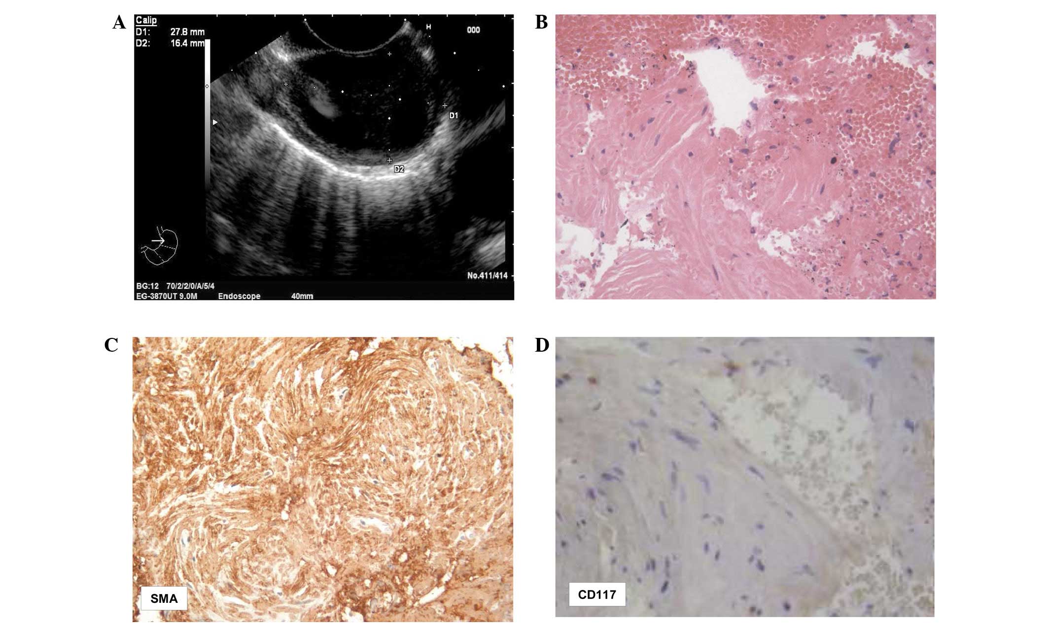
A 39-year-old female presented with dyspepsia and esophageal reflux that had been apparent for 4 weeks. There was no weight loss, but nausea and mild vomiting were occasionally present. Upon physical examination, local peri-gastric discomfort and pain were noted. EUS scanning showed a 27.8×16.4-mm ovoid, homogeneous and hypoechoic well-delimited mass originating from the esophageal sub-mucosa (Fig. 2A). No lesions were evident elsewhere in the abdominal organs or lymph nodes. EUS-FNAC was performed with the same procedure as utilized in case 1; again, adequate cellularity and one cell block were obtained.

Figure 2

Case 2: (A) EUS scanning results revealing a 27.8×16.4-mm, hypoechoic, ovoid, well-delimited lesion originating from the muscle layer of the distal esophagus. (B) Clusters of spindle cells intermingled with red blood cells are indicative of leiomyoma (hematoxylin and eosin staining; magnification, ×160). (C) These elements were reactive for SMA (immunoperoxidase and Mayer’s hemalum counterstain; magnification, ×120), (D) while no immunoreactivity was found with CD117 (immunoperoxidase and Mayer’s hemalum counterstain; magnification, ×160). EUS, endoscopic ultrasound; SMA, smooth muscle actin.

Following the FNAC procedures, the two patients were observed for a period of 48 h for any procedure-related complications.

Cytological and immunocytochemical findings

The smears from the two cases exhibited a hemorrhagic background, with loose clusters or small aggregates of spindle-shaped cells (Figs. 1C and 2B) that had elongated nuclei, occasionally showing finely granular chromatin. No mitotic figures were found. The corresponding cell blocks documented an equivalent morphology characterized by small tissue fragments, with relatively low to moderate cellularity composed of monomorphic-uniform spindle cells, eosinophilic cytoplasm and vesicular nuclei (Figs. 1C and 2B). The nuclear chromatin was finely granular and evenly dispersed, while micronucleoli were inconspicuous. No atypia or mitoses were noted.

Immunohistochemical procedures were carried out on the 3-μm serial sections, utilizing the following commercially obtained antisera (all DakoCytomation, Copenhagen, Denmark): Vimentin [working dilution (w.d.), 1:250], smooth muscle actin (SMA; w.d. 1:200), desmin (w.d., 1:250), CD117 (w.d., 1:150), CD34 (w.d. 1:200), S-100 (w.d., 1:400) and Ki67 (MIB-1; w.d., 1:50). In each of the two cases, strong and diffuse cytoplasmic immunostaining was encountered for vimentin, desmin and SMA (Figs. 1D and 2C). No immunostaining was recorded for S100, CD34 and CD117 (Fig. 2D). The growth fraction, determined using Ki67 as the MIB-1 labeling index, was extremely low and quite inconspicuous, showing <1% positively-labeled nuclei.

In light of the microscopic examination and immunohistochemical findings, the two esophageal lesions were diagnosed as intraparietal true leyomiomas, without atypia. The patients refused surgical procedures, and were lost to follow-up subsequent to a period of 12 months.

Discussion

It is well known that the diagnostic yield of EUS-FNAC greatly depends on the site, size and characteristics of the target tissues, as well as certain procedural aspects (9,15,18). By contrast, although conventional endoscopy and CT scans may identify esophageal lesions, these procedures cannot reveal the nature, size or origin of sub-mucosal neoplasms (7,9). However, the efficacy of EUS-FNAC as a main diagnostic procedure is also largely dependent on the expertise, training and interaction between the endosonographer and cytopathologist (15). In the present study, adequate cellular smears and corresponding cell blocks were obtained using the EUS-FNAC approach that is used on esophageal mesenchymal tumors, particularly true leiomyomas. Even if the observed spindle-shaped cells with elongated nuclei could also be confused with other gastrointestinal non-epithelial tumors, the serial immunohistochemical procedures performed on the cell blocks allowed acquisition of the final diagnosis. In fact, the coexistence of desmin and SMA strongly supported the smooth muscle nature of the observed esophageal neoplastic lesions, while the constant negativity for CD34, CD117 and S-100 excluded other diagnostic hypotheses, including inflammatory fibroid polyps, GISTs and schwannomas. Consequently, the availability of an adequate number of serial sections obtained from tissue blocks appears to be an additional diagnostic aid in order to perform the indicated immunohistochemical algorithm, as described previously (15,19,20). Finally, the low growth fraction, revealed by the Ki67 labeling index in the present study, further indicates the benign nature of leiomyomas, thus discounting the diagnostic hypotheses of highly malignant neoplasms, including leiomyosarcomas, spindle-cell amelanotic melanomas and undifferentiated sarcomatoid carcinomas (14,15).

Esophageal leiomyomas are rare benign tumors, with a frequent asymptomatic occurrence, that do not metastasize (21). In fact, patients with these tumors more commonly seek care due to difficulty in swallowing or as a result of the tumors being detected during the endoscopic workup for other diseases, as documented in case 2 of the present study. Moreover, the progression of these neoplasms shows a slow growing phase and the size of the lesions remains stable during the first year of follow-up. Therefore for those patients who refuse to receive surgical excision, as in the present cases, a periodic follow-up with EUS has been considered preferable and more accepted (22,23). On the other hand, the surgical treatment for esophageal leiomyomas depends on multiple factors, including tumor size, location, gross morphology and the patient’s symptoms and overall condition (21,24,25). Furthermore, indications for surgical treatment include unremitting symptoms, a progressive increase in tumor size, mucosal ulceration or the requirement to achieve the histopathological diagnosis due to an inconclusive EUS-FNAC procedure (25–27).

In summary, the present study provided further indications that EUS-FNAC has great clinico-diagnostic pre-surgical value, also allowing a correct differential diagnosis of other esophageal mesenchymal/stromal neoplasias with unpredictable biological behavior to be generated by immunohistochemistry.

References

1 

Matsui M, Goto H, Niwa Y, Arisawa T, Hirooka Y and Hayakawa T: Preliminary results of fine needle aspiration biopsy histology in upper gastrointestinal submucosal tumors. Endoscopy. 30:750–755. 1998.

2 

Miettinen M, Sarlomo-Rikala M, Sobin LH and Lasota J: Esophageal stromal tumors: a clinicopathologic, immunohistochemical, and molecular genetic study of 17 cases and comparson with esophageal leiomyomas and leiomyosarcomas. Am J Surg Pathol. 24:211–222. 2000.

3 

Miettinen M, Furlong M, Sarlomo-Rikala M, Burke A, Sobin LH and Lasota J: Gastrointestinal stromal tumors, intramural leiomyomas, and leiomyosarcomas in the rectum and anus: a clinicopathologic, immunohistochemical, and molecular genetic study of 144 cases. Am J Surg Pathol. 25:1121–33. 2001.

4 

Wieczorek TJ, Faquin WC, Rubin BP and Cibas ES: Cytologic diagnosis of gastrointestinal stromal tumor with emphasis on the differential diagnosis with leiomyosarcoma. Cancer. 93:276–287. 2001.

5 

Ando N, Goto H, Niwa Y, Hirooka Y, Ohmiya N, Nagasaka T and Hayakawa T: The diagnosis of GI stromal tumors with EUS-guided fine needle aspiration with immunohistochemical analysis. Gastrointest Endosc. 55:37–43. 2002.

6 

Trupiano JK, Stewart RE, Misick C, Appelman HD and Goldblum JR: Gastric stromal tumors: a clinicopathologic study of 77 cases with correlation of features with nonaggressive and aggressive clinical behaviors. Am J Surg Pathol. 26:705–714. 2002.

7 

Stelow EB, Stanley MW, Mallery S, Lai R, Linzie BM and Bardales RH: Endoscopic ultrasound-guided fine-needle aspiration findings of gastrointestinal leiomyomas and gastrointestinal stromal tumors. Am J Clin Pathol. 119:703–708. 2003.

8 

Stelow EB, Jones DR and Shami VM: Esophageal leiomyosarcoma diagnosed by endoscopic ultrasound-guided fine-needle aspiration. Diagn Cytopathol. 35:167–170. 2007.

9 

Jenssen C and Dietrich CF: Endoscopic ultrasound-guided fine-needle aspiration biopsy and trucut biopsy in gastroenterology - An overview. Brest Pract Res Clin Gastroenterol. 23:743–759. 2009.

10 

Fletcher CD, Berman JJ, Corless C, et al: Diagnosis of gastrointestinal stromal tumors: A consensus approach. Hum Pathol. 33:459–465. 2002.

11 

Blay JY, Bonvalot S, Casali P, et al; GIST consensus meeting panelists. Consensus meeting for the management of gastrointestinal stromal tumors. Report of the GIST Consensus Conference of 20–21 March 2004, under the auspices of ESMO. Ann Oncol. 16:566–578. 2005.

12 

Nilsson B, Bümming P, Meis-Kindblom JM, et al: Gastrointestinal stromal tumors: the incidence, prevalence, clinical course, and prognostication in the preimatinib mesylate era - a population-based study in western Sweden. Cancer. 103:821–829. 2005.

13 

Blanke CD, Demetri GD, von Mehren M, et al: Long-term results from a randomized phase II trial of standard- versus higher-dose imatinib mesylate for patients with unresectable or metastatic gastrointestinal stromal tumors expressing KIT. J Clin Oncol. 26:620–625. 2008.

14 

Maheshwari V, Alam K, Varshney M, Jain A, Asif Siddiqui F and Bhargava S: Fine-needle aspiration diagnosis of GIST: a diagnostic dilemma. Diagn Cytopathol. 40:834–838. 2012.

15 

Todaro P, Crinò SF, Pallio S, Fazzari C, Consolo P and Tuccari G: Gastrointestinal stromal tumors of the stomach: Cytological and immunocytochemical diagnostic features of two cases diagnosed by endoscopic ultrasound-guided fine needle aspiration. Oncol Lett. 5:1862–1866. 2013.

16 

Nathan NA, Narayan E, Smith MM and Horn MJ: Cell block cytology. Improved preparation and its efficacy in diagnostic cytology. Am J Clin Pathol. 114:599–606. 2000.

17 

Barresi V, Cerasoli S, Paioli G, Vitarelli E, Giuffrè G, Guiducci G, Tuccari G and Barresi G: Caveolin-1 in meningiomas: expression and clinico-pathological correlations. Acta Neuropathol. 112:617–626. 2006.

18 

Yoshinaga S, Suzuki H, Oda I and Saito Y: Role of endoscopic ultrasound-guided fine needle aspiration (EUS-FNA) for diagnosis of solid pancreatic masses. Dig Endosc. 23(Suppl 1): 29–33. 2011.

19 

Wong DW, Lupton SC, Bhatt L, Gross L, Tanière P, Peake DR, Spooner D and Geh JI: Use of imatinib mesylate in gastrointestinal stromal tumors: Pan-Birmingham Cancer Network experience. Clin Oncol (R Coll Radiol). 20:517–522. 2008.

20 

Reid R, O’Dywer P, MacDuff E, et al: Guidelines for the management of gastrointestinal stromal tumors (GIST) in Scotland. pp. 53–55. 2009, http://www.pathologyscotland.org/download/sgpg/guidelines/gist.pdf. Accessed November 13, 2012

21 

Jiang W, Rice TW and Goldblum JR: Esophageal leiomyoma: experience from a single institution. Dis Esophagus. 26:167–174. 2013.

22 

Choi SH, Kim YT, Han KN, Ra YJ, Kang CH, Sung SW and Kim JH: Surgical management of the esophageal leiomyoma: lessons from a retrospective review. Dis Esophagus. 24:325–329. 2011.

23 

Xu GQ, Qian JJ, Chen MH, Ren GP and Che HT: Endoscopic ultrasonography for the diagnosis and selecting treatment of esophageal leiomyoma. J Gastroenterol Hepatol. 27:521–525. 2012.

24 

Hatch GF 3rd, Wertheimer-Hatch L, Hatch KF, Davis GB, Blanchard DK, Foster RS Jr and Skandalakis JE: Tumors of the esophagus. World J Surg. 24:401–411. 2000.

25 

Lee LS, Singhal S, Brinster CJ, Marshall B, Kochman ML, Kaiser LR and Kucharczuk JC: Current management of esophageal leiomyoma. J Am Coll Surg. 198:136–146. 2004.

26 

Maish M: Esophagus. Sabiston Textbook of Surgery. Townsend CM Jr, Beauchamp RD, Ever BM and Mattox KL: 18th edition. Saunders; Philadelphia, PA: pp. 1087–1088. 2007

27 

Jiang G, Zhao H, Yang F, Li J, Li Y, Liu Y, Liu J and Wang J: Thoracoscopic enucleation of esophageal leiomyoma: a retrospective study on 40 cases. Dis esophagus. 22:279–283. 2009.

Related Articles

  • Abstract
  • View
  • Download
  • Twitter
Copy and paste a formatted citation
Spandidos Publications style
Todaro P, Crinò SF, Ieni A, Pallio S, Consolo P and Tuccari G: Intraparietal esophageal leiomyomas diagnosed by endoscopic ultrasound‑guided fine‑needle aspiration cytology: Cytological and immunocytochemical features in two cases. Oncol Lett 8: 123-126, 2014.
APA
Todaro, P., Crinò, S.F., Ieni, A., Pallio, S., Consolo, P., & Tuccari, G. (2014). Intraparietal esophageal leiomyomas diagnosed by endoscopic ultrasound‑guided fine‑needle aspiration cytology: Cytological and immunocytochemical features in two cases. Oncology Letters, 8, 123-126. https://doi.org/10.3892/ol.2014.2077
MLA
Todaro, P., Crinò, S. F., Ieni, A., Pallio, S., Consolo, P., Tuccari, G."Intraparietal esophageal leiomyomas diagnosed by endoscopic ultrasound‑guided fine‑needle aspiration cytology: Cytological and immunocytochemical features in two cases". Oncology Letters 8.1 (2014): 123-126.
Chicago
Todaro, P., Crinò, S. F., Ieni, A., Pallio, S., Consolo, P., Tuccari, G."Intraparietal esophageal leiomyomas diagnosed by endoscopic ultrasound‑guided fine‑needle aspiration cytology: Cytological and immunocytochemical features in two cases". Oncology Letters 8, no. 1 (2014): 123-126. https://doi.org/10.3892/ol.2014.2077
Copy and paste a formatted citation
x
Spandidos Publications style
Todaro P, Crinò SF, Ieni A, Pallio S, Consolo P and Tuccari G: Intraparietal esophageal leiomyomas diagnosed by endoscopic ultrasound‑guided fine‑needle aspiration cytology: Cytological and immunocytochemical features in two cases. Oncol Lett 8: 123-126, 2014.
APA
Todaro, P., Crinò, S.F., Ieni, A., Pallio, S., Consolo, P., & Tuccari, G. (2014). Intraparietal esophageal leiomyomas diagnosed by endoscopic ultrasound‑guided fine‑needle aspiration cytology: Cytological and immunocytochemical features in two cases. Oncology Letters, 8, 123-126. https://doi.org/10.3892/ol.2014.2077
MLA
Todaro, P., Crinò, S. F., Ieni, A., Pallio, S., Consolo, P., Tuccari, G."Intraparietal esophageal leiomyomas diagnosed by endoscopic ultrasound‑guided fine‑needle aspiration cytology: Cytological and immunocytochemical features in two cases". Oncology Letters 8.1 (2014): 123-126.
Chicago
Todaro, P., Crinò, S. F., Ieni, A., Pallio, S., Consolo, P., Tuccari, G."Intraparietal esophageal leiomyomas diagnosed by endoscopic ultrasound‑guided fine‑needle aspiration cytology: Cytological and immunocytochemical features in two cases". Oncology Letters 8, no. 1 (2014): 123-126. https://doi.org/10.3892/ol.2014.2077
Follow us
  • Twitter
  • LinkedIn
  • Facebook
About
  • Spandidos Publications
  • Careers
  • Cookie Policy
  • Privacy Policy
How can we help?
  • Help
  • Live Chat
  • Contact
  • Email to our Support Team