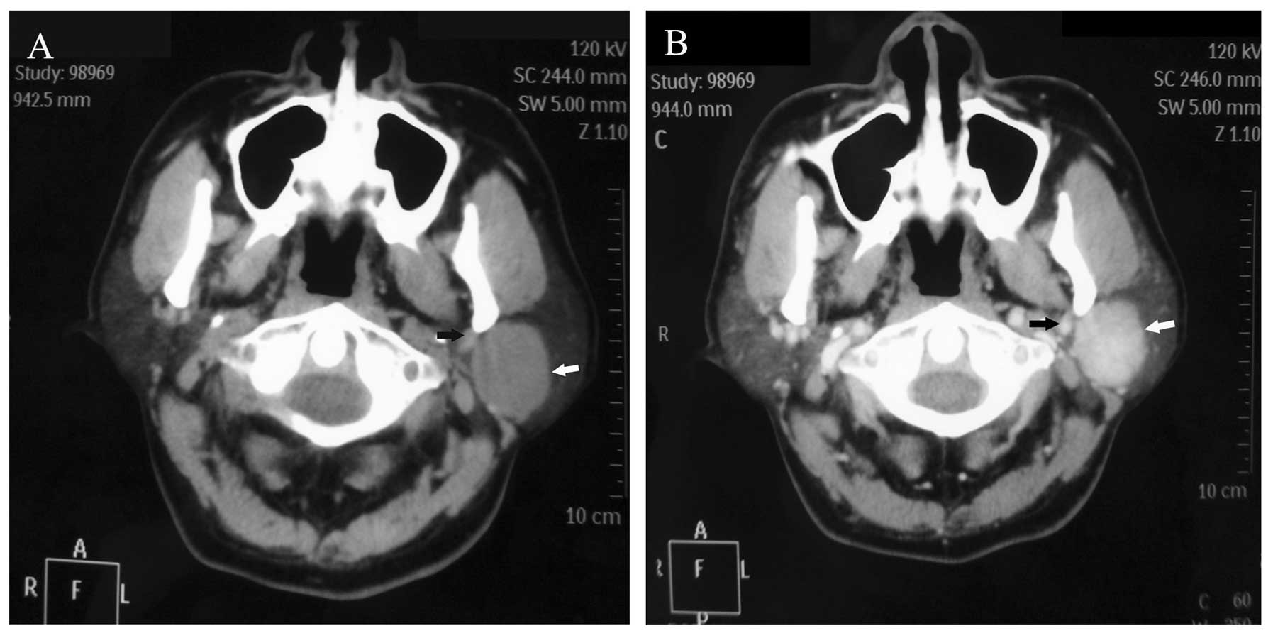
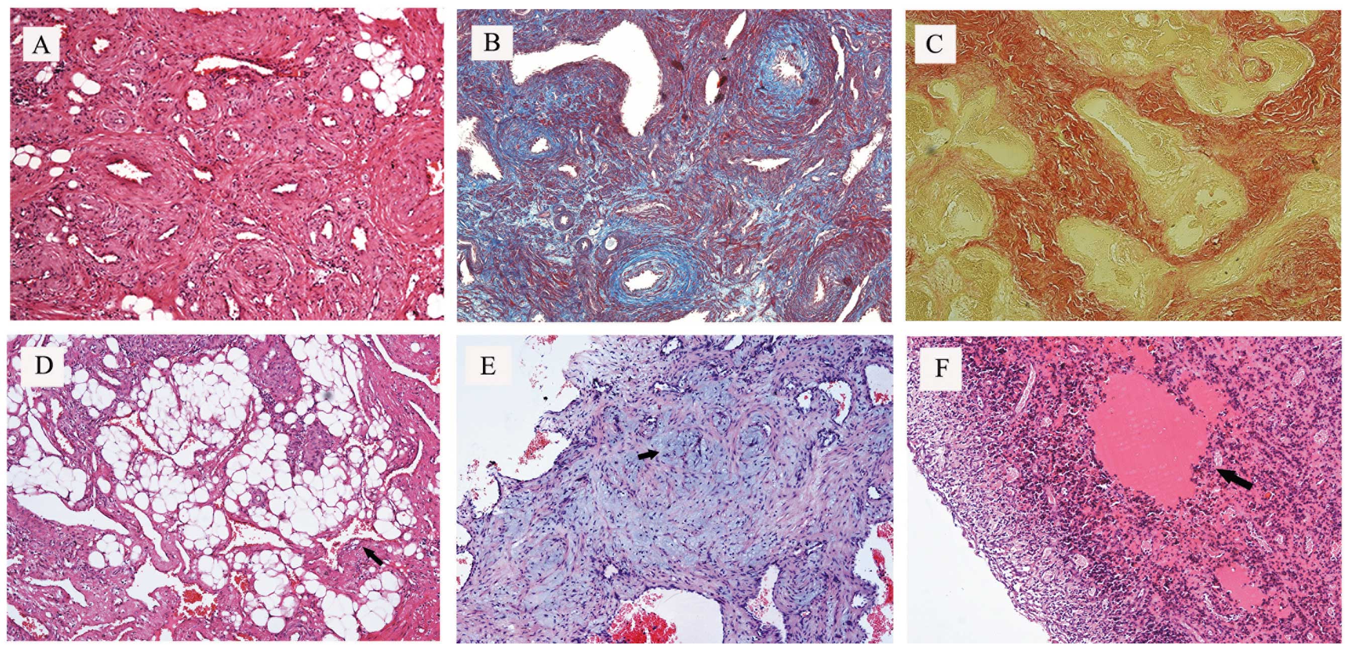
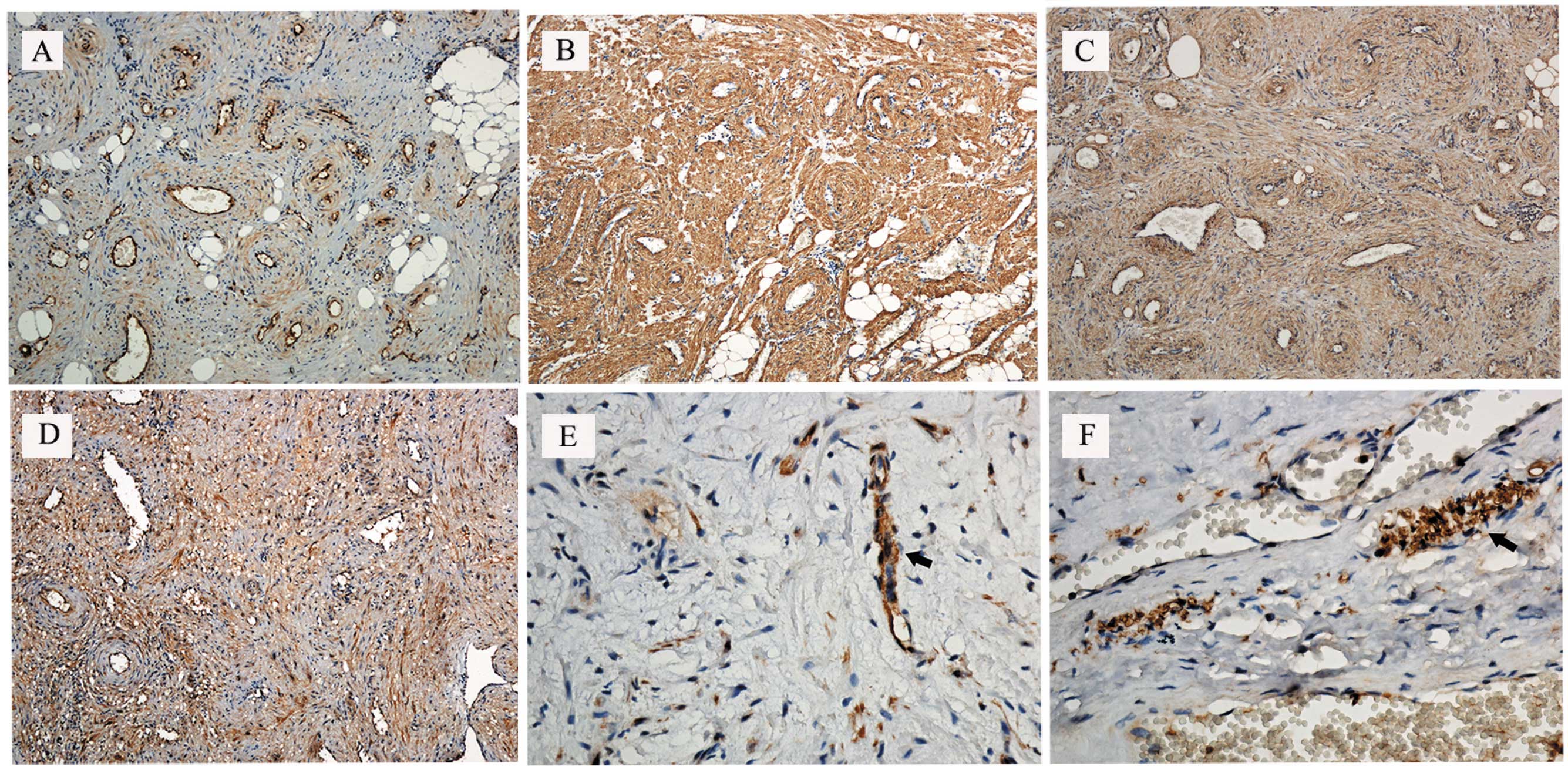
|
1
|
Hachisuga T, Hashimoto H and Enjoji M:
Angioleiomyoma. A clinicopathologic reappraisal of 562 cases.
Cancer. 54:126–130. 1984.
|
|
2
|
Enzinger FM and Weiss SW: Angioleiomyoma
(vascular leiomyoma). Soft Tissue Tumors. 3rd edition. Mosby; St.
Louis, MO: pp. 467–470. 1995
|
|
3
|
Brooks JK, Nikitakis NG, Goodman NJ and
Levy BA: Clinicopathologic characterization of oral
angioleiomyomas. Oral Surg Oral Med Oral Pathol Oral Radiol Endod.
94:221–227. 2002.
|
|
4
|
Wertheimer-Hatch L, Hatch GF 3rd, Hatch
BSK, et al: Tumors of the oral cavity and pharynx. World J Surg.
24:395–400. 2000.
|
|
5
|
Baden E, Doyle JL and Lederman DA:
Leiomyoma of the oral cavity: a light microscopic and
immunohistochemical study with review of the literature from 1884
to 1992. Eur J Cancer B Oral Oncol. 30B:1–7. 1994.
|
|
6
|
Cherrick HM, Dunlap CL and King OH Jr:
Leiomyomas of the oral cavity. Review of the literature and
clinicopathologic study of seven new cases. Oral Surg Oral Med Oral
Pathol. 35:54–66. 1973.
|
|
7
|
Morimoto N: Angiomyoma (vascular
leiomyoma): a clinicopathologic study. Med J Kagoshima Univ.
24:663–683. 1973.
|
|
8
|
MacDonald DM and Sanderson KV:
Angioleiomyoma of the skin. Br J Dermatol. 91:161–168. 1974.
|
|
9
|
Ramesh P, Annapureddy SR, Khan F and
Sutaria PD: Angioleiomyoma: a clinical, pathological and
radiological review. Int J Clin Pract. 58:587–591. 2004.
|
|
10
|
McGuff HS, Jones AC and Ellis E 3rd: Oral
and maxillofacial pathology case of the month. Angiomyoma (vascular
leiomyoma). Tex Dent J. 129:454–455. 2012.
|
|
11
|
Gaitan Cepeda LA, Quezada Rivera D,
Tenorio Rocha F, Leyva Huerta ER and Mendez Sánchez ER: Vascular
leiomyoma of the oral cavity. Clinical, histopathological and
immunohistochemical characteristics Presentation of five cases and
review of the literature. Med Oral Pathol Oral Cir Bucal.
13:E483–E488. 2008.
|
|
12
|
Duhig JT and Ayer JP: Vascular leiomyoma.
A study of sixty one cases. Arch Pathol. 68:424–430. 1959.
|
|
13
|
Wang CP, Chang YL and Sheen TS: Vascular
leiomyoma of the head and neck. Laryngoscope. 114:661–665.
2004.
|
|
14
|
Esguep A and Solar M: Oral vascular
leiomyoma - report of 5 cases and review of the literature. J Oral
Med. 41:126–129. 1986.
|
|
15
|
Kim YH, Jang YW, Pai H and Kim SG:
Congenital angiomyoma of the tongue: case report. Dentomaxillofac
Radiol. 39:446–448. 2010.
|
|
16
|
Epivatianos A, Trigonidis G and
Papanayotou P: Vascular leiomyoma of the oral cavity. J Oral
Maxillofac Surg. 43:377–382. 1985.
|
|
17
|
Wong SK, Ahuja A, Chow J and King WW:
Angioleiomyoma in the submandibular region: an unusual tumor in an
unusual site. Otolaryngol Head Neck Surg. 122:144–145. 2000.
|
|
18
|
Natiella JR, Neiders ME and Greene GW:
Oral leiomyoma. Report of six cases and a review of the literature.
J Oral Pathol. 11:353–365. 1982.
|
|
19
|
Kido T and Sekitani T: Vascular leiomyoma
of the parotid gland. ORL J Otorhinolaryngol Relat Spec.
51:187–191. 1989.
|
|
20
|
Ide F, Mishima K and Saito I: Angiomyoma
in the submandibular gland: a rare location for a ubiquitous
tumour. J Laryngol Otol. 117:1001–1002. 2003.
|
|
21
|
McDaniel RK: Benign mesenchymal neoplasms.
Surgical Pathology of the Salivary Glands. Ellis GL, Auclair P and
Gnepp DR: 2nd edition. WB Saunders; Philadelphia: pp. 489–513.
1991
|
|
22
|
Toida M, Koizumi H and Shimokawa K:
Painful angiomyoma of the oral cavity: report of a case and review
of the literature. J Oral Maxillofac Surg. 58:450–453. 2000.
|
|
23
|
Akizawa S: Angiomyoma: an analysis of 124
cases. Jikeikai Med J. 27:71–82. 1980.
|
|
24
|
Montgomery H and Winkelmann RK:
Smooth-muscle tumours of the skin. AMA Arch Derm. 79:32–40.
1959.
|
|
25
|
Stout AP: Solitary cutaneous and
subcutaneous leiomyoma. Am J Cancer. 29:435–469. 1937.
|
|
26
|
Magner D and Hill DP: Encapsulated
angiomyoma of the skin and subcutaneous tissues. Am J Clin Pathol.
35:137–141. 1961.
|
|
27
|
Fox SB, Heryet A and Khong TY:
Angioleiomyomas: an immunohistological study. Histopathology.
16:495–496. 1990.
|
|
28
|
Stoller DW, Steinkirchner TM and Porter
BA: Bone and soft tissue tumors. Magnetic Resonance Imaging in
Orthopaedics and Sports Medicine. Stoller DW: 2nd edition. JB
Lippincott; Philadelphia: pp. 1092–1093. 1993
|
|
29
|
Eley KA, Alroyayamina S, Golding SJ, Tiam
RN and Watt-Smith SR: Angioleiomyoma of the hard palate: report of
a case and review of the literature and magnetic resonance imaging
findings of this rare entity. Oral Surg Oral Med Oral Pathol Oral
Radiol. 114:e45–e49. 2012.
|
|
30
|
Bouquot JE and Nikai H: Lesions of the
oral cavity. Diagnostic Surgical Pathology of the Head and Neck.
Gnepp DR: 1st edition. WB Saunders; Philadelphia: pp. 141–233.
2001
|
|
31
|
Kawakami T, Hasegawa H and Chino T: A
transmission electron microscopic study of two cases of oral smooth
muscle neoplasm. J Oral Maxillofac Surg. 45:551–555. 1987.
|
|
32
|
Nikitakis NG, Lopes MA, Bailey JS,
Blanchaert RH Jr, Ord RA and Sauk JJ: Oral leiomyosarcoma: review
of the literature and report of two cases with assessment of the
prognostic and diagnostic significance of immunohistochemical and
molecular markers. Oral Oncol. 38:201–208. 2002.
|
|
33
|
Keerthi R, Nanjappa M, Deora SS and
Kumaraswamy SV: Angioleiomyoma of cheek: report of two cases. J
Maxillofac Oral Surg. 8:298–300. 2009.
|
|
34
|
Svane TJ, Smith BR, Cosentino BJ, Cundiff
EJ and Ceravolo JJ Jr: Oral leiomyomas. Review of the literature
and report of a case of palatal angioleiomyoma. J Periodontol.
57:433–435. 1986.
|
|
35
|
Hirshoren N, Weinberger JM, Neuman T, Ilan
O and Ben-Yaakov A: Recurrent vascular leiomyoma of the larynx:
clinical and histopathologic characteristics and treatment. Ear
Nose Throat J. 89:382–386. 2010.
|