Evaluation of gastric submucosal tumors using endoscopically visualized features with submucosal endoscopy

  • Authors:
    • Hideki Kobara
    • Hirohito Mori
    • Kazi Rafiq
    • Tae Matsunaga
    • Shintaro Fujihara
    • Noriko Nishiyama
    • Maki Ayaki
    • Tatsuo Yachida
    • Johji Tani
    • Hisaaki Miyoshi
    • Kiyohito Kato
    • Hideki Kamada
    • Hirohito Yoneyama
    • Asahiro Morishita
    • Kunihiko Tsutsui
    • Hisakazu Iwama
    • Reiji Haba
    • Tsutomu Masaki
  • View Affiliations

  • Published online on: May 8, 2014     https://doi.org/10.3892/ol.2014.2126
  • Pages: 161-168
Metrics: Total Views: 0 (Spandidos Publications: | PMC Statistics: )
Total PDF Downloads: 0 (Spandidos Publications: | PMC Statistics: )


Abstract

Although the macroscopic characteristics of submucosal tumors (SMTs), such as gastrointestinal stromal tumors (GISTs), have been characterized, the assessment of SMTs by their endoscopically visualized features (EVF; which are observed by endoscopic imaging under direct view) remains unevaluated. The aim of the present study was to investigate the potential of endoscopic diagnostics for SMTs using EVF. The EVF of 26 gastric SMT cases, in which the final pathological diagnosis was obtained by core biopsy using the submucosal endoscopy with mucosal flap method, were retrospectively reviewed. Each type of SMT was classified according to the following five EVF: Color, clarity, shape, tumor coating and solidity. Additionally, the EVF of 13 low‑risk GISTs and 13 benign submucosal tumors (BSTs) were comparatively evaluated for the five abovementioned EVF. Similar trends were identified between the low‑risk GISTs, granular cell tumors and the schwannoma with regard to EVF. However, while these tumors exhibited cloudy EVF, the leiomyomas tended to exhibit clear EVF. Among SMTs of the heterotopic pancreas type, the EVF demonstrated particularly small nodules of the pancreatic tissue itself. Although the sample size included in the present study is small, a classification system for gastric SMTs was proposed according to the EVF. When compared with the BST group, the GIST group demonstrated a significantly higher frequency of tumors that exhibited a combination of three EVF (white, cloudy and rigid) that are consistent with all gastric GISTs (P<0.05). Gastric SMTs may be classified based on the EVF, which indicates that the EVF possess potential diagnostic value for the differentiation of GISTs from BSTs.
View Figures
View References

Related Articles

Journal Cover

July-2014
Volume 8 Issue 1

Print ISSN: 1792-1074
Online ISSN:1792-1082

Sign up for eToc alerts

Recommend to Library

Copy and paste a formatted citation
x
Spandidos Publications style
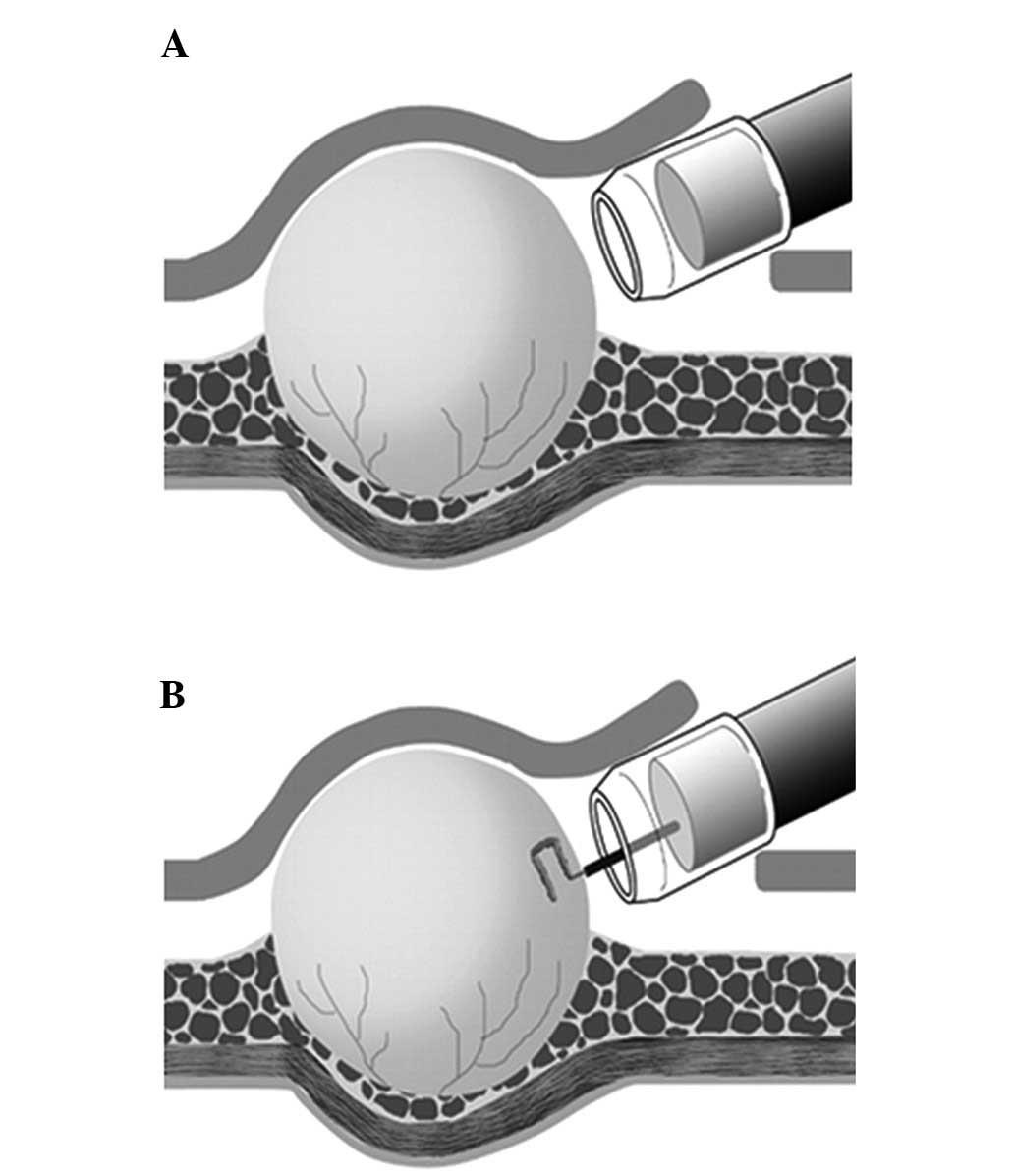
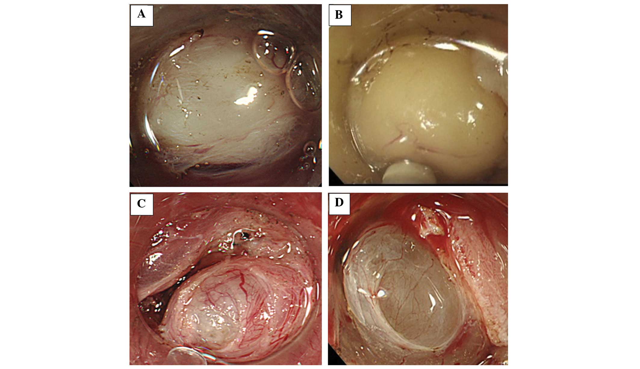
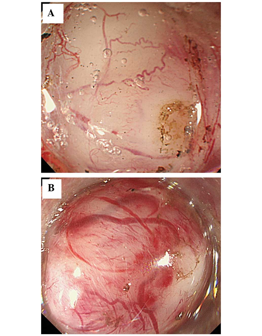
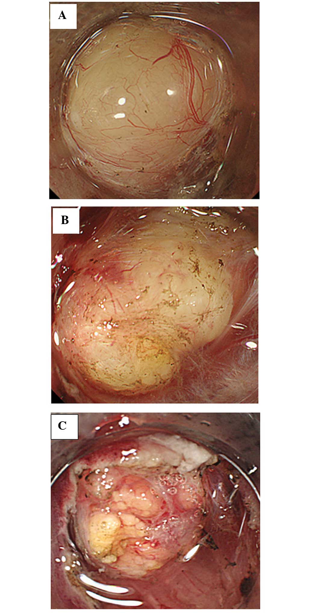
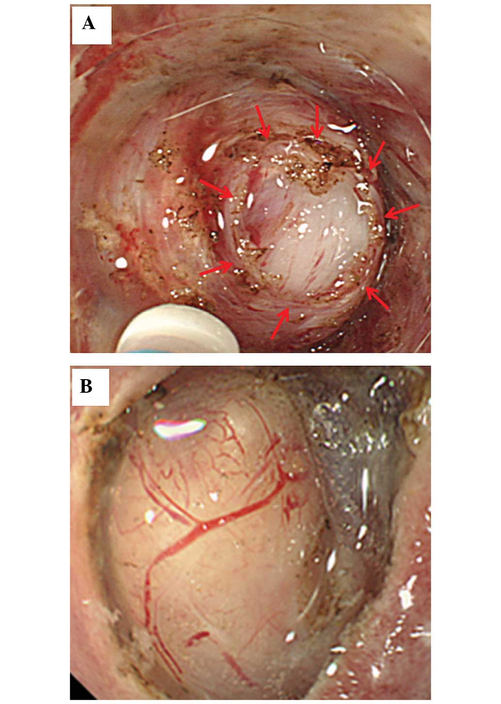
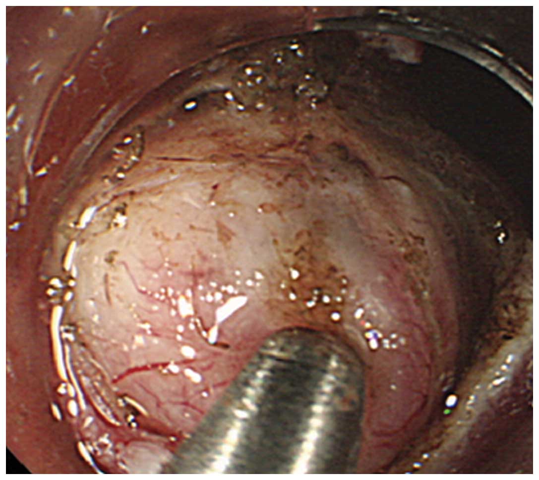
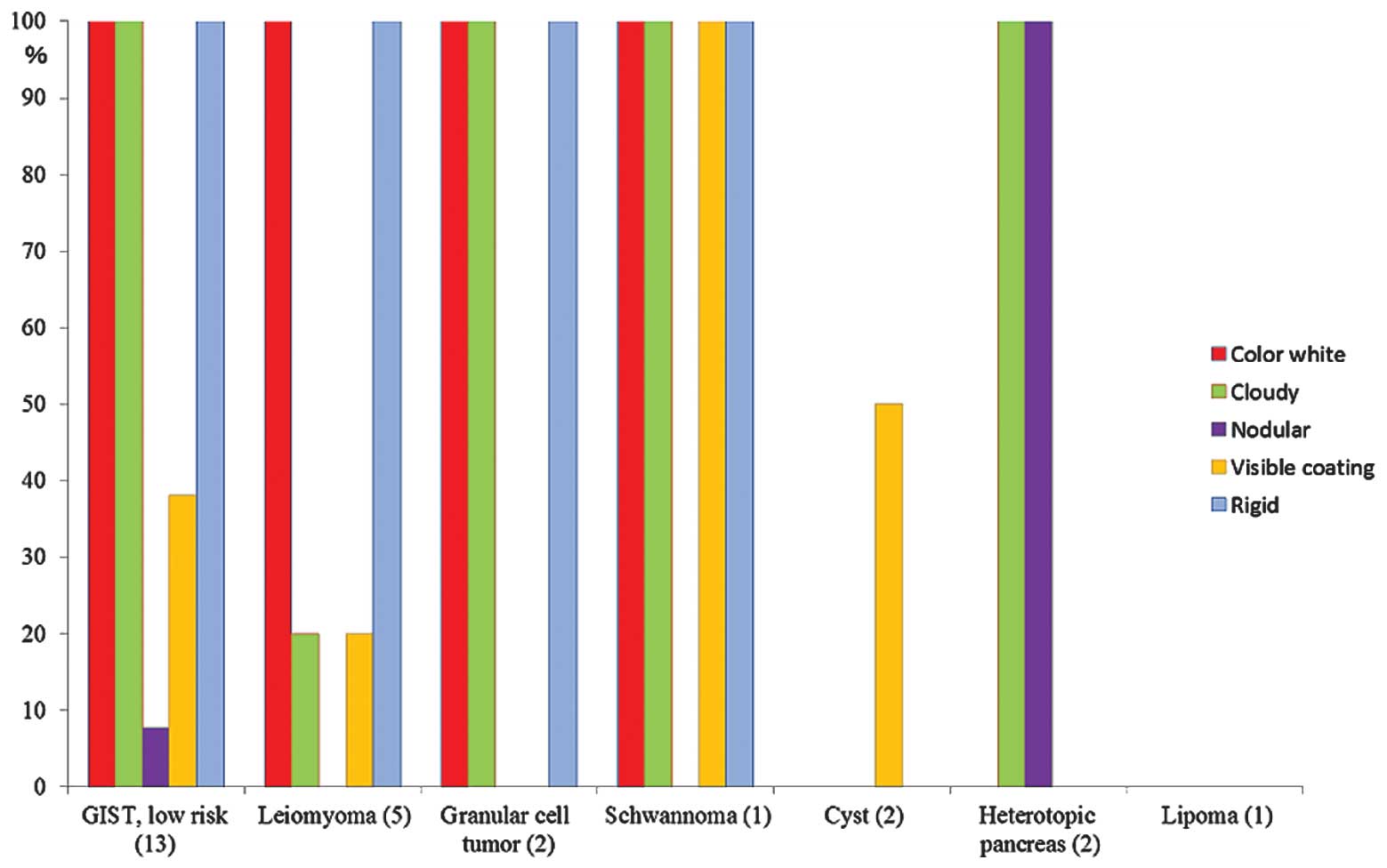
Kobara H, Mori H, Rafiq K, Matsunaga T, Fujihara S, Nishiyama N, Ayaki M, Yachida T, Tani J, Miyoshi H, Miyoshi H, et al: Evaluation of gastric submucosal tumors using endoscopically visualized features with submucosal endoscopy. Oncol Lett 8: 161-168, 2014
APA
Kobara, H., Mori, H., Rafiq, K., Matsunaga, T., Fujihara, S., Nishiyama, N. ... Masaki, T. (2014). Evaluation of gastric submucosal tumors using endoscopically visualized features with submucosal endoscopy. Oncology Letters, 8, 161-168. https://doi.org/10.3892/ol.2014.2126
MLA
Kobara, H., Mori, H., Rafiq, K., Matsunaga, T., Fujihara, S., Nishiyama, N., Ayaki, M., Yachida, T., Tani, J., Miyoshi, H., Kato, K., Kamada, H., Yoneyama, H., Morishita, A., Tsutsui, K., Iwama, H., Haba, R., Masaki, T."Evaluation of gastric submucosal tumors using endoscopically visualized features with submucosal endoscopy". Oncology Letters 8.1 (2014): 161-168.
Chicago
Kobara, H., Mori, H., Rafiq, K., Matsunaga, T., Fujihara, S., Nishiyama, N., Ayaki, M., Yachida, T., Tani, J., Miyoshi, H., Kato, K., Kamada, H., Yoneyama, H., Morishita, A., Tsutsui, K., Iwama, H., Haba, R., Masaki, T."Evaluation of gastric submucosal tumors using endoscopically visualized features with submucosal endoscopy". Oncology Letters 8, no. 1 (2014): 161-168. https://doi.org/10.3892/ol.2014.2126