Introduction
The incidence of penile cutaneous horn has been
particularly low since the first case was reported (1). It is a lesion that exhibits
hyperkeratotic features on the penile glans. These hyperkeratoses
are superimposed on a wide variety of benign, premalignant or
malignant lesions (2). In the
current report, a case of a 43-year-old male is presented. The
patient underwent surgical excision of the lesion that indicated a
penile cutaneous horn, this subsequently progressed to squamous
cell carcinoma in <1.5 months. Finally, a partial penectomy was
performed. Patient provided written informed consent.
Case report
In February 2013, a 43-year-old male presented to
the Department of Urology, The Third Xiangya Hospital of Central
South University (Changsha, China) with a six-month history of a
progressive growth in the glans penis. The patient had undergone a
circumcision for phimosis six months previously and subsequently
identified a yellow lesion projecting out from the left aspect of
the glans penis. The patient experienced no pain and disregarded
the lesion, which grew rapidly and prevented the patient from
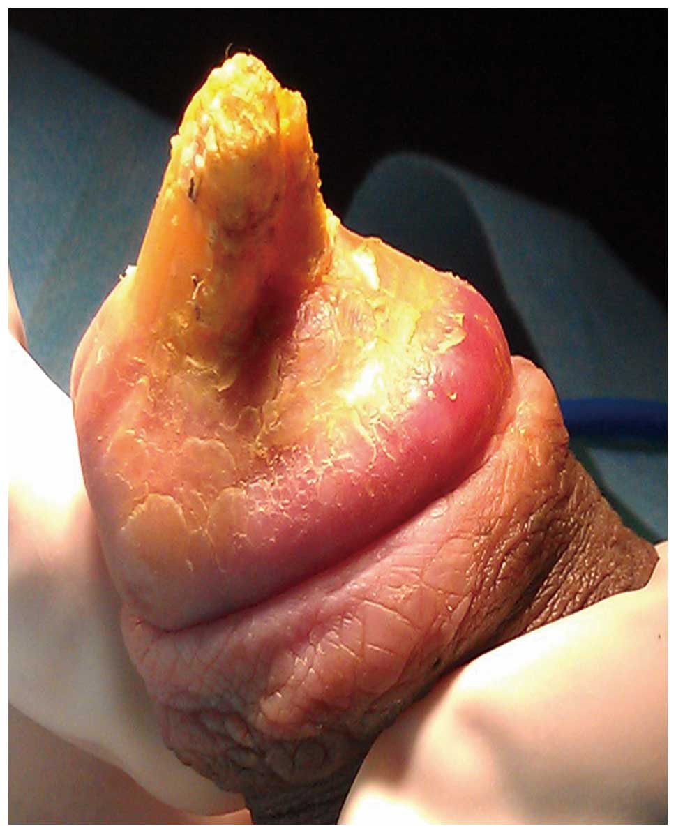
engaging in sexual intercourse. Examination of the patient revealed
a horn-like keratinized lesion of ~2.0×1.0×0.6 cm in the glans
penis with no inguinal lymphadenopathy (Fig. 1). The International Index of
Erectile Function 5 (IIEF-5) questionnaire was used for assessment
and the patient’s score was 18 (mild) (3). As the patient was initially reluctant
to undergo a partial penectomy, a surgical excision of the lesion
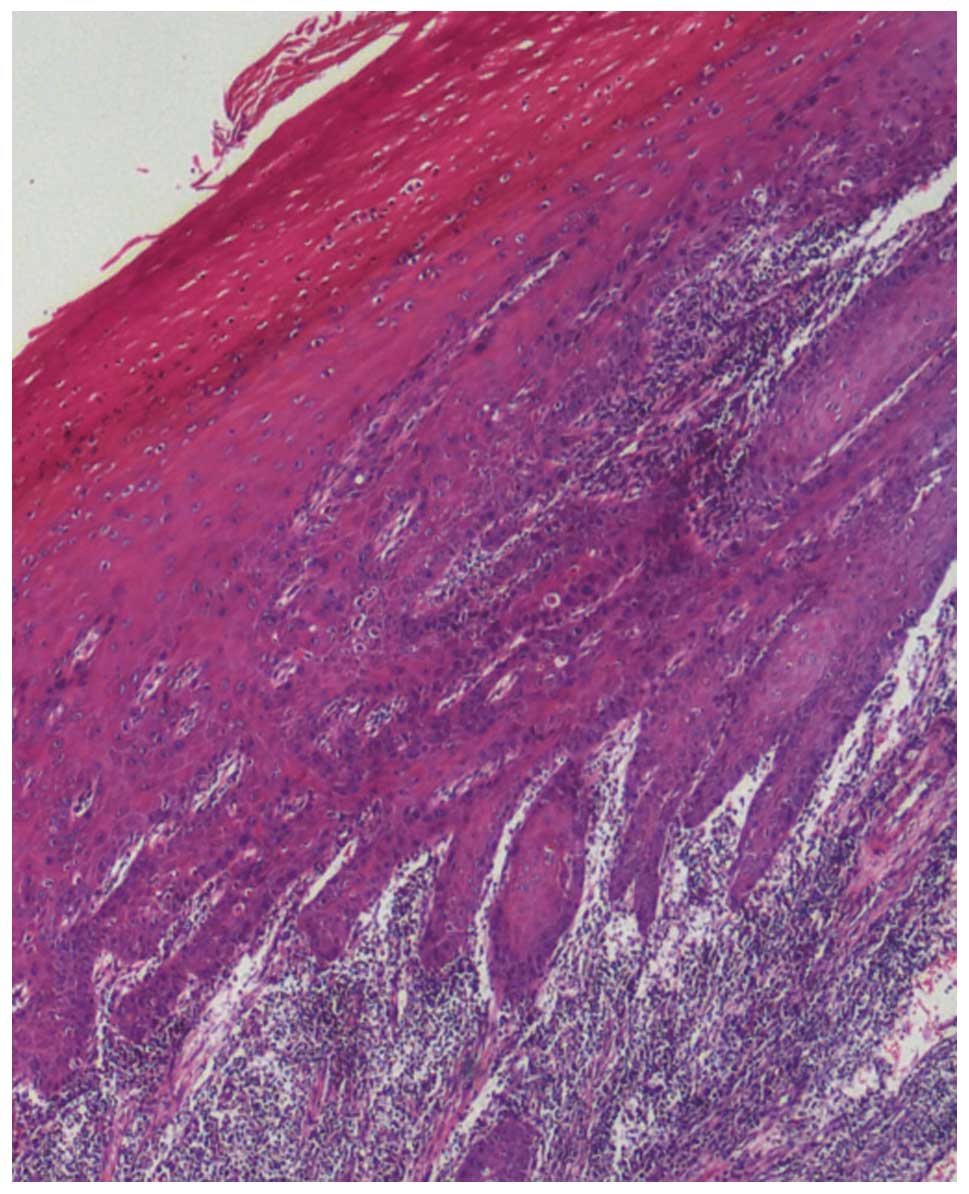
was performed. Histopathology the lesion demonstrated extreme
hyperkeratosis, dyskeratosis and epithelial hyperplasia (Fig. 2). The patient’s postoperative
recovery was uneventful.
The first postoperative visit was conducted on day
39 following surgery, the patient presented with a painful swelling
over the glans penis and purulent discharge of the urethra. On
examination, the glans penis was stiff and there was a ~5-mm
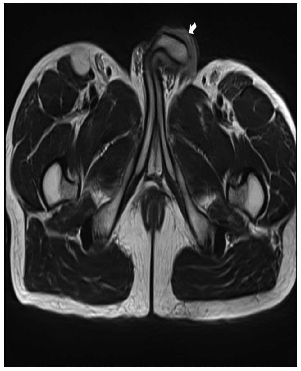
nodular lesion on the distal aspect of the penile shaft. Pelvic
magnetic resonance imaging (MRI) revealed a lesion with an unclear
boundary measuring 0.6 cm in diameter (Fig. 3). A biopsy was performed and
histopathology indicated squamous atypia and suspected squamous
cell carcinoma. Finally, the patient consented to a partial
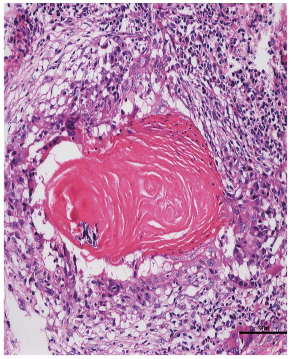
penectomy and histopathology revealed a well differentiated
squamous cell carcinoma (Fig. 4). A
six-month postoperative follow-up was conducted and the patient was
able to urinate whilst standing and the IIEF-5 score was 15
(moderate). Follow-up of the patient is ongoing.
Discussion
The etiology of penile cutaneous horn remains
uncertain since the first case was reported in 1854 (1). Viral and non-viral factors may be
implicated in penile cutaneous horn formation. Solivan et al
(4) identified a positive human
papillomavirus (HPV) reaction for HPV 16 using in situ DNA
hybridization; furthermore, Zhu et al (5) demonstrated that the HPV 16/18
infection was positive in a patient exhibiting a penile cutaneous
horn. These findings indicate that the HPV infection may be one of
the pathogens involved. In addition, the presentation of a penile
cutaneous horn may be associated with phimosis, warts and trauma,
amongst other conditions (6,7). To
the best of our knowledge, penile cutaneous horn usually occurs
following an adult circumcision (2). However in the present case, the
formation of the penile cutaneous horn preceded the patient’s
circumcision, as it was during the circumcision that the lesion was
identified. It was hypothesized in the present study that
continuous, chronic preputial inflammation aggravates the formation
of a penile cutaneous horn and the trauma of circumcision
accelerates its development.
The European Association of Urology guidelines on
penile cancer classify penile cutaneous horn as a premalignant
lesion (8) and approximately
one-third of penile cutaneous horns are associated with an
underlying malignancy (2). It has
previously been reported that MRI is helpful when there is
uncertainty regarding the depth of infiltration or proximal
extension (9). When the patient in
the present case was hospitalized for the second time, a physical
examination presented a nodular lesion on the distal aspect of the
penile shaft and the MRI result provided the basis of the nature of
the lesion.
As instances of penile cutaneous horn are
particularly rare and the majority of studies regarding them are
case reports, various treatment methods have been adopted,
including laser therapy, administration of keratolytic agents and
surgical excision (10,11). In the present report, a malignant
lesion did not exist when the patient was first diagnosed with a
penile cutaneous horn. However, a malignancy was present less than
1.5 months following surgery, thus a partial penectomy was
conducted. The IIEF-5 questionnaire, which has previously been used
to evaluate sexual function and satisfaction following a partial
penectomy (12), was used in the
present case; the patient’s IIEF-5 score in the preoperative period
was greater than the score six months postoperatively. Based on
these findings, undergoing a partial penectomy when a penile
cutaneous horn is initially diagnosed should be considered in order
to conserve a greater quantity of penile tissue and improve the
postoperative quality of life.
In conclusion, to the best of our knowledge, such a
rapid progression of a penile cutaneous horn to squamous cell
carcinoma has not previously been described. In addition, a decline
in the patient’s quality of life was noted. Thus, performance of a
partial penectomy should be considered as soon as a diagnosis of
penile cutaneous horn is determined.
References
|
1
|
Jewett PA: A case of horn on the glans
penis. Med Times. 3:91854.
|
|
2
|
Lowe FC and McCullough AR: Cutaneous horns
of the penis: an approach to management. Case report and review of
the literature. J Am Acad Dermatol. 13:369–373. 1985.
|
|
3
|
Rosen RC, Cappelleri JC, Smith MD, Lipsky
J and Peña BM: Development and evaluation of an abridged, 5-item
version of the International Index of Erectile Function (IIEF-5) as
a diagnostic tool for erectile dysfunction. Int J Impot Res.
11:319–326. 1999.
|
|
4
|
Solivan GA, Smith KJ and James WD:
Cutaneous horn of the penis: its association with squamous cell
carcinoma and HPV-16 infection. J Am Acad Dermatol. 23:969–972.
1990.
|
|
5
|
Zhu JW, Luo D, Li CR, Lu Y, Ji X, Zhu J,
Ming YL and Shen CH: A case of penile verrucous carcinoma
associated with cutaneous horn. Clin Exp Dermatol. 32:213–214.
2007.
|
|
6
|
Karthikeyan, Thappa DM, Jaisankar TJ,
Balamourougane, Ananthakrishnan N and Ratnakar C: Cutaneous horn of
glans penis. Sex Transm Infect. 74:456–457. 1998.
|
|
7
|
Agarwalla A, Agrawal CS, Thakur A, Garg
VK, Joshi A, Agrawal S and Jacob M: Cutaneous horn on condyloma
acuminatum. Acta Derm Venereol. 80:1592000.
|
|
8
|
Pizzocaro G, Algaba F, Horenblas S,
Solsona E, Tana S, Van Der Poel H and Watkin NA; European
Association of Urology (EAU) Guidelines Group on Penile Cancer. EAU
penile cancer guidelines 2009. Eur Urol. 57:1002–1012. 2010.
|
|
9
|
Lynch DF and Pettaway CA: Tumors of the
penis. Campbell’s Urology. Walsh PC, Retik AB, Vaughan ED, et al:
4. 8th edition. W.B. Saunders; Philadelphia, PA: pp. 2453–2485.
2002
|
|
10
|
Mastrolorenzo A, Tiradritti L, Locunto U,
Carini M, Massi D and Zuccati G: Incidental finding: a penile
cutaneous horn. Acta Derm Venereol. 85:283–284. 2005.
|
|
11
|
Schellhammer PF, Jordan GH, Robey EL and
Spaulding JT: Premalignant lesions and nonsquamous malignancy of
the penis and carcinoma of the scrotum. Urol Clin North Am.
19:131–142. 1992.
|
|
12
|
Romero FR, Romero KR, Mattos MA, Garcia
CR, de Fernandes RC and Perez MD: Sexual function after partial
penectomy for penile cancer. Urology. 66:1292–1295. 2005.
|