Introduction
Claudins are tight junctional proteins which are
present at epithelial and endothelial cell membranes (1,2). Tight
junctions form the primary barrier to the paracellular transport of
solutes across the cells, and also play a critical role in
establishing and maintaining epithelial cell polarity (3,4).
Claudins are the major integral membrane proteins forming the
backbone of tight junctions. The claudin family consists of 23
transmembrane proteins exhibiting distinct tissue- and
development-specific distribution patterns (5).
Modulations in tight junction structure and function
have been shown in epithelial tumorigenesis (6,7). A
previous tissue microarray study showed that claudin-1, -3 and -4
are markedly expressed in the majority of intestinal-type gastric
cancers, but are less frequently expressed in diffuse-type gastric
cancer (8). Using cDNA microarray
and immunohistochemistry analysis, our previous studies have shown
that the expression of claudin-4 is significantly higher in
intestinal-type than in diffuse-type gastric cancer (9,10).
Other previous studies have also shown that claudin-2 expression
gradually increases in the multistage process of gastric
carcinogenesis (11,12). In addition, several other previous
studies have reported aberrant claudin expression in various types
of cancer. Specific examples include increased expression of
claudin-3 and -4 in types of prostate and uterine cancer (13,14),
high claudin-4 expression in pancreatic cancer (15), downregulation of claudin-7 in head
and neck (16) and metastatic
breast (17) cancer, and an
increase in claudin-3 and -4 in breast cancer (18). However, the exact role of claudin
overexpression and the functional importance of these proteins in
the development of gastric cancer remain unclear.
Gastric cancer is one of the most common malignant
tumors of the alimentary tract and is characterized by late
clinical presentation, rapid progression and poor survival
(19). The reason for this poor
prognosis is that, at the time of diagnosis, gastric cancer usually
shows extensive local tumor invasion and frequent spread to
metastatic sites, particularly lymph nodes. Spread of malignant
tumors is a multistep process and numerous stages of tumor invasion
require degradation or breakdown of the extracellular matrix and
connective tissue surrounding tumor cells (20,21).
The matrix metalloproteinases (MMPs) are a family of
zinc-containing enzymes which are involved in the degradation of
various components of the extracellular matrix. In addition, there
is considerable evidence to indicate that individual MMPs have
important roles in tumor invasion and spread (22–27).
Previous specific studies have suggested a major role for MMP-2 and
-9 in the digestion of basement membrane type IV collagen, as an
important mechanism for vessel invasion and metastasis in gastric
cancer (28,29).
Previous studies have indicated modulatory effects
of claudins on MMP activation. Agarwal et al showed that
claudin-3 and -4 expression in ovarian epithelial cells enhanced
invasion and was associated with increased MMP-2 activity (30). Oku et al showed that
claudin-1 enhanced the invasive activity of oral squamous cell
carcinoma cells by promoting cleavage of the laminin-5 γ2 chain via
MMP-2 and membrane-type MMP-1 (31). Takehara et al demonstrated
that the overexpression of claudin-4 specifically stimulated the
invasive activity of colonic cancer cells and increased MMP-2 and
-9 activity (32). Yoon et
al found that claudin-1 is necessary and sufficient to induce
cellular invasion in human hepatocellular carcinoma. In addition,
the authors showed that activation of the C-Abl-PKCδ signaling
pathway is critical for the expression and activation of MMP-2 and
the subsequent induction of cellular invasion in response to
claudin-1 expression (33). Using
immunohistochemical analysis, the present study showed that the
expression of claudin-4 was found to correlate with tumor invasion
and MMP-2 and -9 expression in gastric cancer (34). AGS cells constitutively expressing
wild-type claudin-4 were generated, and their effects on cell
invasion and migration were studied. In addition, overexpression of
claudin-4 in gastric cancer cells was shown to lead to increased
expression of MMP-2 and -9, thus, suggesting a mechanism for the
increased invasive potential of claudin-4-expressing gastric cancer
cells.
Materials and methods
Cell culture and overexpression of
claudin-4
AGS cells were purchased from the Food Industry
Research and Development Institute (Hsinchu, Taiwan) and cultured
in Ham’s F-12 medium containing 10% fetal bovine serum. Cells
(2×10⁵ per well in a 24-well plate) were cultured for 24
h and used in the experiments. A full-length human claudin-4 cDNA
was PCR-amplified from the cDNA of AGS cells and cloned into
pcDNA3.1(+) (Invitrogen Life Technologies, Carlsbad, CA, USA).
Transfection of AGS cells with plasmids was performed using
Lipofectamine 2000 (Invitrogen Life Technologies), according to the
manufacturer’s instructions. The stable transfectants expressing
claudin-4 were selected by G418 (Sigma-Aldrich, St. Louis, MO, USA)
and confirmed by immunoblotting analysis. Positive clones were
maintained in the presence of 300 μg/ml G418.
Expression construct
Full-length cDNAs for claudin-4 were PCR-amplified
from AGS cells following RNA isolation and reverse transcription.
For amplification of claudin-4, the following primers were used:
Forward, CGGGATCCCTGA CAATGGCCTCCATGGGGCT; and reverse, GCTCTAGAT
TACACGTAGTTGCTGGCAGC (35). The
resulting PCR fragments of claudin-4 were cloned into BamHI
and XbaI restriction sites of the expression vector
pcDNA3.1(+) (Invitrogen Life Technologies), and the sequence of all
the constructs was verified by sequencing. The restriction enzyme
sites recognized on the primers by BamHI and Xba1
were forward, CGGGATCCCTGACAATGGCCTCCATGG GGCT and reverse, GCTCTA
GATTACACGTAGTTGCTG GCAGC, respectively.
Immunoblotting and densitometry
Confluent cell cultures were washed with Hank’s
balanced salt solution (Invitrogen Life Technologies) and whole
cell lysates were produced using lysis buffer: 62.5 mM Tris-HCl (pH
6.8), 10% glycerol and 2% SDS. Protein concentration was determined
using the bicinchoninic acid kit (Pierce Biotechnology, Inc.,
Rockford, IL, USA). In total, 20 μg total proteins were separated
by 10–20% SDS-PAGE on Tris-glycine gels (Invitrogen Life Sciences)
and transferred to polyvinylidene difluoride membranes (Millipore,
Bedford, MA, USA). The membranes were blocked with 5% non-fat dry
milk, washed in Tris buffered saline and Tween-20 buffer (Pierce
Biotechnology, Inc.) and probed with the primary antibody at the
following dilutions: Rabbit polyclonal anti-human claudin-4, 1:500
(Abcam, Cambridge, UK); rabbit polyclonal anti-human MMP-2,
1:1,000; and rabbit polyclonal anti-human MMP-9, 1:1,000 (both
purchased from Cell Signaling Technology, Inc., Beverly, MA, USA).
The blots were then washed and incubated in horseradish
peroxidase-conjugated secondary antibody (rabbit polyclonal
anti-mouse IgG; 1:10,000; Amersham Pharmacia Biotech, Piscataway,
NJ, USA). Enhanced chemiluminescence was performed using the
enhanced chemiluminescence kit (Amersham Pharmacia Biotech) for
detection. Scanning densitometry was performed using the Kodak 1D
3.6 program (Eastman Kodak, Rochester, NY, USA).
Invasion and cell migration assay
The cell invasion capabilities of the
claudin-4-overexpressing clone and AGS cells were determined using
a modified Boyden chamber invasion (36). The ECMatrix™ insert (Chemicon,
Temecula, CA, USA) of 8-μm pore size was coated with 25 μg/filter
of Matrigel basement membrane matrix extracted from
Engelbreth-Holm-Swarm mouse tumor (Chemicon, Temecula, CA, USA).
Cells were cultured to ~80% confluency and serum-starved overnight.
On the first day of the invasion experiment, cells were trypsinized
and a viable cell count was obtained. In total, 3×10⁵
cells were plated into the top of each of the coated filters in
serum-free medium. An equal volume of the same medium containing
10% FBS was placed in the lower chamber (the well beneath the
filter) to act as a chemoattractant. The assay plate was incubated
at 37°C for 48 h. Following incubation, the filters were fixed with
3% glutaraldehyde in phosphate-buffered saline and stained with
crystal violet. Cells on the upper surface of the filter were
gently scraped off, and those that had penetrated through the
Matrigel to the lower surface of the filter were counted using a
microscope (Olympus CX21LED; Olympus Corporation, Tokyo, Japan).
Three independent experiments were performed with triplicate
measurements. For assessing cell migration, the assay was performed
essentially as described above, with the exception that the cells
were plated on top of uncoated ECMatrix inserts.
Statistical analysis
Data are presented as the mean ± standard error,
calculated from at least three repeated groups in all experiments.
The differences between groups were assessed by Student’s t-test,
and P<0.05 was considered to indicate a statistically
significant difference.
Results
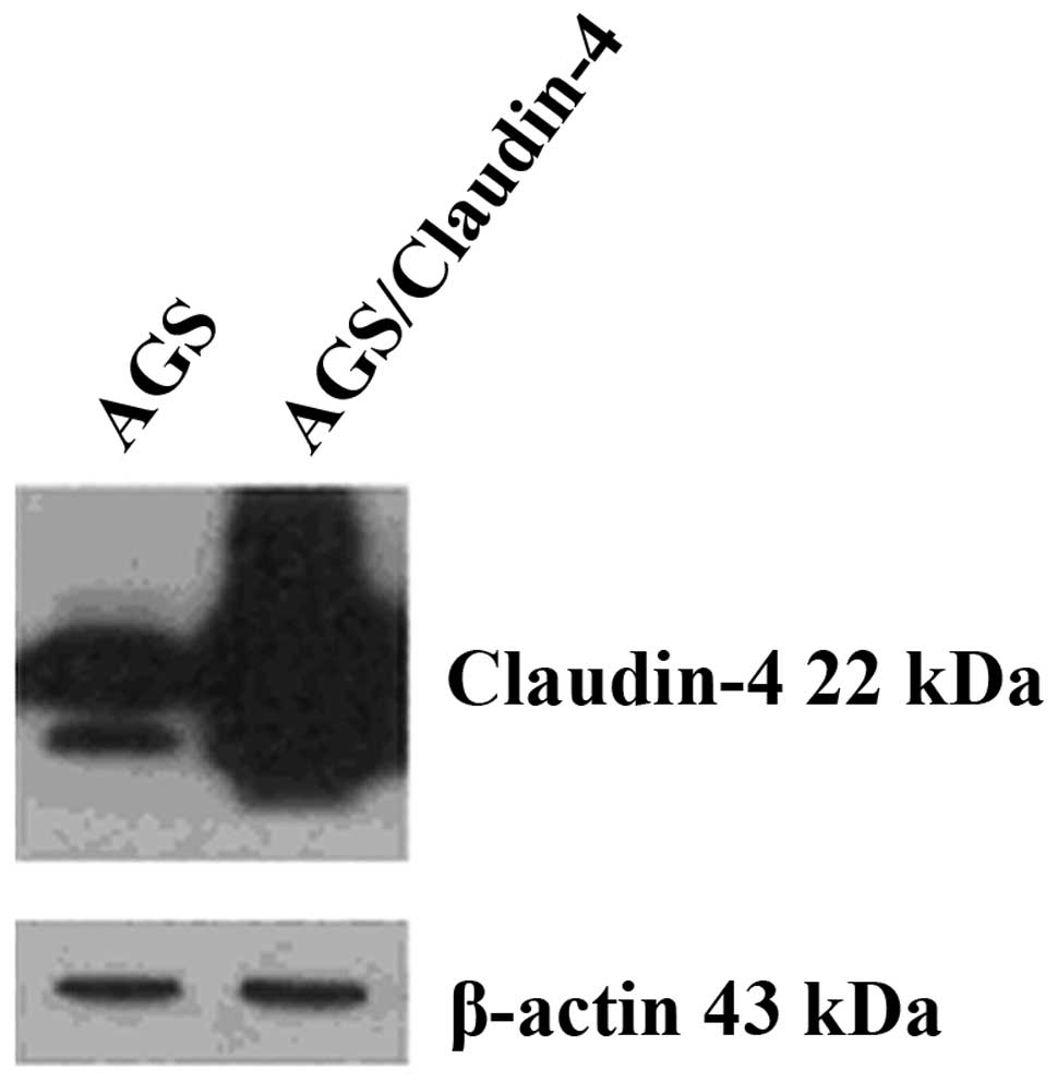
Claudin-4 expression in AGS cells
To study the roles of claudin-4 protein
overexpression in gastric cancer cells, the human adenocarcinoma
cell line, AGS, was transfected with expression vector pcDNA3.1(+)
encoding wild-type claudin-4 cDNA. Immunoblot analysis of the
selected stable clones using claudin-4 specific antibody showed
that the clone, AGS/claudin-4, expressed high levels of claudin-4
(Fig. 1).
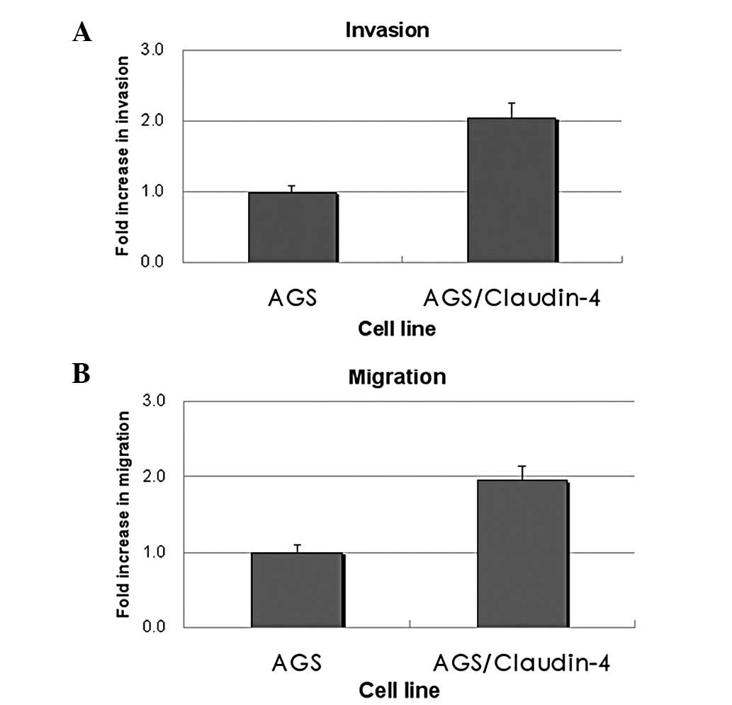
Claudin-4 expression enhances invasion
and cell migration of AGS cells
Differences in invasion between claudin-4-expressing
cells and control AGS cells were evaluated using a modified Boyden
chamber invasion assay. Claudin-labeled cells were placed on
Matrigel-coated ECMatrix inserts and the cells invading through
Matrigel were analyzed after 48 h. The results showed that
claudin-4-overexpressing AGS cells were significantly more invasive
(2-fold increase) than the control AGS cells (P<0.05; Fig. 2A). In addition, the rate of cell
migration between these two cells was also compared using the
two-chamber assay with an uncoated insert. AGS cells overexpressing
claudin-4 showed increased migration (2-fold increase) compared
with the control AGS cells (P<0.05; Fig. 2B).
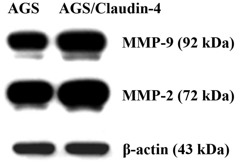
Effects of claudin-4 overexpression on
MMP-2 and -9 expression
A previous study indicated the modulatory effects of
claudins on MMP-2 activation in ovarian cancer cells (30). To understand the underlying
mechanism of claudin-induced increased invasion of AGS cells,
changes in MMP-2 and -9 expression were examined by immunoblotting
analysis. AGS cells stably transfected with full-length wild-type
claudin-4, expressed higher levels of MMP-2 and -9 than the control
AGS cells (Fig. 3).
Discussion
In our previous study, immunohistochemistry was used
to examine the expression levels of claudin-4 and MMP-2 and -9 in
189 gastric cancer samples, and their correlation with tumor
invasion and clinicopathological parameters was analyzed (34). It was found that the expression of
claudin-4 was significantly higher in gastric cancer cases with
advanced depth of wall invasion, lymph node metastasis, lymphatic
invasion and high tumor node metastasis stage. Further analysis
revealed that claudin-4 expression was found to significantly
correlate with the expression of MMP-2 and -9. In the current
study, human gastric adenocarcinoma cells, AGS, constitutively
expressing claudin-4 were generated. Overexpression of claudin-4 in
AGS cells was found to increase cell invasion and migration.
Moreover, the claudin-4-expressing AGS cells were found to have
increased MMP-2 and -9 expression. The results indicated that
claudin-mediated invasion may be mediated through the activation of
the MMP protein.
A similar effect has been observed in other types of
cancer cells (30,32). Previously, Agarwal et al
showed that claudin-3 and -4 expression in ovarian epithelial cells
enhanced invasion and was associated with increased MMP-2 activity
(30). In addition, Takehara et
al demonstrated that overexpression of claudin-4 specifically
stimulated the invasive activity of colonic cancer cells and
increased MMP-2 and -9 activity (32). It is known that claudins affect cell
physiology through recruiting signal transduction-related molecules
at tight junctions (37). The
carboxylic terminal region of claudin proteins contain a PDZ
domain-binding motif that potentially interacts with a number of
PDZ domain-containing proteins, such as ZO proteins (38,39).
These interactions also serve as adapters for other proteins
involved in cell signaling. A number of other cytosolic and nuclear
proteins, including regulatory proteins (e.g. Rab3b), protein
kinases (e.g. c-Abl-PKCδ) and transcription factors (e.g. ZONAB),
have also been shown to interact directly or indirectly with tight
junction complexes (33,40,41).
To further confirm the role of claudin
overexpression on invasive properties of ovarian cancer cells,
Agarwal et al previously performed siRNA-mediated knockdown
of claudin-3 and -4 expression in the ovarian cancer cell line,
OVCAR-5. Inhibition of claudin-3 and -4 expression in OVCAR-5 cells
significantly reduced the invasive potential of these cells
(30). However, siRNA-mediated
knockdown of claudin-3 and -4 in OVCAR-5 cells did not lead to a
decrease in the large amount of MMP-2 activity present in the
cells. These results implied that the malignant ovarian cells may
have additional or alternative pathways to active MMP-2 activity
(30). Previously, Surgucheva et
al and Nothnick investigated the importance of microRNA in
MMP-9 regulation (42,43). Surgucheva et al identified
targets for microRNAs in the 3′-untranslated region of MMP-9
involved in the regulation of MMP-9 expression (42). The authors then isolated microRNAs
from the optic nerve A7 astrocytes and 293T cells and confirmed the
role of mi340 in regulation using specific inhibitors and mimics.
The results obtained showed a novel microRNA-mediated mechanism of
MMP-9 expression regulation.
However, the opposite effect has been previously
observed in pancreatic cancer cells (44). Michl et al showed that
claudin-4 was overexpressed in pancreatic cancer and associated
with decreased invasiveness in vitro and in vivo. In
the authors’ ultrastructal studies, an increase in tight junctions
was found between neighboring claudin-4-overexpressing tumor cells.
This led to the conclusion that an increase in the density of
cell-cell adhesions formed by tight junctions may present a crucial
impediment against the dissociation of pancreatic cancer cells from
the original tumor. This, in turn, is likely to prevent the
invasion into neighboring tissues or formation of distant
metastasis. A similar mechanism has previously been proposed for
the E-cadherin-mediated development of epithelial polarity and
suppression of the invasiveness of cancer cells, which has been
associated with increased cell contact formation. Thus, an
alteration in claudin-4 expression appears to play a role in the
invasiveness of cancer cells, by modulating the barrier function of
tight junctions or by mediating MMP-2 and -9 activity. However, the
overall correlation between claudin-4 overexpression and the
invasive capacity have not been fully elucidated. Additional
studies have been warranted to investigate the correlation between
claudin-4 overexpression and cell invasion in various cancer
cells.
Previously, Surgucheva et al showed that an
additional protein, γ-synuclein, also upregulated MMP-2 and -9 in
retinoblastoma cells (45).
γ-synuclein is a member of a family of small soluble proteins,
which is involved in tumorigenesis since it is overexpressed in
advanced infiltrating carcinomas of the breast (46). Notably, γ-synuclein stimulates
metastasis, being a key positive regulator for cancer invasion and
metastasis and a marker for malignant progression (47). The present study showed that
claudin-4 is overexpressed in human gastric cancer cells and
associated with increased cell invasiveness. Furthermore, the
claudin-4-expressing gastric cancer cells were found to increase
MMP-2 and MMP-9 expression, indicating that claudin-mediated
increased cell invasion may be the result of MMP protein
activation.
Acknowledgements
The present study was supported by grants from the
National Science Council (no. NSC 98-2314-B-238-001) and the Vanung
University, Taiwan, R.O.C. (no. VIT-98-CM-01).
References
|
1
|
Tsukita S and Furuse M: Pores in the wall:
claudins constitute tight junction strands containing aqueous
pores. J Cell Biol. 149:13–16. 2000.
|
|
2
|
Tsukita S and Furuse M: Claudin-based
barrier in simple and stratified cellular sheets. Curr Opin Cell
Biol. 14:531–536. 2002.
|
|
3
|
Anderson JM: Molecular structure of tight
junctions and their role in epithelial transport. News Physiol Sci.
16:126–130. 2001.
|
|
4
|
Cereijido M, Valdes J, Shoshani L and
Contreras RG: Role of tight junctions in establishing and
maintaining cell polarity. Annu Rev Physiol. 60:161–177. 1998.
|
|
5
|
Tsukita S, Furuse M and Itoh M:
Multifunctional strands in tight junctions. Nat Rev Mol Cell Biol.
2:285–293. 2001.
|
|
6
|
Mullin JM: Potential interplay between
luminal growth factors and increased tight junction permeability in
epithelial carcinogenesis. J Exp Zool. 279:484–489. 1997.
|
|
7
|
Soler AP, Miller RD, Laughlin KV, Carp NZ,
Klurfeld DM and Mullin JM: Increased tight junctional permeability
is associated with the development of colon cancer. Carcinogenesis.
20:1425–1431. 1999.
|
|
8
|
Resnick MB, Gavilanez M, Newton E, Konkin
T, Bhattacharya B, Britt DE, Sabo E and Moss SF: Claudin expression
in gastric adenocarcinomas: a tissue microarray study with
prognostic correlation. Hum Pathol. 36:886–892. 2005.
|
|
9
|
Wu CM, Lee YS, Wang TH, Lee LY, Kong WH,
Chen ES, Wei ML, Liang Y and Hwang TL: Identification of
differential gene expression between intestinal and diffuse gastric
cancer using cDNA microarray. Oncol Rep. 15:57–64. 2006.
|
|
10
|
Kuo WL, Lee LY, Wu CM, Wang CC, Yu JS,
Liang Y, Lo CH, Huang KH and Hwang TL: Differential expression of
claudin-4 between intestinal and diffuse-type gastric cancer. Oncol
Rep. 16:729–734. 2006.
|
|
11
|
Xin S, Huixin C, Benchang S, Aiping B,
Jinhui W, Xiaoyan L, Yu WB and Minhu C: Expression of Cdx2 and
claudin-2 in the multistage tissue of gastric carcinogenesis.
Oncology. 73:357–365. 2007.
|
|
12
|
Song X, Li X, Tang Y, Chen H, Wong B, Wang
J and Chen M: Expression of claudin-2 in the multistage process of
gastric carcinogenesis. Histol Histopathol. 23:673–682. 2008.
|
|
13
|
Long H, Crean CD, Lee WH, Cummings OW and
Gabig TG: Expression of Clostridium perfringens enterotoxin
receptors claudin-3 and claudin-4 in prostate cancer epithelium.
Cancer Res. 61:7878–7881. 2001.
|
|
14
|
Santin AD, Zhan F, Cane S, et al: Gene
expression fingerprint of uterine serous papillary carcinoma:
identification of novel molecular markers for uterine serous cancer
diagnosis and therapy. Br J Cancer. 92:1561–1573. 2005.
|
|
15
|
Nichols LS, Ashfaq R and Iacobuzio-Donahue
CA: Claudin 4 protein expression in primary and metastatic
pancreatic cancer: support for use as a therapeutic target. Am J
Clin Pathol. 121:226–230. 2004.
|
|
16
|
Al Moustafa AE, Alaoui-Jamali MA, Batist
G, Hernandez-Perez M, Serruya C, Alpert L, Black MJ, Sladek R and
Foulkes WD: Identification of genes associated with head and neck
carcinogenesis by cDNA microarray comparison between matched
primary normal epithelial and squamous carcinoma cells. Oncogene.
21:2634–2640. 2002.
|
|
17
|
Kominsky SL, Argani P, Korz D, Evron E,
Raman V, Garrett E, Rein A, Sauter G, Kallioniemi OP and Sukumar S:
Loss of the tight junction protein claudin-7 correlates with
histological grade in both ductal carcinoma in situ and invasive
ductal carcinoma of the breast. Oncogene. 22:2021–2033. 2003.
|
|
18
|
Kominsky SL, Vali M, Korz, Gabig TG,
Weitzman SA, Argani P and Sukumar S: Clostridium perfringens
enterotoxin elicits rapid and specific cytolysis of breast
carcinoma cells mediated through tight junction proteins claudin 3
and 4. Am J Pathol. 164:1627–1633. 2004.
|
|
19
|
Morson BC, Dawson IMP and Day DW: Morson
and Dawson’s gastrointestinal pathology. 3rd edition. Blackwell
Science; Oxford: pp. 53–70. 1990
|
|
20
|
Hart IR and Saini A: Biology of tumour
metastasis. Lancet. 339:1453–1457. 1992.
|
|
21
|
Kohn EC and Liotta LA: Molecular insights
into cancer invasion: strategies for prevention and intervention.
Cancer Res. 55:1856–1862. 1995.
|
|
22
|
Murphy G and Docherty AJ: The matrix
metalloproteinases and their inhibitors. Am J Respir Cell Mol Biol.
7:120–125. 1992.
|
|
23
|
Stetler-Stevenson WG, Liotta LA and
Kleiner DE Jr: Extracellular matrix 6: role of matrix
metalloproteinases in tumor invasion and metastasis. FASEB J.
7:1434–1441. 1993.
|
|
24
|
Davies B, Waxman J, Wasan H, Abel P,
Williams G, Krausz T, Neal D, Thomas D, Hanby A and Balkwill F:
Levels of matrix metalloproteases in bladder cancer correlate with
tumor grade and invasion. Cancer Res. 53:5365–5369. 1993.
|
|
25
|
Boag AH and Young ID: Increased expression
of the 72-kd type IV collagenase in prostatic adenocarcinoma.
Demonstration by immunohistochemistry and in situ hybridization. Am
J Pathol. 144:585–591. 1994.
|
|
26
|
Muller D, Wolf C, Abecassis J, Millon R,
Engelmann A, Bronner G, Rouyer N, Rio MC, Eber M, Methlin G, et al:
Increased stromelysin 3 gene expression is associated with
increased local invasiveness in head and neck squamous cell
carcinomas. Cancer Res. 53:165–169. 1993.
|
|
27
|
Urbanski SJ, Edwards DR, Hershfield N,
Huchcroft SA, Shaffer E, Sutherland L and Kossakowska AE:
Expression pattern of metalloproteinases and their inhibitors
changes with the progression of human sporadic colorectal
neoplasia. Diagn Mol Pathol. 2:81–89. 1993.
|
|
28
|
Sakurai Y, Otani Y, Kameyama K, Hosoda Y,
Okazaki I, Kubota T, Kumai K and Kitajima M: Expression of
interstitial collagenase (matrix metalloproteinase-1) in gastric
cancers. Jpn J Cancer Res. 88:401–406. 1997.
|
|
29
|
Torii A, Kodera Y, Uesaka K, Hirai T,
Yasui K, Morimoto T, Yamamura Y, Kato T, Hayakawa T, Fujimoto N and
Kito T: Plasma concentration of matrix metalloproteinase 9 in
gastric cancer. Br J Surg. 84:133–136. 1997.
|
|
30
|
Agarwal R, D’Souza T and Morin PJ:
Claudin-3 and claudin-4 expression in ovarian epithelial cells
enhances invasion and is associated with increased matrix
metalloproteinase-2 activity. Cancer Res. 65:7378–7385. 2005.
|
|
31
|
Oku N, Sasabe E, Ueta E, Yamamoto T and
Osaki T: Tight junction protein claudin-1 enhances the invasive
activity of oral squamous cell carcinoma cells by promoting
cleavage of laminin-5 gamma2 chain via matrix metalloproteinase
(MMP)-2 and membrane-type MMP-1. Cancer Res. 66:5251–5257.
2006.
|
|
32
|
Takehara M, Nishimura T, Mima S, Hoshino T
and Mizushima T: Effect of claudin expression on paracellular
permeability, migration and invasion of colonic cancer cells. Biol
Pharm Bull. 32:825–831. 2009.
|
|
33
|
Yoon CH, Kim MJ, Park MJ, Park IC, Hwang
SG, An S, Choi YH, Yoon G and Lee SJ: Claudin-1 acts through
c-Abl-protein kinase Cdelta (PKCdelta) signaling and has a causal
role in the acquisition of invasive capacity in human liver cells.
J Biol Chem. 285:226–233. 2010.
|
|
34
|
Hwang TL, Lee LY, Wang CC, Liang Y, Huang
SF and Wu CM: Claudin-4 expression is associated with tumor
invasion, MMP-2 and MMP-9 expression in gastric cancer. Exp Ther
Med. 1:789–797. 2010.
|
|
35
|
Mima S, Tsutsumi S, Ushijima H, Takeda M,
Fukuda I, Yokomizo K, Suzuki K, Sano K, Nakanishi T, Tomisato W,
Tsuchiya T and Mizushima T: Induction of claudin-4 by nonsteroidal
anti-inflammatory drugs and its contribution to their
chemopreventive effect. Cancer Res. 65:1868–1876. 2005.
|
|
36
|
Albini A, Iwamoto Y, Kleinman HK, Martin
GR, Aaronson SA, Kozlowski JM and McEwan RN: A rapid in vitro assay
for quantitating the invasive potential of tumor cells. Cancer Res.
47:3239–3245. 1987.
|
|
37
|
Matter K and Balda MS: Signalling to and
from tight junctions. Nat Rev Mol Cell Biol. 4:225–236. 2003.
|
|
38
|
Morita K, Furuse M, Fujimoto K and Tsukita
S: Claudin multigene family encoding four-transmembrane domain
protein components of tight junction strands. Proc Natl Acad Sci
USA. 96:511–516. 1999.
|
|
39
|
Itoh M, Furuse M, Morita K, Kubota K,
Saitou M and Tsukita S: Direct binding of three tight
junction-associated MAGUKs, ZO-1, ZO-2, and ZO-3, with the COOH
termini of claudins. J Cell Biol. 147:1351–1363. 1999.
|
|
40
|
Yamamoto Y, Nishimura N, Morimoto S,
Kitamura H, Manabe S, Kanayama HO, Kagawa S and Sasaki T: Distinct
roles of Rab3B and Rab13 in the polarized transport of apical,
basolateral, and tight junctional membrane proteins to the plasma
membrane. Biochem Biophys Res Commun. 308:270–275. 2003.
|
|
41
|
Balda MS, Garrett MD and Matter K: The
ZO-1-associated Y-box factor ZONAB regulates epithelial cell
proliferation and cell density. J Cell Biol. 160:423–432. 2003.
|
|
42
|
Surgucheva I, Chidambaram K, Willoughby DA
and Surguchov A: Matrix metalloproteinase 9 expression: new
regulatory elements. J Ocul Biol Dis Infor. 3:41–52. 2010.
|
|
43
|
Nothnick WB: Regulation of uterine matrix
metalloproteinase-9 and the role of microRNAs. Semin Reprod Med.
26:494–499. 2008.
|
|
44
|
Michl P, Barth C, Buchholz M, Lerch MM,
Rolke M, Holzmann KH, Menke A, Fensterer H, Giehl K, Löhr M, Leder
G, Iwamura T, Adler G and Gress TM: Claudin-4 expression decreases
invasiveness and metastatic potential of pancreatic cancer. Cancer
Res. 63:6265–6271. 2003.
|
|
45
|
Surgucheva IG, Sivak JM, Fini ME, Palazzo
RE and Surguchov AP: Effect of gamma-synuclein overexpression on
matrix metalloproteinases in retinoblastoma Y79 cells. Arch Biochem
Biophys. 410:167–176. 2003.
|
|
46
|
Ji H, Liu YE, Jia T, Wang M, Liu J, Xiao
G, Joseph BK, Rosen C and Shi YE: Identification of a breast
cancer-specific gene, BCSG1, by direct differential cDNA
sequencing. Cancer Res. 57:759–764. 1997.
|
|
47
|
Jia T, Liu YE, Liu J and Shi YE:
Stimulation of breast cancer invasion and metastasis by synuclein
gamma. Cancer Res. 59:742–747. 1999.
|