|
1
|
Rojo MG, Bueno G and Slodkowska J: Review
of imaging solutions for integrated quantitative
immunohistochemistry in the Pathology daily practice. Folia
Histochem Cytobiol. 47:349–354. 2009.
|
|
2
|
Lloyd MC, Allam-Nandyala P, Purohit CN, et
al: Using image analysis as a tool for assessment of prognostic and
predictive biomarkers for breast cancer: How reliable is it? J
Pathol Inform. 1:292010.
|
|
3
|
Chabot-Richards DS, Martin DR, Myers OB,
Czuchlewski DR and Hunt KE: Quantitative image analysis in the
assessment of diffuse large B-cell lymphoma. Mod Pathol.
24:1598–1605. 2011.
|
|
4
|
Klapczynski M, Gagne GD, Morgan SJ, et al:
Computer-assisted imaging algorithms facilitate histomorphometric
quantification of kidney damage in rodent renal failure models. J
Pathol Inform. 3:202012.
|
|
5
|
Laurinavicius A, Laurinaviciene A,
Ostapenko V, et al: Immunohistochemistry profiles of breast ductal
carcinoma: factor analysis of digital image analysis data. Diagn
Pathol. 7:272012.
|
|
6
|
Singh R, Stockard CR, Grizzle WE, Lillard
JW Jr and Singh S: Expression and histopathological correlation of
CCR9 and CCL25 in ovarian cancer. Int J Oncol. 39:373–381.
2011.
|
|
7
|
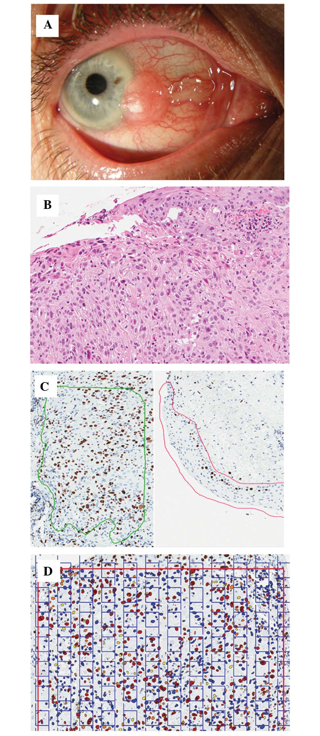
Yang J and Foster CS: Squamous cell
carcinoma of the conjunctiva. Int Ophthalmol Clin. 37:73–85.
1997.
|
|
8
|
Kiire CA, Srinivasan S and Karp CL: Ocular
surface squamous neoplasia. Int Ophthalmol Clin. 50:35–46.
2010.
|
|
9
|
McKelvie PA, Daniell M, McNab A, Loughnan
M and Santamaria JD: Squamous cell carcinoma of the conjunctiva: a
series of 26 cases. Br J Ophthalmol. 86:168–173. 2002.
|
|
10
|
Tunc M, Char DH, Crawford B and Miller T:
Intraepithelial and invasive squamous cell carcinoma of the
conjunctiva: analysis of 60 cases. Br J Ophthalmol. 83:98–103.
1999.
|
|
11
|
Jung SM, Lin HC, Chu PH, et al: Expression
of cell cycle-regulatory proteins, MIB-1, p16, p53, and p63, in
squamous cell carcinoma of conjunctiva: not associated with human
papillomavirus infection. Virchows Arch. 448:301–305. 2006.
|
|
12
|
Schlüter C, Duchrow M, Wohlenberg C, et
al: The cell proliferation-associated antigen of antibody Ki-67: a
very large, ubiquitous nuclear protein with numerous repeated
elements, representing a new kind of cell cycle-maintaining
proteins. J Cell Biol. 123:513–522. 1993.
|
|
13
|
Alvarenga AW, Coutinho-Camillo CM,
Rodrigues BR, et al: A comparison between manual and automated
evaluations of tissue microarray patterns of protein expression. J
Histochem Cytochem. 61:272–282. 2013.
|
|
14
|
Brazdziute E and Laurinavicius A: Digital
pathology evaluation of complement C4d component deposition in the
kidney allograft biopsies is a useful tool to improve
reproducibility of the scoring. Diagn Pathol. 6(Suppl 1):
S52011.
|
|
15
|
Prasad K and Prabhu GK: Image analysis
tools for evaluation of microscopic views of immunohistochemically
stained specimen in medical research - a review. J Med Syst.
36:2621–3261. 2012.
|
|
16
|
Fasanella S, Leonardi E, Cantaloni C, et
al: Proliferative activity in human breast cancer: Ki-67 automated
evaluation and the influence of different Ki-67 equivalent
antibodies. Diagn Pathol. 6(Suppl 1): S72011.
|
|
17
|
Słodkowska J, Filas V, Buszkiewicz E, et
al: Study on breast carcinoma Her2/neu and hormonal receptors
status assessed by automated images analysis systems: ACIS III
(Dako) and ScanScope (Aperio). Folia Histochem Cytobiol. 48:19–25.
2010.
|
|
18
|
Salek D, Vesela P, Boudova L, et al:
Retrospective analysis of 235 unselected patients with mantle cell
lymphoma confirms prognostic relevance of Mantle Cell Lymphoma
International Prognostic Index and Ki-67 in the era of rituximab:
long-term data from the Czech Lymphoma Project Database. Leuk
Lymphoma. 55:802–810. 2014.
|
|
19
|
Liu Y, Yin W, Yan T, et al: The clinical
significance of Ki-67 as a marker of prognostic value and
chemosensitivity prediction in hormone-receptor-positive breast
cancer: a meta-analysis of the published literature. Curr Med Res
Opin. 29:1453–1461. 2013.
|
|
20
|
Otto W, Denzinger S, Fritsche HM, et al:
Introduction and first clinical application of a simplified
immunohistochemical validation system confirms prognostic impact of
KI-67 and CK20 for stage T1 urothelial bladder carcinoma:
single-center analysis of eight biomarkers in a series of three
hundred six patients. Clin Genitourin Cancer. 11:537–544. 2013.
|
|
21
|
Ohara M, Sotozono C, Tsuchihashi Y and
Kinoshita S: Ki-67 labeling index as a marker of malignancy in
ocular surface neoplasms. Jpn J Ophthalmol. 48:524–529. 2004.
|
|
22
|
Wolberg WH, Street N, Heisey DM and
Mangasarian OL: Computer-derived nuclear features distinguish
malignant from benign breast cytology. Hum Pathol. 26:792–796.
1995.
|