Introduction
Schwannomas are derived from Schwann cells (1). More than 90% of schwannomas are
benign. They often have a single place of origin and 10% have
multiple locations of origin. Schwannomas often arise in the head
and neck (~25–40%), but are rarely located in the retroperitoneum
(6% of primary retroperitoneal tumors) (1,2). These
tumors, although rare, should be considered in the differential
diagnosis of slow-growing masses of an anal location, such as
lipomyomas. The growth of these masses occasionally causes
displacement and compression of the nerve of origin, resulting in
clinical signs and symptoms (3).
The tumors can develop in either of the two genders and there is no
age predilection (4). Surgical
excision is the treatment of choice for schwannomas (3). In the present study, the case of a
patient with an anal schwannoma is reported, where the
pre-operative clinical diagnosis was inconclusive and the final
diagnosis was established based on radiographic and
histopathological examination. Written informed consent was
obtained from the patient.
Case report
A 66-year-old female was admitted to the Third
People’s Hospital of Dalian (Dalian, China) due to a right anal
mass that had been present for several weeks. The patient
experienced no pain, difficulty in defecation or other symptoms. A
physical examination revealed no unusual findings, with the
exception of 10×7-cm, tenacious, mobile and smooth mass that could
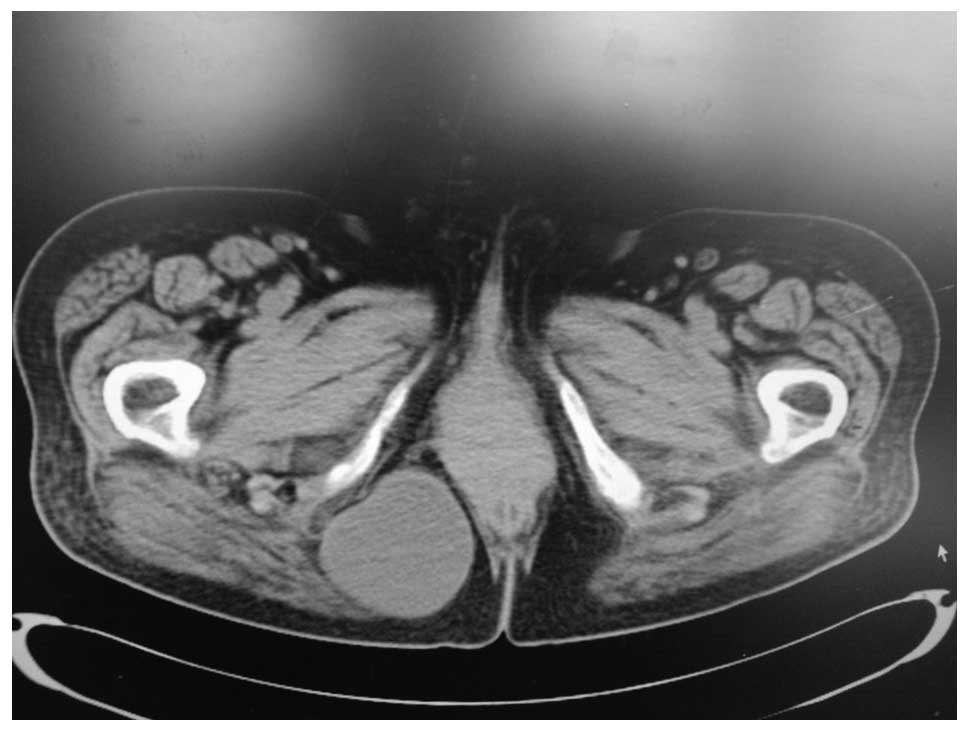
be palpated when in the knee-chest decubitus position. Computed
tomography (CT) scans showed that the tumor, which had uniform
density and a clear tunica, was located under a fat layer and
inside of the gluteus maximus (Fig.
1). The results of blood and kidney function tests were normal
and the patient underwent surgery to completely excise the tumor,
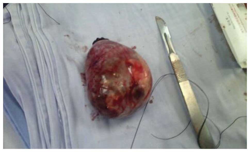
with no postoperative complications. The pathological analysis
reported a 6×8-cm cystic tumor, which was full of a gelatinous
material (Fig. 2). Microscopically,
the tumor was diagnosed as a schwannoma with cystic change.
Immunohistochemistry revealed that the tumor was S-100-positive.
The patient was followed-up for one year and no disease recurrence
was identified.
Discussion
Schwannomas are nerve sheath tumors that arise from
almost any anatomical site, but particularly in the peripheral,
cranial or visceral nerves (1).
Schwannomas can be found not only in children, but in adults
also.
The majority of schwannomas are benign, and few are
malignant. Malignant schwannomas are usually large, infiltrating
and characterized histologically by perineural and intraneural
spread; in the present study, mainly benign schwannomas are
discussed. Schwannomas have the characteristics of a slow growth
rate, no invasion and a low rate of transformation or recurrence
following resection (1).
The site of the schwannoma may vary, however, the
tumors mostly occur in the neck, head and tendon sheaths of the
limbs. There has also been a study reporting schwannoma of the
abdomen (3).
Unusual locations, such as the anus, are rarely
reported (6). In the present study,
a rare mass is reported, which was believed to originate from the
cranial nerves due to its location under the deep skin layer.
CT and magnetic resonance imaging (MRI) were more
beneficial for the identification of characteristics of the mass,
although obviously distinct in clinical features and histological
appearance; the pre-operative diagnosis of a schwannoma is not easy
owing to a lack of distinguishing features. It is possible to
distinguish between benign and malignant schwannomas by imaging
techniques, such as CT, ultrasound or MRI. Additionally, it is
possible to distinguish between schwannomas and other tumors, for
example, fibrosarcomas or liposarcomas (7), via imaging, and clinical data, such as
the size, exact location, association with other organs and
invasion can be obtained (8). It
has been reported that, to a certain extent, MRI is more sensitive
than CT, although they each have the same difficulty in
distinguishing bladder schwannoma from carcinoma (3).
In the present case, CT was used to find the exact
location of the mass beside the rectum, which was compressed as a
result, although there were no symptoms of this. A clear tunica
could also be observed, therefore the surgical risk and procedural
design could be determined.
During the surgery, incisions were made in the skin
and subcutaneous tissue. The mass was confirmed to be in the
subcutaneous tissue, as previously shown by CT. Due to the
pre-operative examination and assessment, the mass could be
completely resected, and the duration of the surgery was not
excessive.
Histologically, schwannomas consist of compact
cellular lesions. Antoni A (interlacing and cellular fascicles) and
Antoni B (less cellular and myxoid) areas, combined with positive
uniform S-100 staining characterize the histological appearance of
typical schwannomas (4). Therefore,
S-100 immunostaining is extremely useful in the differential
diagnosis of schwannoma. The mass reported in the present study
exhibited the characteristics typical of a schwannoma (9) and positive S-100 immunostaining.
In conclusion, surgical excision, which can be
diagnostic and curative, is the optimal treatment for schwannomas,
whether they are benign or malignant. In the present case, a large
mass developed, however, there were no resultant symptoms. This was
unexpected, as changes, such as altered defecation habits, were
anticipated with the development of the tumor. This was just one of
the reasons that the nature of the tumor could not be precisely
determined prior to the surgery.
References
|
1
|
Machairiotis N, Zarogoulidis P, Stylianaki
A, et al: Pelvic schwannoma in the right parametrium. Int J Gen
Med. 6:123–126. 2013.
|
|
2
|
Lira RB, Gonçalves Filho J, Carvalho GB,
et al: Lingual schwannoma: case report and review of the
literature. Acta Otorhinolaryngol Ital. 33:137–140. 2013.
|
|
3
|
Samarakoon L, Weerasekera A, Sanjeewa R
and Kollure S: Giant presacral schwannoma presenting with
constipation: a case report. J Med Case Rep. 6:2852012.
|
|
4
|
Canbay S, Hasturk AE, Markoc F and Caglar
S: Schwannoma of the conus medullaris: a rare case. Chin J Cancer.
30:867–870. 2011.
|
|
5
|
Fenoglio L, Severini S, Cena P, et al:
Common bile duct schwannoma: a case report and review of
literature. World J Gastroenterol. 13:1275–1278. 2007.
|
|
6
|
Łabędź W, Kubaszewski L and Adamek J:
Operative treatment of Schwannoma, the primary sacral tumor – case
presentation. Polish Orthop Traumatol. 77:11–15. 2012.
|
|
7
|
Hughes MJ, Thomas JM, Fisher C and
Moskovic EC: Imaging features of retroperitoneal and pelvic
schwannomas. Clin Radiol. 60:886–893. 2005.
|
|
8
|
Ke Z, Yi M, Li J, et al: The management of
retroperitoneal giant schwannomas in AIDS patients: A case report.
Oncol Lett. 5:1430–1432. 2013.
|
|
9
|
Schindler OS, Dixon JH and Case P:
Retroperitoneal giant schwannomas: report on two cases and review
of the literature. J Orthop Surg (Hong Kong). 10:77–84. 2002.
|