Introduction
Breast-conserving surgery may eradicate macroscopic
diseases that have been detected and palpated in females with
early-stage breast cancer (1,2).
However, certain microscopic tumor foci may remain in the conserved
breast, leading to local recurrence and/or life-threatening distant
metastases. The administration of adjuvant radiotherapy following
breast-conserving surgery is effective in reducing the risk of
locoregional recurrence and distant metastases in patients with
early-stage breast cancer (1,2).
Postoperative radiation treatment in patients with breast cancer is
conventionally delivered using external beam radiation therapy,
which is determined by rectangular tangential fields. With this
radiotherapy technique, an appreciable dose within the irradiated
volume may be administered, and the dose delivered to the lung and
heart may be higher than predicted (3).
Over the past decade, there has been a rapid
increase in the utilization of advanced radiation delivery
technologies for the curative management of numerous types of solid
cancer. Radiation patterns have shifted from conventional
two-dimensional (2D) radiotherapy to a more developed
three-dimensional (3D) approach in treating breast cancer (4,5).
However, whether intensity-modulated radiotherapy (IMRT) is
superior to traditional 3D radiation delivery remains unknown.
In recent years, the use of IMRT has been greatly
improved, as the beam intensity profile has been conformed to the
chest wall or delineated target volume, resulting in reduced
radiation dose variations and sparing of organs at risk
(OAR)(6–9). The shape of the IMRT plan can be
optimized on the basis of geometrical parameters, including the
shape of the breast and thoracic wall, or dosimetric parameters
using inverse planning. Consequently, it has not always been
possible to establish a satisfactory compromise between the dose
delivered to the target volume or the clinical tumor volume (CTV)
and the dose delivered to the OARs (10,11).
Compared with conventional rectangular tangential fields, the dose
distribution conforms more to the target volume when 3D data are
available and conformal treatment fields are used. This approach
reduces the dose to OARs. Intensity modulation may be considered as
an additional step and allows for greater freedom in improving the
dose distribution compared with the combination of open and wedged
beams. This may result in a further improvement of the dose
distribution in the target volume and the OARs. Inverse planning
provides a method for minimizing the dose to OARs, whilst
maintaining adequate target coverage.
The majority of IMRT studies have shown a potential
clinical benefit in sparing OARs and improving dose homogeneity
over the target volume, as compared with rectangular tangential
fields without conformal blocks (12–14).
However, the implementation of IMRT in clinical practice requires
additional resources for patients with breast cancer in the
adjuvant setting, as the IMRT plan is more time-consuming and
complex. Therefore, it is useful to identify patients with a
medical necessity for the IMRT plan. Several studies have compared
a number of forms of IMRT, including forward and inverse methods,
with conventional radiotherapy (CR) (15–17).
However, the relative improvement attributable to intensity
modulated irradiated fields compared with conformed fields is not
yet known for the irradiation of patients with early-stage breast
cancer. Therefore, a clinical trial was initiated to investigate
radiation dosimetry of IMRT, assessed by changes in breast
appearance and discomfort in patients with early stage breast
cancer.
Patients and methods
Eligibility
A total of 20 patients under the care of the
Department of Radiation Oncology, the First Affiliated Hospital of
Anhui Medical University (Hefei, China), with early-stage breast
cancer (T1–2N0M0; stage I or
IIA), according to 7th edition of American Joint
Committee on Cancer (18), between
July 2008 and October 2009 were included in this study. Patients
with no previous malignancies, complete microscopic excision of
tumors and histological confirmation of breast cancer underwent
breast-conserving surgery (16 patients with invasive ductal
carcinoma, two with invasive lobular carcinoma, one with
intraductal carcinoma, and one with medulla carcinoma).
Radiotherapy was prescribed for the whole breast, and written
informed consent was obtained prior to the trial. The study was
approved by the ethics committee of The First Affiliated Hospital
of Anhui Medical University, and was conducted in accordance with
the declaration of Helsinki.
Patient positioning and localization
Localization of the treatment volume and the field
geometry was conducted by an Acuity™ simulator (Varian Medical
Systems, Inc., Palo Alto, CA, USA) according to laser mark points
on the bodies of the patients. The computed tomography (CT) results
were applied to this retrospective treatment planning study. CT
slices were acquired every 5 mm, with the patient lying in a supine
position. Patients were positioned in the supine position on an
angled board with the arms abducted to 90°, such that the sternum
was horizontal. All patients were positioned with the arms resting
on an armrest placed above the head equivalent to the treatment
position. The patients were immobilized about the shoulders and
upper arms, by applying a vacuum air cushion across the shoulders.
This position has been demonstrated to facilitate treatment
planning of tangential fields without the arms extending into the
treatment fields and changing the shape of the breast significantly
(19). The CT scan included the
complete left and right lung, breast, heart and liver. The median
separation distance between the most medial and lateral aspects of
the breast was 21.1 cm (range, 18.0–26.5 cm), which was confirmed
in the current study. All graphic files of the CT scans were
transmitted to the Topslane treatment planning system (Xops 2.0;
Shanghai Topslane Medical Technology Co., Ltd., Shanghai, China)
for further analysis.
Target volume outline
The CTV was delineated by a radiation oncologist
according to surface mark points and CT scan files, which were
optimized to visualize the glandular tissue of patients that had
received breast-conserving surgery. The radiation oncologist
contoured the CTV based on the CT scan. The CTV was assumed to
start 3–5 mm below the skin and delineated smoothly with an
anterior margin of 0.5 cm beneath the skin, a posterior margin of
the chest wall surface, a medial margin of the body midline and a
lateral margin of midaxillary line. A planning target volume (PTV)
was generated by expanding the CTV by 7 mm isotropically, with the
exception of the direction towards the skin surface, where
expansion stopped at 5 mm below the skin, to account for the
uncertainty in the patient set-up and CTV delineation. The cranial
extent of the heart included the infundibulum of the right
ventricle, right atrium and right atrium auricle, but excluded the
pulmonary trunk, ascending aorta and superior vena cava. The lowest
external contour of the heart was the caudal border of the
myocardium, with the pericardium excluded. The contours of the
lungs and skin were automatically outlined.
CR plan
The gantry and collimator angles of the tangential
fields were selected using the beam’s eye view option on the 3D
treatment planning system (3DTPS; Xops 2.0). The edges of the
tangential fields were non-divergent, to minimize the irradiated
lung volume to a margin of 1.5–2.0 cm. In the conformal plans, an
automatically generated conformal wedge block around the PTV, with
a margin of 1.5–2.0 cm, was added to these fields. The treatment
planning was performed using the 3DTPS (Xops 2.0). The optimum
wedge angle and beam weights were calculated using the optimization
module. In the context of an equivalent beam weight of the
tangential fields, the goal of the optimization module was to
obtain a homogeneous dose, while maintaining a low dose in the
lungs and heart.
IMRT plan
Using the same gantry angles as applied in the CR
plans, tangential 6-MV photon beam intensity profiles were
calculated using the inverse planning program (radio-SOFT 1.0;
Apache Technologies Inc., Dayton, OH, USA). Following sequencing,
the segment weights were refined with the optimization module using
the same objective score function as the CR plan. From the PTV to
normal tissues, the final dose distribution was calculated to
inversely optimize the beam intensity profiles, which in turn was
converted to a segmental sequence. Thus, the same dose calculation
algorithm was used for all rectangular, conformal and
step-and-shoot IMRT plans. The maximal dose in the IMRT plan was
≤105% of the prescribed dose; the ipsilateral lung (V20,
volume of lung receiving >20 Gy) was ≤25% and the heart was
V50 ≤50% in patients with left breast cancer. The
prescription doses for both plans are as follows: total dose 5000
cGy/25,200 cGy/times, five times per week, a total of 25 times.
Statistical analysis
Dosimetric comparisons between the tumors and OARs
were completed based on the following parameters from a dose-volume
histogram (DVH): D95, maximal dose at 95%;
Dmax, dose received by ≤1% volume of the PTV;
Dmin, dose received by ≥99% volume of the PTV; and
Dmean of the PTV, V30, V20,
V10 and V5 of the lung (fraction of the lung
volume receiving >30, 20, 10 or 5 Gy, respectively) and
V30, V40 and V50 of the heart
(fraction of the heart volume receiving >30, 40 or 50 Gy,
respectively) in patients with left breast cancer. Statistical
analyses were performed using SPSS, version 16.0 (SPSS. Inc.,
Chicago, IL, USA). Student’s paired, two-tailed t-tests were used
to determine statistical significance. P≤0.05 was considered to
indicate a statistically significant difference.
Results
Dosimetric comparison between the PTV in
IMRT and CR
An adequate dose coverage of the mammary glands and
lymph nodes in the IMRT and CR plans was achieved in the majority
of patients. For example, 95% of the PTV of the mammary glands was
delivered by ≥95.4% of the prescribed dose. Similarly, the CR PTV
was 95% in the CR plan, as the partial PTV was located in a
low-dose region of the tangential fields. Of note, the volume of
the low-dose region in the breast PTV was observed to correlate
with the breast tissue thickness at the medial beam edge of the
tangential fields.
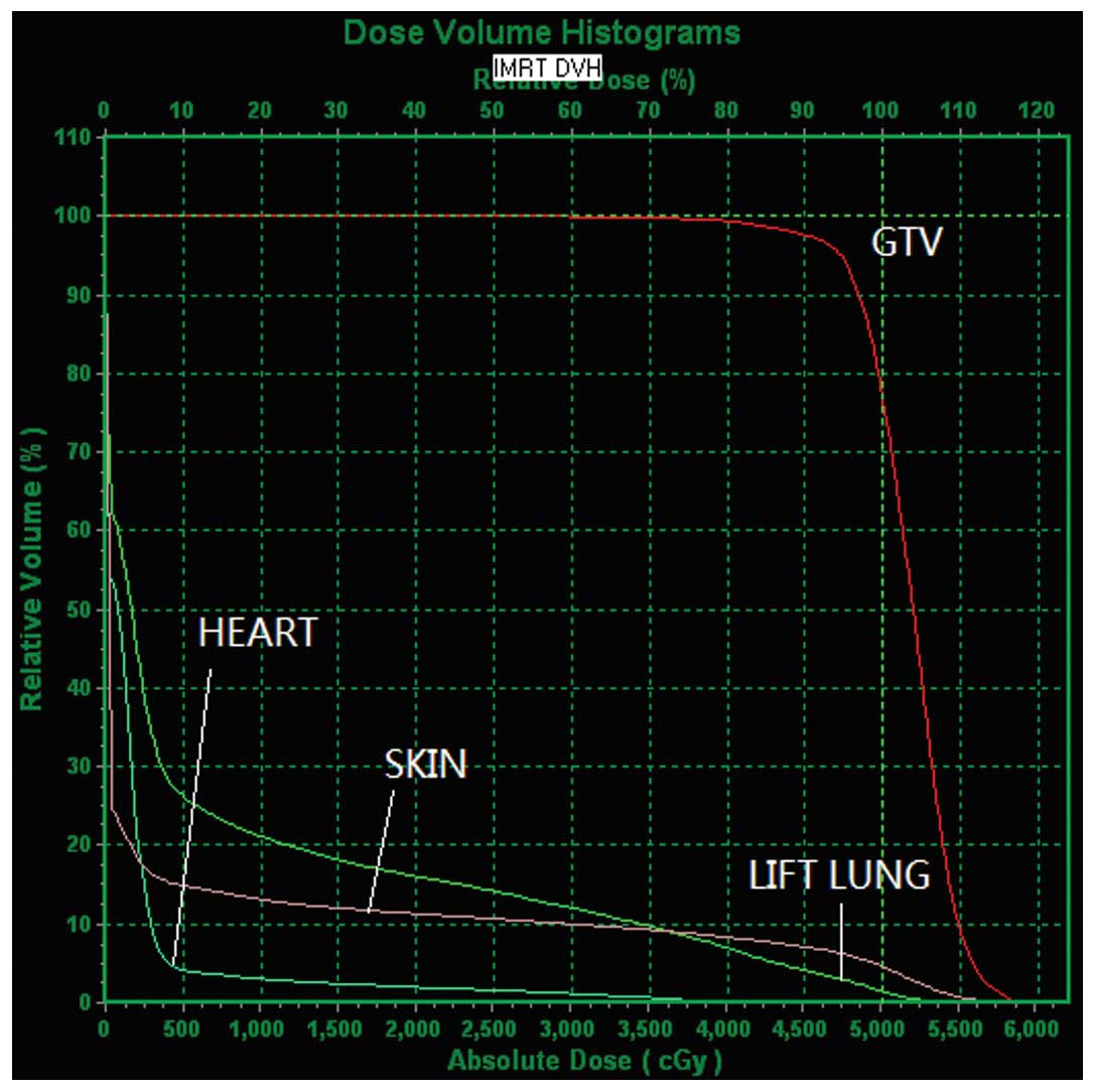
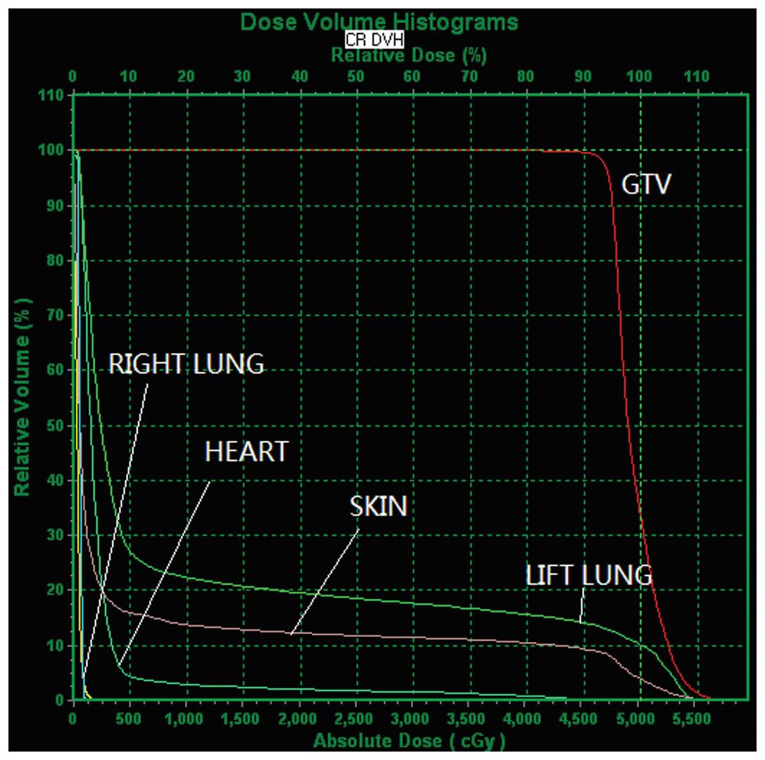
DVH plots of the two modalities for the PTV of a
typical patient are illustrated in Figs. 1 and 2. D95, Dmax,
Dmin and Dmean values are presented in
Table I. No significant difference
in Dmean was observed between the IMRT and CR (P=0.326).
Furthermore, IMRT acquired a significantly lower Dmax
(P=0.015), but a higher Dmin (P=0.031), as compared with
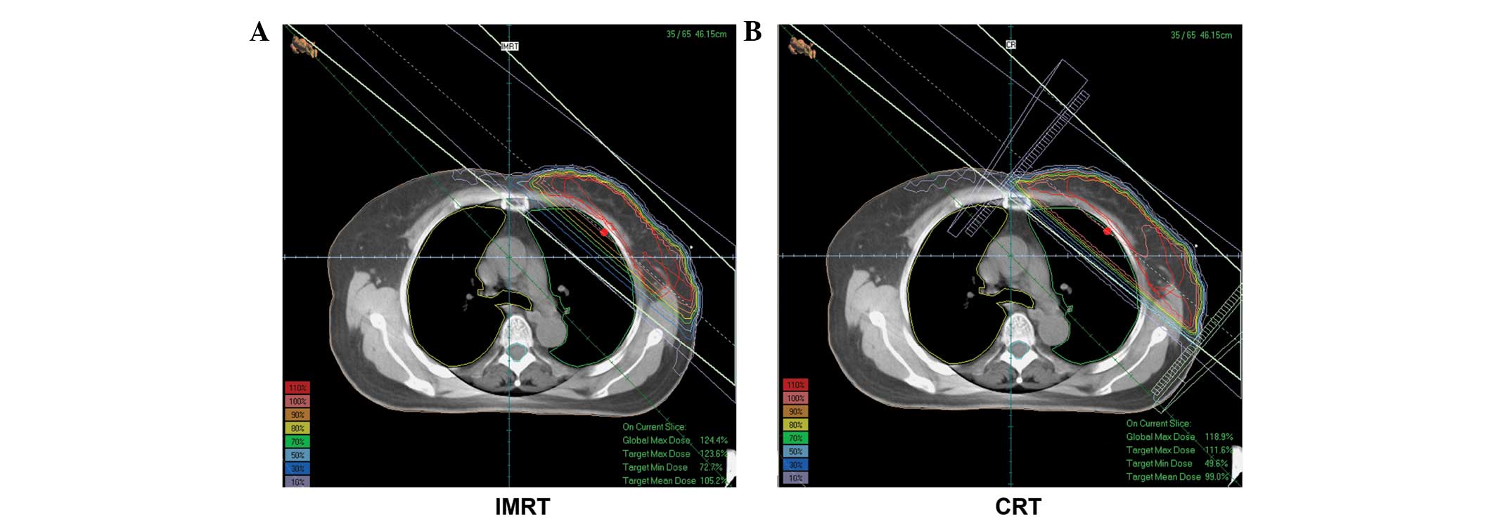
the CR. Accordingly, the IMRT improved dosimetric homogeneity more
efficiently, without dosimetric hot and cold spots (Fig. 3).
 | Table IComparison between the D95,
Dmin, Dmax and Dmean of the PTV in
IMRT and CR. |
Table I
Comparison between the D95,
Dmin, Dmax and Dmean of the PTV in
IMRT and CR.
| Variables | D95 | Dmin,
cGy | Dmax,
cGy | Dmean,
cGy |
|---|
| CR | 4518.3±60.4 | 3807.9±243.6 | 5832.2±61.4 | 5086.9±49.0 |
| IMRT | 4541.4±35.4 | 3868.4±248.3 | 5795.0±54.5 | 5075.8±47.3 |
| P-value | 0.009 | 0.031 | 0.016 | 0.326 |
Comparison between the dosimetric
parameters of OARs
The dosimetric parameters of OARs, including
V5, V10, V20 and V30 of
the ipsilateral lung and V30, V40,
V50 and Dmean of the heart are listed in
Tables II and III, respectively. DVH plots for OARs of
the two modalities are depicted in Figs
1. and 2. V5,
V10, V20 and V30 of the
ipsilateral lung were significantly reduced by 10.8, 8.4, 6.9 and
6.4%, (P=0.000, P=0.001, P=0.003 and P=0.002, respectively) in
IMRT, as compared with the CR. Additionally, V30 of the
heart was significantly reduced (P=0.046) and decreasing trends for
V40 and V50 were observed in the IMRT.
Collectively, the IMRT improved OAR protection by decreasing the
irradiation dose and volume sparing the OARs, as compared with the
CR.
| Table IIComparison between the dosimetric
parameters of the ipsilateral lung. |
Table II
Comparison between the dosimetric
parameters of the ipsilateral lung.
| Ipsilateral lung |
|---|
|
|
|---|
| Variables | V5,
% | V10,
% | V20,
% | V30,
% |
|---|
| CR | 38.3±0.8 | 31.8±0.8 | 27.7±0.9 | 24.9±1.0 |
| IMRT | 27.5±1.7 | 23.4±2.0 | 20.8±2.0 | 18.5±2.0 |
| P-value | 0.000 | 0.001 | 0.003 | 0.002 |
| Table IIIComparison between the dosimetric
parameters of the heart in patients with left breast cancer. |
Table III
Comparison between the dosimetric
parameters of the heart in patients with left breast cancer.
| Heart |
|---|
|
|
|---|
| Variables | V30,
% | V40,
% | V50,
% | Dmean,
cGy |
|---|
| CR | 10.06±1.7 | 4.13±1.0 | 1.3±0.5 | 598.4±118.2 |
| IMRT | 5.3±1.4 | 1.9±0.5 | 0.0±0.0 | 348.3±91.6 |
| P-value | 0.046 | 0.095 | 0.076 | 0.004 |
Discussion
Over the past decade, there has been a rapid rise in
the application of advanced radiation delivery technologies for the
curative management of numerous types of solid cancer. Clinical
irradiation patterns have shifted from conventional 2D therapy to a
more developed 3D therapy based on CT (4,19).
Furthermore, 3D conformal radiation, including IMRT, exhibits
accurate information with regard to the radiation dose to the
affected breast, regional nodes and adjacent normal tissues.
Therefore, this approach may reduce morbidity and improve long-term
cosmesis, while maintaining local tumor control (20–22).
In the three published randomized trials of IMRT in breast cancer
(2,23,24),
the focus was on early-stage breast cancer treatment and, thus, the
radiation target volume was only the breast. In these trials, IMRT
only improved the radiation dose homogeneity, which was due to the
elimination of significant hotspots in the breast that presented in
wedge-based 2D plans (23,24). Improved radiation dosimetry is
associated with improvements in acute radiation reactions,
including skin dermatitis and overall breast cosmesis (12,25,26).
IMRT may be beneficial for the treatment of node-positive breast
cancer, particularly when the internal mammary nodal regions
require treatment (17,27,28).
At the Department of Tumor Radiotherapy, The First Affiliated
Hospital of Anhui Medical University, CR is the routine technique
for patients receiving breast-conserving surgery. Larger irradiated
volumes of the ipsilateral lung and heart have been identified in
CR compared with IMRT, as well as larger fields of rectangular
shape, formed by jaws in the X and Y direction. However, intensity
gradients in CR are only generated in a single direction (?).
The advantages of IMRT have been discussed in
several studies; however, the results remain contradictory
(17,19). Dogan et al (28) demonstrated the benefits of the IMRT
plan as compared with a standard treatment plan using optimized
beam weights and wedges. However, the treatment plans were not
compared with the rectangular tangential fields commonly used in
clinical practice. Additionally, it has been demonstrated that
standard dose optimization, also known as two-step IMRT (21,27,29),
does not generate the optimum intensity distributions, due to
degradation during the segmentation process. Therefore, the
clinical application of IMRT for breast cancer remains unknown.
In the current study, although no significant
difference in Dmean was identified between IMRT and CR
(P=0.326), IMRT acquired a significantly lower Dmax
(P=0.015) and a higher Dmin (P=0.031), which is
consistent with previous reports (6,28,30,31).
This suggested that IMRT improves dosimetric homogeneity and
uniformity without dosimetric hot and cold spots. Furthermore,
compared with CR, IMRT decreased the OAR volumes receiving high
doses and increased the volumes receiving low doses. Additionally,
CR increased the volumes exposed to the ipsilateral lung and heart
compared with the IMRT. Accordingly, in order to obtain clinically
accepted plans, the IMRT plan requires clinical planners with
advanced treatment skills to achieve different combinations of
wedge angles, collimator angles and beam weights.
IMRT has been used to avoid late toxicity, including
pneumonitis, lung fibrosis and coronary heart disease (15). However, the probability for
radiation-induced secondary malignancies may increase when larger
volumes of normal tissue are exposed to lower doses. In the
outcomes presented in this study, IMRT yielded a smaller proportion
of irradiated volume in high-dose areas when compared with CR, but
a larger proportion in low-dose areas. This was likely to be due to
increased leakage from more segments in IMRT. To date, only skin
reactions from reverse IMRT have been reported (26).
With regard to set-up uncertainty during the
treatment process, the boundary of the irradiation fields may be
expanded far enough to ensure the PTV is completely included when
using CR and IMRT techniques. However, set-up accuracy in IMRT must
be the focus, which may be improved through breath gating and
imaging guidance techniques. Notably, the lymph nodes were not
included in the treatment volume in this study. In addition, with
the limited sample size, clinically meaningful improvements with
the use of IMRT require further, large randomized trials.
In conclusion, CR has exhibited satisfactory results
in breast cancer patients treated with breast-conserving surgery in
our department at The First Affiliated Hospital of Anhui Medical
University. However, the present dosimetric analyses demonstrated
that IMRT provides improved uniformity and coverage of target
volumes and an associated reduction of dose delivery to critical
organs when compared with CR. In addition, IMRT decreased the OAR
volumes receiving higher doses and increased the volumes receiving
lower doses. Clinical trials and long-term follow-up may be
required to evaluate the clinical significance of the dosimetric
characteristics associated with IMRT.
References
|
1
|
Early Breast Cancer Trialists’
Collaborative Group (EBCTCG). Darby S, McGale P, Correa C, et al:
Effect of radiotherapy after breast-conserving surgery on 10-year
recurrence and 15-year breast cancer death: meta-analysis of
individual patient data for 10,801 women in 17 randomised trials.
Lancet. 378:1707–1716. 2011. View Article : Google Scholar : PubMed/NCBI
|
|
2
|
Veronesi U, Cascinelli N, Mariani L, et
al: Twenty-year follow-up of a randomized study comparing
breast-conserving surgery with radical mastectomy for early breast
cancer. N Engl J Med. 347:1227–1232. 2002. View Article : Google Scholar : PubMed/NCBI
|
|
3
|
Haviland JS, Yarnold JR and Bentzen SM:
Hypofractionated radiotherapy for breast cancer. N Engl J Med.
362:1843–1844. 2010. View Article : Google Scholar : PubMed/NCBI
|
|
4
|
Haffty BG, Buchholz TA and McCormick B:
Should intensity-modulated radiation therapy be the standard of
care in the conservatively managed breast cancer patient? J Clin
Oncol. 26:2072–2074. 2008. View Article : Google Scholar : PubMed/NCBI
|
|
5
|
Zhou GX, Xu SP, Dai XK, et al: Clinical
dosimetric study of three radiotherapy techniques for postoperative
breast cancer: Helical Tomotherapy, IMRT, and 3D-CRT. Technol
Cancer Res Treat. 10:15–23. 2011.PubMed/NCBI
|
|
6
|
Zhang F and Zheng M: Dosimetric evaluation
of conventional radiotherapy, 3-D conformal radiotherapy and direct
machine parameter optimisation intensity-modulated radiotherapy for
breast cancer after conservative surgery. J Med Imaging Radiat
Oncol. 55:595–602. 2011. View Article : Google Scholar : PubMed/NCBI
|
|
7
|
Rudat V, Alaradi AA, Mohamed A, Ai-Yahya K
and Altuwaijri S: Tangential beam IMRT versus tangential beam
3D-CRT of the chest wall in postmastectomy breast cancer patients:
a dosimetric comparison. Radiat Oncol. 6:262011. View Article : Google Scholar : PubMed/NCBI
|
|
8
|
Ercan T, Iğdem S, Alço G, et al:
Dosimetric comparison of field in field intensity-modulated
radiotherapy technique with conformal radiotherapy techniques in
breast cancer. Jpn J Radiol. 28:283–289. 2010. View Article : Google Scholar : PubMed/NCBI
|
|
9
|
Askoxylakis V, Jensen AD, Häfner MF, et
al: Simultaneous integrated boost for adjuvant treatment of breast
cancer - intensity modulated vs. conventional radiotherapy: the
IMRT-MC2 trial. BMC Cancer. 11:2492011. View Article : Google Scholar
|
|
10
|
Bhatnagar AK, Beriwal S, Heron DE, et al:
Initial outcomes analysis for large multicenter integrated cancer
network implementation of intensity modulated radiation therapy for
breast cancer. Breast J. 15:468–474. 2009. View Article : Google Scholar : PubMed/NCBI
|
|
11
|
McDonald MW, Godette KD, Butker EK, Davis
LW and Johnstone PA: Long-term outcomes of IMRT for breast cancer:
a single-institution cohort analysis. Int J Radiat Oncol Biol Phys.
72:1031–1040. 2008. View Article : Google Scholar : PubMed/NCBI
|
|
12
|
Smith BD, Pan IW, Shih YC, et al: Adoption
of intensity-modulated radiation therapy for breast cancer in the
United States. J Nat Cancer Inst. 103:798–809. 2011. View Article : Google Scholar : PubMed/NCBI
|
|
13
|
Selvaraj RN, Beriwal S, Pourarian RJ, et
al: Clinical implementation of tangential field intensity modulated
radiation therapy (IMRT) using sliding window technique and
dosimetric comparison with 3D conformal therapy (3DCRT) in breast
cancer. Med Dosim. 32:299–304. 2007. View Article : Google Scholar : PubMed/NCBI
|
|
14
|
Guerrero M, Li XA, Earl MA, Sarfaraz M and
Kiggundu E: Simultaneous integrated boost for breast cancer using
IMRT: a radiobiological and treatment planning study. Int J Radiat
Oncol Biol Phys. 59:1513–1522. 2004. View Article : Google Scholar : PubMed/NCBI
|
|
15
|
Tan W, Liu D, Xue C, et al: Anterior
myocardial territory may replace the heart as organ at risk in
intensity-modulated radiotherapy for left-sided breast cancer. Int
J Radiat Oncol Biol Phys. 82:1689–1697. 2012. View Article : Google Scholar
|
|
16
|
Peulen H, Hanbeukers B, Boersma L, et al:
Forward intensity-modulated radiotherapy planning in breast cancer
to improve dose homogeneity: feasibility of class solutions. Int J
Radiat Oncol Biol Phys. 82:394–400. 2012. View Article : Google Scholar
|
|
17
|
White JR and Meyer JL: Intensity-modulated
radiotherapy for breast cancer: advances in whole and partial
breast treatment. Front Radiat Ther Oncol. 43:292–314. 2011.
View Article : Google Scholar : PubMed/NCBI
|
|
18
|
Edge SB and Compton CC: The American Joint
Committee on Cancer: the 7th edition of the AJCC cancer staging
manual and the future of TNM. Ann Surg Oncol. 17:1471–1474. 2010.
View Article : Google Scholar : PubMed/NCBI
|
|
19
|
Yu JM, YWB, et al: Tumor Accurate
Radiotherapy Treating Learning. 1st edition. Shandong Science and
Technology Press; China: pp. 815–824. 2004
|
|
20
|
Kachnic LA and Powell SN: IMRT for breast
cancer - balancing outcomes, patient selection, and resource
utilization. J Nat Cancer Inst. 103:777–779. 2011. View Article : Google Scholar
|
|
21
|
Coles CE, Moody AM, Wilson CB and Burnet
NG: Reduction of radiotherapy-induced late complications in early
breast cancer: the role of intensity-modulated radiation therapy
and partial breast irradiation. Part II - Radiotherapy strategies
to reduce radiation-induced late effects. Clin Oncol (R Coll
Radiol). 17:98–110. 2005. View Article : Google Scholar
|
|
22
|
Vicini FA, Sharpe M, Kestin L, et al:
Optimizing breast cancer treatment efficacy with
intensity-modulated radiotherapy. Int J Radiat Oncol Biol Phys.
54:1336–1344. 2002. View Article : Google Scholar : PubMed/NCBI
|
|
23
|
Owen JR, Ashton A, Bliss JM, et al: Effect
of radiotherapy fraction size on tumour control in patients with
early-stage breast cancer after local tumour excision: long-term
results of a randomised trial. Lancet Oncol. 7:467–471. 2006.
View Article : Google Scholar : PubMed/NCBI
|
|
24
|
Barnett GC, Wilkinson JS, Moody AM, et al:
Randomized controlled trial of forward-planned intensity modulated
radiotherapy for early breast cancer: interim results at 2 years.
Int J Radiat Oncol Biol Phys. 82:715–723. 2012. View Article : Google Scholar
|
|
25
|
Donovan E, Bleakley N, Denholm E, et al;
Breast Technology Group. Randomised trial of standard 2D
radiotherapy (RT) versus intensity modulated radiotherapy (IMRT) in
patients prescribed breast radiotherapy. Radiother Oncol.
82:254–264. 2007. View Article : Google Scholar : PubMed/NCBI
|
|
26
|
Freedman GM, Anderson PR, Li J, et al:
Intensity modulated radiation therapy (IMRT) decreases acute skin
toxicity for women receiving radiation for breast cancer. Am J Clin
Oncol. 29:66–70. 2006. View Article : Google Scholar : PubMed/NCBI
|
|
27
|
Pignol JP, Olivotto I, Rakovitch E, et al:
A multicenter randomized trial of breast intensity-modulated
radiation therapy to reduce acute radiation dermatitis. J Clin
Oncol. 26:2085–2092. 2008. View Article : Google Scholar : PubMed/NCBI
|
|
28
|
Dogan N, Cuttino L, Lloyd R, Bump EA and
Arthur DW: Optimized dose coverage of regional lymph nodes in
breast cancer: the role of intensity-modulated radiotherapy. Int J
Radiat Oncol Biol Phys. 68:1238–1250. 2007. View Article : Google Scholar : PubMed/NCBI
|
|
29
|
Jagsi R, Moran J, Marsh R, Masi K,
Griffith KA and Pierce LJ: Evaluation of four techniques using
intensity-modulated radiation therapy for comprehensive
locoregional irradiation of breast cancer. Int J Radiat Oncol Biol
Phys. 78:1594–1603. 2010. View Article : Google Scholar : PubMed/NCBI
|
|
30
|
Krueger EA, Fraass BA and Pierce LJ:
Clinical aspects of intensity-modulated radiotherapy in the
treatment of breast cancer. Semin Radiat Oncol. 12:250–259. 2002.
View Article : Google Scholar : PubMed/NCBI
|
|
31
|
Tan W, Wang X, Qiu D, et al: Dosimetric
comparison of intensity-modulated radiotherapy plans, with or
without anterior myocardial territory and left ventricle as organs
at risk, in early-stage left-sided breast cancer patients. Int J
Radiat Oncol Biol Phys. 81:1544–1551. 2011. View Article : Google Scholar : PubMed/NCBI
|