Introduction
Synovial sarcoma originates from mesenchymal tissue
that undergoes sufficient differentiation to exhibit the
histological appearance of the synovium. Synovial sarcomas
constitute 10% of all primary soft-tissue malignant tumors
(1,2) and occur in a variety of locations,
including the head and neck, retroperitoneum and mediastinum. The
majority (80–95%) of the tumors are reported in the extremities,
with two-thirds being located in the lower limbs, close to the
knees, and being closely associated with the tendon sheath, bursae
and articular space. An increasing number of synovial sarcomas have
been reported in other locations (1,2).
The lack of characteristic clinical manifestations
and specific imaging features leads to a high rate of misdiagnosis
for synovial sarcoma (3). The
present study retrospectively reviewed 24 cases of pathologically
confirmed synovial sarcomas and studied the clinical
manifestations, magnetic resonance imaging (MRI) and computed
tomography (CT) results to improve the accuracy of the
pre-operative diagnosis and enhance the differential diagnosis of
synovial sarcoma.
Materials and methods
In total, 24 cases of pathologically proven synovial
sarcoma treated at The First Affiliated Hospital of Xinxiang
Medical University (Henan, China) between 2004 and 2011 were
included in the present study. The subjects consisted of 15 females
and nine males, with a median age of 35 years (range, 22–56 years).
The patients typically presented with a palpable mass or pain that
had persisted for three weeks up to six years. In total, 12
patients presented with local tenderness upon physical examination
and six patients possessed distant metastasis at the time of
diagnosis. No redness or swelling was observed in any patient.
In total, 18 patients underwent MRI, including 15
who underwent contrast-enhanced MRI. Nine patients underwent CT and
another nine patients underwent contrast-enhanced CT scans. MRI
fast-spin echo T2-weighted imaging [T2WI; repetition time (TR),
2,800 msec; echo time (TE), 105 msec], T1WI (TR, 400 msec; TE, 20
msec) and short T1 inversion recovery (TR, 8,000 msec; TE, 204
msec; inversion time, 150 msec) were performed with the GE Signa
1.5T or 3.0T MR scanner (GE Healthcare, Cleveland, OH, USA). The
contrast-enhanced T1WI MR imaging was performed with 0.2 ml/kg
meglumine gadopentetate (Gd-DTPA; Bayer, Guangzhou, China). Scans
on the horizontal, coronal and sagittal planes were recorded.
CT scanning was performed with the GE LightSpeed RT
16 CT Scanner (GE Healthcare). Contrast-enhanced CT scans were
performed in the arterial phase using non-ionic iodine (Jiangsu
Henghui Medicine Co., Ltd., Lianyungang, Jiangsu, China) and
contrast medium (concentration, 320 mg/ml), with 1.5 ml/kg being
administered intravenously at a rate of 3.0 ml/sec.
Written informed consent was obtained from the
families of patients. The study was approved by the ethics
committee of The First Affiliated Hospital of Zhengzhou University,
Zhengzhou, China
Results
Location and size of lesions
Out of the 24 cases of synovial sarcoma, nine were
located in the spine, consisting of three sarcomas located in the
cervical spine and six in the thoracic spine. Three tumors were
located in the ankle, three in the knee, three in the subclavian
area around the shoulder joint, three in the groin area and three
at the upper thigh. All the lesions were deeply located, with nine
lesions located near the joints of the extremities. The lesions
observed were between 6.2 and 15.0 cm in size. Six patients
exhibited signs of recurrence at one to two years post-surgery.
CT findings
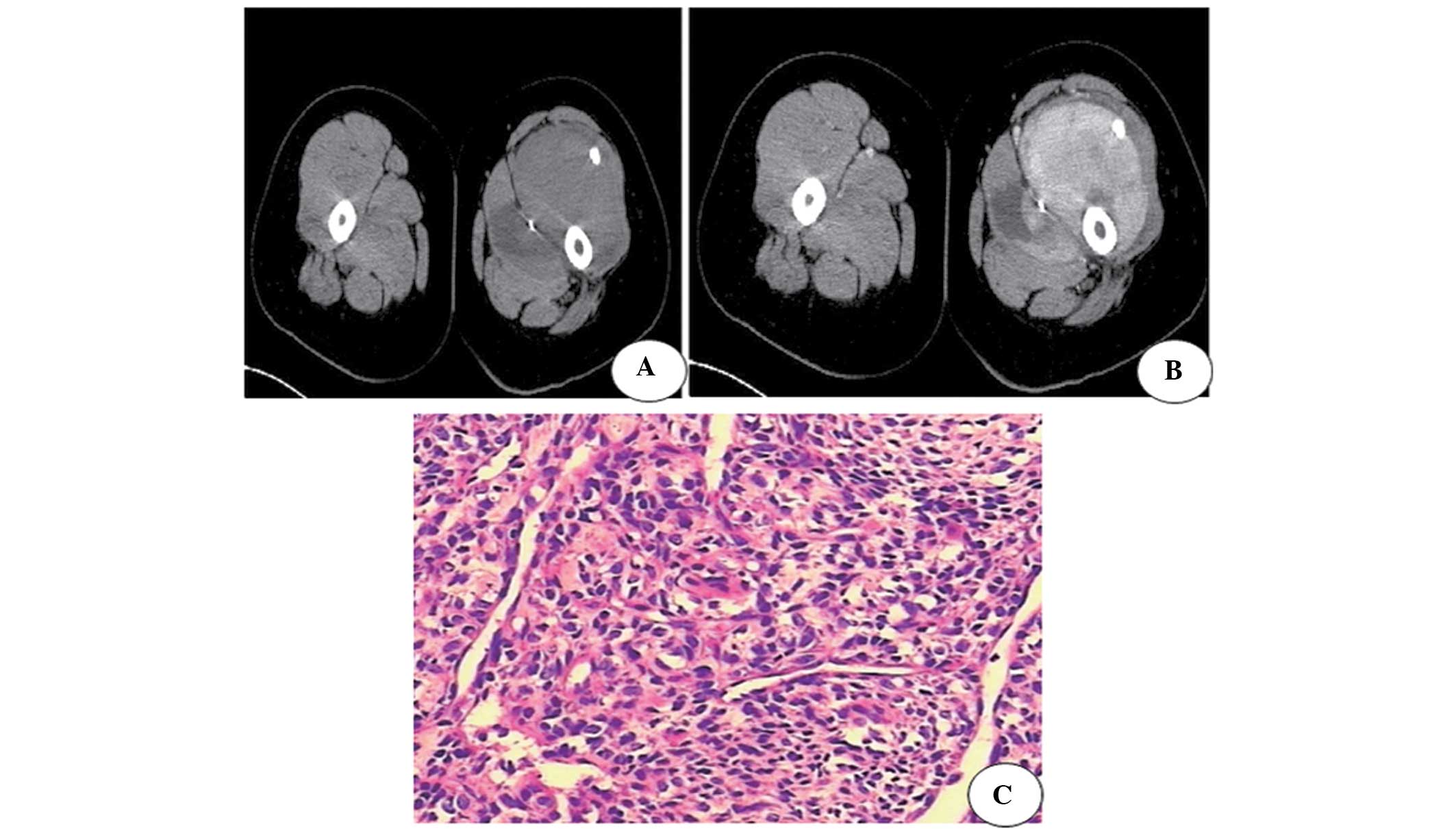
The images of nine tumors in the shoulder, groin and
upper thigh revealed lobulated masses deep in the intramuscular
space, with a density slightly lower than that of muscle. Six
lesions were unclearly defined and three were well-defined. The
masses were heterogeneous, with six lesions exhibiting punctate or
lobular calcification (Fig. 1) and
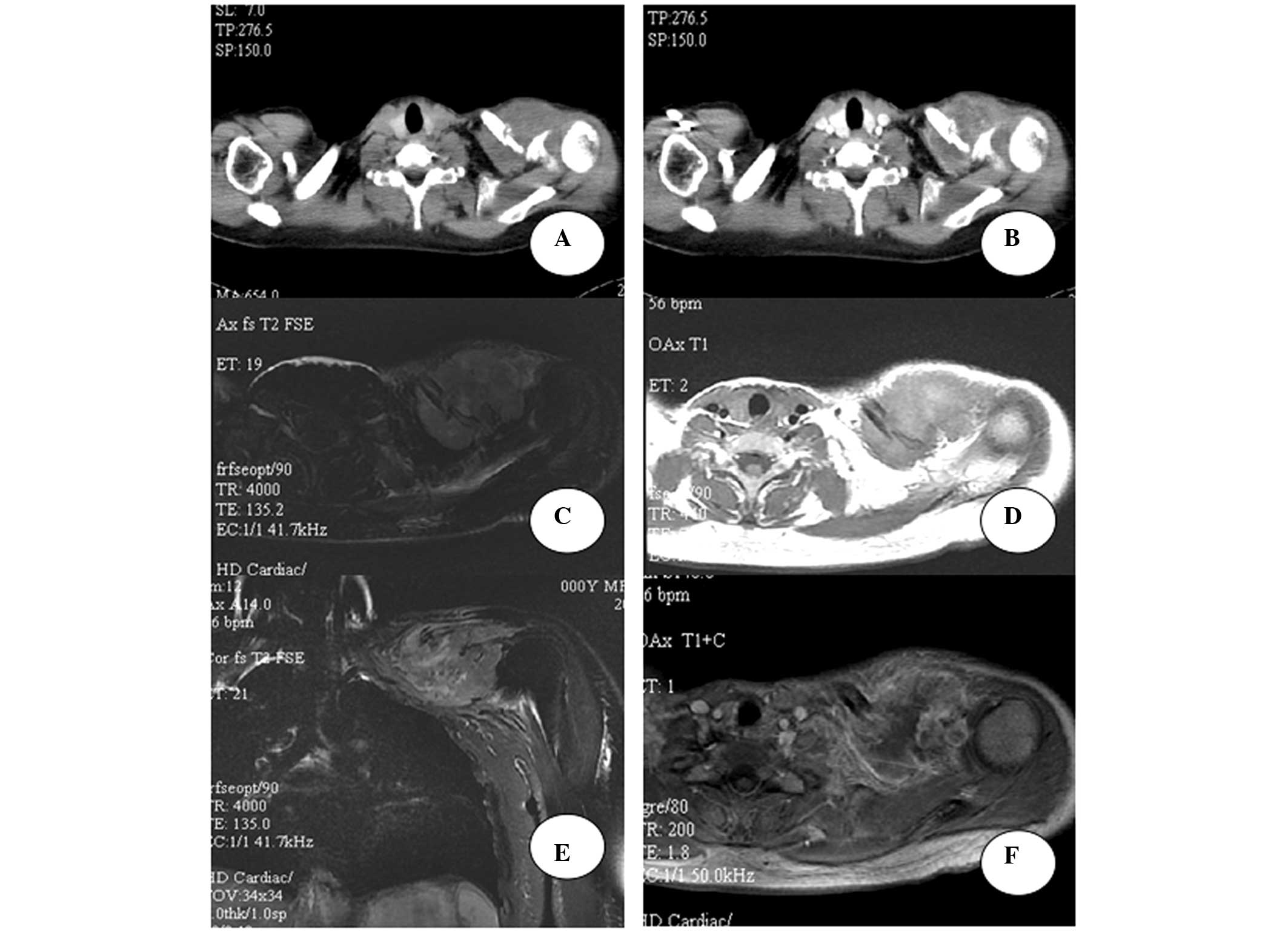
six lesions being in contact with the bone and causing osseous
destruction (Fig. 2A and B).
Contrast-enhanced scans revealed nine heterogeneously enhanced
lesions. No enhancement was observed in necrotic or cystic areas
(Figs. 1A and B and 2A and B).
Features of MRI
Images of the 18 lesions located on the spine, knee,
ankle and subclavian area close to the shoulder joint revealed that
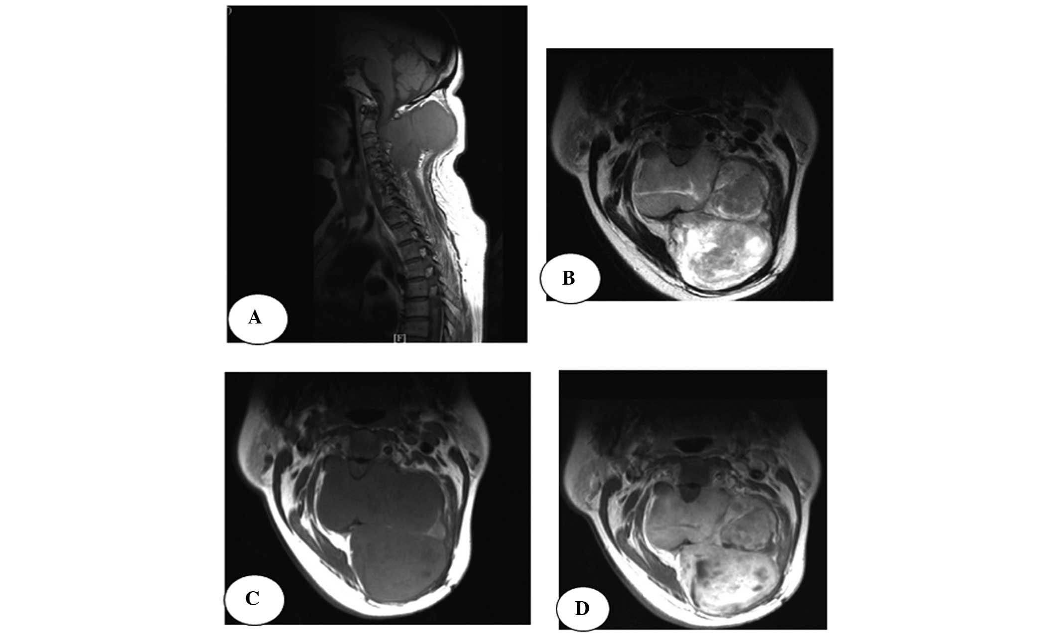
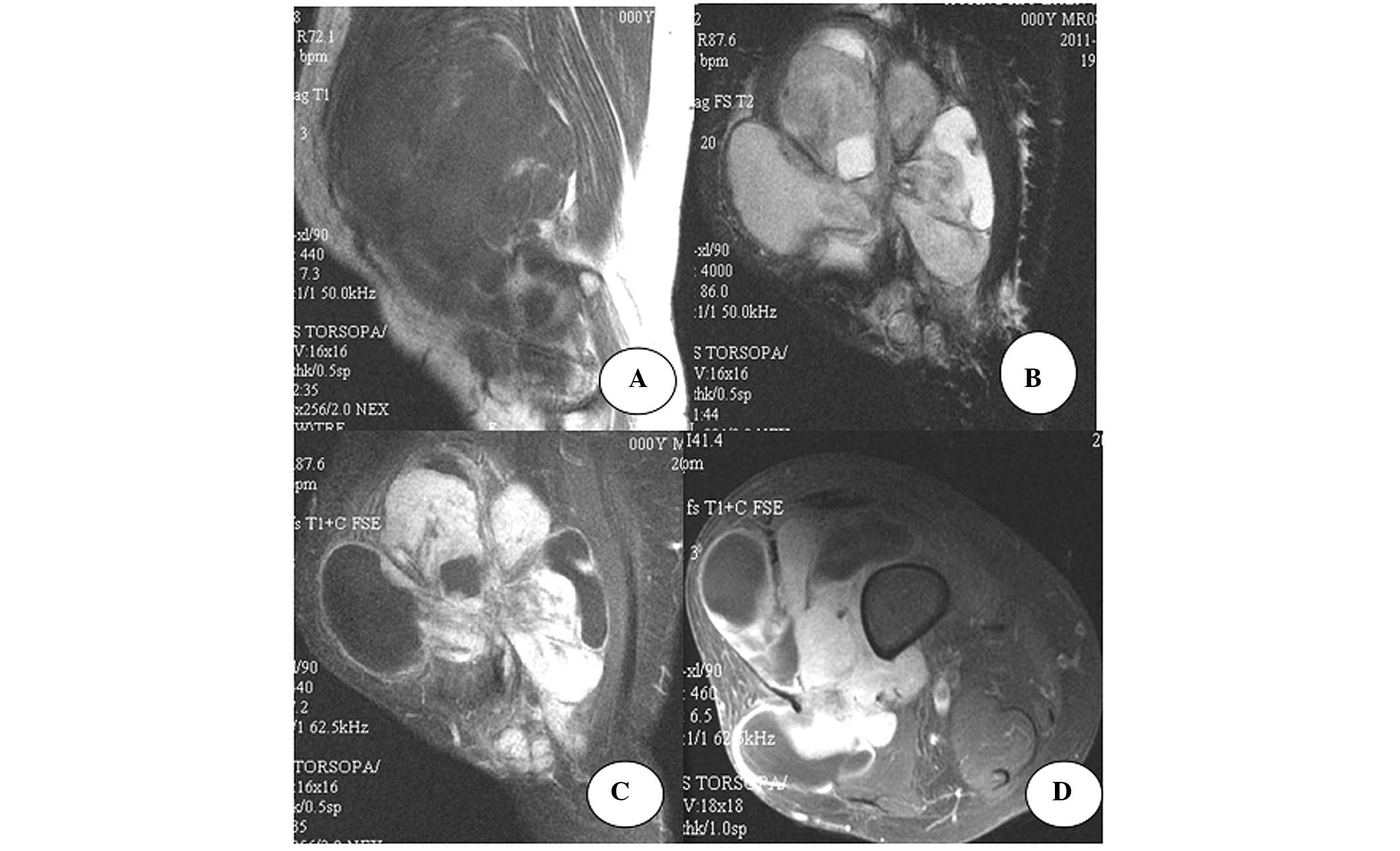
15 lesions were lobulated masses (Figs.
3 and 4) and deeply located,
with 12 well-defined lesions and three unclearly defined round
lesions. Overall, 15 lesions caused osseous destruction of
neighboring bones (Fig. 3). All the
lesions on the spine had compressed the spinal cord. All cases
demonstrated areas of hyperintensity, isointensity and
hypointensity relative to the muscle. Hyperintensive signals on
T1WI and T2WI were observed in necrotic and cystic areas. Nine
cases demonstrated a hypointensive T1 signal, suggesting
hemorrhage. Three cases with hypointensive T1 signals were
considered to possess fibrous tissue. In total, 12 lesions
exhibited a hypointensive T2 signal with internal septations.
Contrast-enhanced images revealed heterogeneous lesions (Figs. 2–4),
with no enhancement in the areas containing cysts, necrosis or
septation (Fig. 3D).
Discussion
Synovial sarcoma is a mesenchymal spindle cell
neoplasm, arising from mesenchymal tissues, with the histological
appearance of the synovium. Classic biphasic synovial sarcoma
exhibits differentiation of the tumor cells into epithelial cells
and fibroblasts, and is categorized into three subtypes, including
fibroblastic, epithelial and mixed-differentiation tumors (4). The current World Health Organization
classification includes synovial sarcomas under the ‘Tumors of
Uncertain Differentiation’ classification (5), which accounts for ~10% of all
soft-tissue sarcomas (1,2).
Synovial sarcoma is more common in young adults, but
can occur at any age, with half of the lesions occurring in
individuals between 20 and 40 years old. Males and females are
equally affected. Synovial sarcoma is considered the most common
malignant non-rhabdomyosarcomatous soft-tissue sarcoma in children
and adolescents (6–8). The lesions are commonly located in the
extremities, most often in the lower limbs, accounting for
two-thirds of synovial sarcomas. Extremity lesions typically occur
either in periarticular locations or close to a bursa or tendon
sheath. The most common locations are close to the knee and in
extra-articular positions, but rarely in an intra-articular
position (<10%) (9). Certain
studies have demonstrated that these lesions are associated with a
trauma that led to injury of the soft tissue around the joint
(10). Lesions are rarely observed
in the head, neck, mediastinum or peritoneum. Synovial sarcoma
grows slowly over two or three years. Patients often present with a
palpable, deeply located and painless soft-tissue mass. The lesion
generally does not cause significant dysfunction, however, certain
patients present with pain, tenderness on palpation and dysfunction
of the neighboring joint. In certain cases, pain may be the only
symptom in the early stage of the lesion (9). The primary treatment for synovial
sarcoma is surgery, which has a 50% post-operative recurrence rate,
usually within two years. Overall, ~40% of lesions metastasize to
the lungs, bones and lymph nodes (11).
In the current study, nine lesions were located on
the spine, which is a rarely affected site according to the
literature, indicating that synovial sarcoma may easily be
misdiagnosed as another type of tumor, if it is not located in the
extremities. Additionally, six patients presented with signs of
recurrence within one to two years post-surgery, and six other
patients presented with metastasis involving the lung and other
sites.
Based on the present results and those reported in
the literature, the imaging features of synovial sarcoma can be
summarized as follows: Firstly, the lesions are usually in
periarticular locations or deeply located within other sites, with
a well-defined, round or lobulated soft-tissue mass. Certain
lesions present with poorly-defined margins, with a tendency to
grow as a diffuse tumor mass along the tendon, tendon sheath and
interstitial space, enveloping neighboring tissues and joints, and
leading to osseous destruction of the neighboring bones. In
general, the size of the lesions tends to be large, with 85% of
tumors being >5 cm in size (12). In the current study, all 24 lesions
were deeply located within tissues. Morphologically, three lesions
were round and 21 lesions were lobulated soft-tissue masses. In
total, 15 lesions had well-defined margins, and nine were poorly
defined. All lesions were >5 cm in size, and 18 lesions
demonstrated neighboring bone destruction.
Secondly, images obtained from CT scans demonstrate
areas of isointensity relative to muscle and areas of
hypointensity, indicating necrosis and cysts. Areas of
calcification are observed in 20–30% of synovial sarcomas (8), with plaque or punctuate calcification
mainly located in the periphery of the lesion, termed peripheral
calcification. Rare cases of extensive calcification, resembling an
osteoid matrix or bone, and a case with a central punctate
calcification have been observed. This can aid in differentiating
synovial sarcoma from other types of soft-tissue sarcomas (13). In the present study, nine patients
underwent CT scans, revealing six tumors with peripheral
calcification, six tumors causing osseous destruction of the
neighboring bones and three patients who presented with a
pathological fracture. CT aids in the identification of subtle
soft-tissue calcifications and local bony changes.
Furthermore, MRI is one of the most common imaging
examinations for soft-tissue tumors and is considered the modality
of choice for the detection and staging of soft-tissue tumors. In
the present study, 18 patients underwent MRI examination, revealing
15 tumors that consisted of a heterogeneous mass. T1WI revealed
areas of isointensity or hyperintensity relative to muscle in the
tumor masses. Areas exhibiting a high signal intensity indicated
hemorrhage inside the tumor and a low signal intensity indicated
areas of necrosis or calcification. Cystic necrosis has been
reported to occur in poorly-differentiated lesions and lesions with
a large diameter (14). The tumors
in the present study all exhibited a diameter >5 cm and
hemorrhage was indicated in 12 cases. Jones et al studied
the T2WI results of synovial sarcoma in 34 patients and reported
that synovial sarcomas were frequently heterogeneous, with a
triple-signal intensity that depicted areas of high signal
intensity as fluid, isointensity or hyperintensity relative to fat
and hypointensity relative to fibrous tissue (15). Combined with pathohistological
studies, T2WI reveals that areas of hemosiderosis caused by
hemorrhage, calcification and fibrous tissue demonstrate low signal
intensity, the solid tumor mass exhibits a slightly higher signal
intensity, while areas of necrosis and hemorrhage reveal a
significantly higher signal intensity. However, not all synovial
sarcoma demonstrate a typical triple signal. In the present study,
only 12 cases (50%) exhibited this typical feature, which is
consistent with other studies (6,9,15–17).
Notably, 18–73% of cases present with fluid-fluid levels, which is
considered to be a specific imaging feature of synovial sarcoma
(6,8,13,15).
On T2WI, 12 cases in the present study exhibited hypointensive
internal septation, commonly inside the tumor or in multiple
nodules. Although internal septation is not considered to be a
specific feature of synovial sarcoma, its presentation with
septation often suggests a malignant tumor (18).
Finally, a contrast-enhanced imaging study revealed
that tumors of a large size often demonstrate significantly
heterogeneous enhanced signals on CT and MRI images, while tumors
of a smaller size tend to be homogeneously enhanced (19). Synovial sarcomas in locations other
than the extremities have similar imaging features to those in the
extremities (20–22). In the present study, 21 cases
exhibited significant heterogeneous enhancement, while no
enhancement was observed in areas of either cystic necrosis or
internal septations. The three patients that presented with
significant enhancement similar to that of the arteries were
misdiagnosed with aneurysms.
In the literature, all soft-tissue sarcomas, with
the exception of liposarcoma, lack specific imaging characteristics
(23). The accuracy of the
pre-operative diagnosis of soft-tissue sarcomas is only 25%
(24). A synovial sarcoma located
close to the joint must be distinguished from pigmented
villonodular synovitis, which is a slow-growing, homogeneous and
well-defined tumor that often exhibits high signal intensity on
T1WI and low signal intensity on T2WI, usually homogeneously
enhanced and rarely featuring calcification or osteolytic
destruction (9). Synovial sarcoma
must be distinguished from fibrosarcoma, malignant fibrous
histiocytoma, invasive fibroma, leiomyosarcoma and rhabdomyosarcoma
(25). Fibrosarcoma often occurs in
older patients and tends to be extremely large in size, with less
osseous destruction and no significant calcification. Malignant
fibrous histiocytoma often occurs in patients aged 50–70 years, is
often located in the thigh and is poorly-defined, with a low degree
of calcification. Contrast-enhanced scans frequently reveal
significant enhancement.
Invasive fibroma most often occurs in middle-aged
patients and is frequently located in the thigh, abdominal wall and
retroperitoneal space. Generally, these tumors are well-defined and
homogeneous, demonstrating hypointensity relative to muscle. On
T1WI and T2WI, invasive fibroma tends to present with a
low-intensity signal due to the high content of fibrous tissues and
is gradually enhanced on contrast-enhanced scans. Leiomyosarcoma is
more commonly observed in the uterus and gastrointestinal tract.
Necrosis, hemorrhage and cystic change often occur within the
tumor. On T1WI, leiomyosarcoma often exhibits the same isointensity
as that of muscle. Rhabdomyosarcoma is a common malignant tumor in
children and is often a poorly-defined, painless and deeply located
tumor mass. On T1WI, the tumor exhibits an intensity similar to
that of muscle. Well-defined, slow-growing synovial sarcoma without
infiltrative osseous destruction may be difficult to distinguish
from benign lesions.
In conclusion, the following characteristics of
synovial sarcoma, taken together, may aid in its diagnosis: i) A
lobulated or round soft-tissue mass that develops near the joints
of extremities, particularly in the lower limbs; ii) the tumor can
be well-defined or poorly-defined; iii) the neighboring bones are
affected due to infiltration or compression by the tumor; iv)
calcification or bone structure can be observed in the periphery of
the tumor; v) a high-intensity signal exhibited by the tumor on
T1WI, suggesting hemorrhage, and a triple signal demonstrated on
T2WI; vi) contrast-enhanced imaging reveals significant
heterogeneous enhancement, and areas of cystic necrosis and
internal septations can be observed on enhancement; and vii) tumors
often occur in young or middle-aged adults. However, due to the
rarity of synovial sarcoma in sites other than the extremities,
pre-operative diagnosis must rely on other pathological
examinations apart from imaging examinations. CT and MRI each have
their own advantages in evaluating synovial sarcoma. The
combination of the two approaches can improve the accuracy of the
pre-operative diagnosis. However, the final diagnosis relies on
pathological investigation.
Acknowledgements
The authors would like to thank Professor Fen-bao Li
(Director of the Interventional Ward, The First Affiliated Hospital
of Xinxiang Medical College of Interventional Medicine, Xinxiang,
China) and colleagues from other departments for their
contributions and selfless assistance in writing this study.
References
|
1
|
Fisher C, de Brujin DRH and van Kessel AG:
Synovial sarcoma. World Health Organization Classification of
Tumours. Pathology and Genetics of Tumours of Soft Tissue and Bone.
Fletcher CDM, Unni KK and Mertens F: IARC Press; Lyon: pp. 200–204.
2002
|
|
2
|
Wu Z and Huang XZ: Clinical study of 72
cases of synovial sarcoma. Chin Clin Oncol. 9:84–85. 2004.(In
Chinese).
|
|
3
|
Fisher C: Synovial sarcoma. Ann Diagn
Pathol. 2:401–421. 1998. View Article : Google Scholar
|
|
4
|
Liu GR, Huang YS, Lan BW and He ZH:
Imaging diagnosis of synoviosarcoma (report of 10 cases). J Diagn
Imaging Interv Radiol. 10:83–85. 2001.
|
|
5
|
Zhu XZ: Current WHO classification of soft
tissue tumor. Diagn Interv Imaging. 19:94–96. 2003.
|
|
6
|
Zhang ZH, Meng QF and Zhang XL: MRI
diagnosis of synovial sarcoma of extremities. J Clin Radiol.
25:941–944. 2006.
|
|
7
|
McCarville MB, Spunt SL, Skapek SX and
Pappo AS: Synovial sarcoma in pediatric patients. AJR Am J
Roentgenol. 179:797–801. 2002. View Article : Google Scholar : PubMed/NCBI
|
|
8
|
O’Sullivan PJ, Harris AC and Munk PL:
Radiological features of synovial cell sarcoma. Br J Radiol.
81:346–356. 2008. View Article : Google Scholar
|
|
9
|
Li F, Wang RF, Qi L, et al: Synovial
sarcoma of soft tissues: a CT and MRI study. Radiologic Practice.
25:1396–1399. 2010.
|
|
10
|
Van Hul E, Vanhoenacker F, Van Dyck P, De
Schepper A and Parizel PM: Pseudotumoural soft tissue lesions of
the foot and ankle: a pictorial review. Insights into Imaging.
2:439–452. 2011. View Article : Google Scholar
|
|
11
|
Ren XH, Wu XM, Jin C and Cui YA: Diagnosis
and treatment progress of synovial sarcoma. Medical Recapitulate.
15:541–542. 2009.
|
|
12
|
Kransdorf MJ: Malignant soft-tissue tumors
in a large referral population: distribution of diagnoses by age,
sex, and location. AJR Am J Roentgenol. 164:129–134. 1995.
View Article : Google Scholar : PubMed/NCBI
|
|
13
|
Tateishi U, Hasegawa T, Beppu Y, Satake M
and Moriyama N: Synovial sarcoma of the soft tissues: prognostic
significance of imaging features. J Comput Assist Tomogr.
28:140–148. 2004. View Article : Google Scholar : PubMed/NCBI
|
|
14
|
Bhosale P, Balachandran A and Tamm E:
Imaging of benign and malignant cystic pancreatic lesions and a
strategy for follow up. World J Radiol. 2:345–353. 2010. View Article : Google Scholar : PubMed/NCBI
|
|
15
|
Jones BC, Sundaram M and Kransdorf MJ:
Synovial sarcoma: MR imaging findings in 34 patients. AJR Am J
Roentgenol. 161:827–830. 1993. View Article : Google Scholar : PubMed/NCBI
|
|
16
|
Chen JY, Liu QY, Ye RX, Zhong JL and Liang
BL: Correlation of MR imaging features and histopathology of
synovial sarcoma. Ai Zheng. 24:87–90. 2005.PubMed/NCBI
|
|
17
|
Marzano L, Failoni S, Gallazzi M and
Garbagna P: The role of diagnostic imaging in synovial sarcoma. Our
experience. Radiol Med. 107:533–540. 2004.PubMed/NCBI
|
|
18
|
Fang TS, Xu YK, Peng JY and Fan CS:
Imaging analysis of synovial sarcoma of extremities. J Chin Clin
Med Imaging. 2:106–109. 2009.
|
|
19
|
Rangheard AS, Vanel D, Viala J, Schwaab G,
Casiraghi O and Sigal R: Synovial sarcomas of the head and neck: CT
and MR imaging findings of eight patients. AJNR Am J Neuroradiol.
22:851–857. 2001.PubMed/NCBI
|
|
20
|
Koehler SM, Beasley MB, Chin CS, Wittig
JC, Hecht AC and Qureshi SA: Synovial sarcoma of the thoracic
spine. Spine J. 9:e1–e6. 2009. View Article : Google Scholar : PubMed/NCBI
|
|
21
|
Shaariyah MM, Mazita A, Masaany M, Razif
MY, Isa MR and Asma A: Synovial sarcoma: a rare presentation of
parapharyngeal mass. Chin J Cancer. 29:631–633. 2010. View Article : Google Scholar : PubMed/NCBI
|
|
22
|
Huang ZT, Chen WG, Jia M, et al: One case:
synovial sarcoma of adnexal bone in the third thoracic vertebra. J
Pract Radiol. 12:1563–1564. 2006.
|
|
23
|
Brisse HJ, Orbach D and Klijanienko J:
Soft tissue tumours: imaging strategy. Pediatr Radiol.
40:1019–1028. 2010. View Article : Google Scholar : PubMed/NCBI
|
|
24
|
Cormier JN and Pollock RE: Soft tissue
sarcomas. CA Cancer J Clin. 54:94–109. 2004. View Article : Google Scholar : PubMed/NCBI
|
|
25
|
Vander Salm TJ: Unusual primary tumors of
the heart. Semin Thorac Cardiovasc Surg. 12:89–100. 2000.
View Article : Google Scholar : PubMed/NCBI
|


