Introduction
Lung cancer is the most common cause of
cancer-related mortality worldwide, accounting for 17% of all
cancer mortalities (1). Non-small
cell lung cancer (NSCLC) is the predominant type of lung cancer,
which mainly includes squamous cell carcinoma, large cell carcinoma
and adenocarcinoma (2). Surgery is
the first choice of treatment for early-stage NSCLC, while
chemotherapy and radiotherapy are often administered to advanced
NSCLC patients (3). However, the
majority of advanced-stage NSCLC patients face unsatisfactory
outcomes. Targeted molecular therapy has attained good effects in
the treatment of NSCLC. However, the major challenges are variable
responsiveness and the development of drug resistance (4). Therefore, there is an urgent
requirement to find new therapeutic targets for the treatment of
NSCLC.
When DNA is damaged, the G2 cell cycle checkpoint
prevents cells from entering mitosis, allowing DNA repair to occur
and halting the proliferation of damaged cells (5). Additionally, the role of the G2
checkpoint in facilitating the maintenance of genomic stability
indicates that it is important in understanding the molecular
mechanism of lung cancer. Ataxia telangiectasia mutated (ATM)
kinase, and ataxia telangiectasia and Rad3-related (ATR) kinase are
two serine/threonine kinases that regulate cell cycle checkpoints
and DNA repair in response to exposed DNA double-stranded breaks
(6,7). ATM and ATR kinase act upstream of
checkpoint kinases (Chk) 1 and 2; ATM/ATR phosphorylates Chk1 at
Ser317 and Ser345 (8), and Chk2 at
Thr68 and other sites in the amino-terminal domain, in response to
blocked DNA replication, particularly when caused by DNA
double-stranded breaks (9).
Activated Chk1/2 then exerts its checkpoint mechanism on the cell
cycle, in part, by regulating the cell division cycle 25 (Cdc25)
family of phosphatases, inactivating Cdc25C via phosphorylation at
Ser216, thus preventing the activation of cyclin-dependent kinase 1
(Cdk1) and the transition of the cell into mitosis (10). The entry of all eukaryotic cells
into mitosis is regulated by the activation of Cdk1 at the G2/M
transition. Cdk1 activation is a multi-step process that is
initiated by the binding of the regulatory subunit, cyclin B1, to
Cdk1 to form the mitosis-promoting factor (MPF) (11). MPF remains in an inactive state
until the phosphorylation of Cdk1 at Thr161 by Cdk activating
kinase (CAK) (12) and the
dephosphorylation of Cdk1 at Thr14/Tyr15 by phosphatase Cdc25C
(13); thus, active Cdk1 refers to
dephospho-Cdk1 (Tyr15) and phospho-Cdk1 (Thr161). Furthermore,
active Cdk1 facilitates the smooth transition of lung cancer cells
from the G2 phase to the M phase, and promotes cell growth and
proliferation. Therefore, it has been proposed that the
ATM/ATR-Chk1/2-Cdc25C-Cdk1/cyclin B1 signaling pathway is important
in G2/M arrest in response to DNA damage in lung cancer. The
present study was performed to retrospectively assess the effects
of the expression levels of G2/M signaling pathway proteins in
NSCLC tissues, as determined by immunohistochemical (IHC) methods,
on the prediction of the overall survival (OS) of advanced-stage
NSCLC patients.
Patients and methods
Patients and tissue specimens
Consecutive patients with pathological stage III and
IV squamous cell carcinoma (SCC) or adenocarcinoma (ADC) were
retrospectively enrolled in the present study from two university
hospitals (Xijing Hospital and Shaanxi Provincial Second People’s
Hospital) in Xi’an, China. Patients who received docetaxel and
cisplatin doublet chemotherapy for >2 cycles and standard
supportive care between June 2008 and July 2011 were qualified for
involvement in the present study. Tumors were staged according to
the seventh edition of the International System for Staging Lung
Cancer developed by the American Joint Committee for Cancer
(14), and histopathological tumor
types were determined according to the classification of the World
Health Organization (15). The
inclusion criteria were as follows: i) Tumor unsuitable for
surgical removal; ii) availability of formalin-fixed,
paraffin-embedded primary lung cancer tissue blocks; and iii)
availability of OS data. Patients who had previously received <2
cycles of docetaxel and cisplatin doublet chemotherapy, had
previously experienced severe liver and kidney dysfunction or had a
history of other types of cancer were excluded from the present
study. To increase the quality of the data obtained, the reports of
the tissue pathology, the radiological examinations, including
chest computed tomography scans and positron emission tomography,
and the clinical information for all patients were independently
reviewed. The scientific study committee of Xijing Hospital (Xi’an,
China) for pulmonary diseases reviewed and approved the database.
Consent was obtained from the families of the patients.
IHC staining
IHC analysis was conducted using BOND-MAX™ (cat. no.
M 211-64; Leica Microsystems Ltd., Wetzlar, Germany), a fully
automatic IHC and in situ hybridization machine. From the
prepared sections, one section containing the maximum amount tumor
tissue and minimal or absent necrosis and hemorrhage was selected
for each patient. The selected sections were deparaffinized via a
graduated alcohol and xylene series followed by rehydration in
distilled water. Antigen retrieval was performed by adding citrate
buffer (pH 6.0) and heating in a microwave oven for 20 min at
100°C. The sections were subsequently incubated in a 3% hydrogen
peroxide solution to block endogenous peroxidase activity and then
washed with a phosphate-buffered saline solution. Following
incubation with blocking solution for 20 min, the sections were
incubated again with primary and then secondary antibodies at
appropriate dilutions. Cdk1 requires Thr14/Tyr15 dephosphorylation
by phosphatase Cdc25C and Thr161 phosphorylation by CAK to
transition from an inactive to active protein (16), therefore, the present study used a
rabbit monoclonal anti-Cdk1 antibody (dephospho Cdk1 Tyr15; cat no.
ab32384; dilution, 1:200) to stain the Cdk1 protein without
phosphorylation of Tyr15, a rabbit polyclonal anti-Cdk1 antibody
(phospho-Thr161; cat no. ab47329; dilution, 1:100) to stain
phospho-Cdk1, and a mouse anti-Cdk1 monoclonal antibody (total
Cdk1; cat no. ab8040; dilution, 1:200) to stain the total Cdk1
protein (all Abcam, Cambridge, UK). The reaction was visualized
using a 3,3′-diaminbenzidine substrate system (cat. no. 08102;
Leica Microsystems Ltd.) and counterstaining was performed using
Mayer’s hematoxylin. All of the aforementioned procedures were
performed in accordance with the antibody manufacturer’s
instructions.
Evaluation of protein expression
level
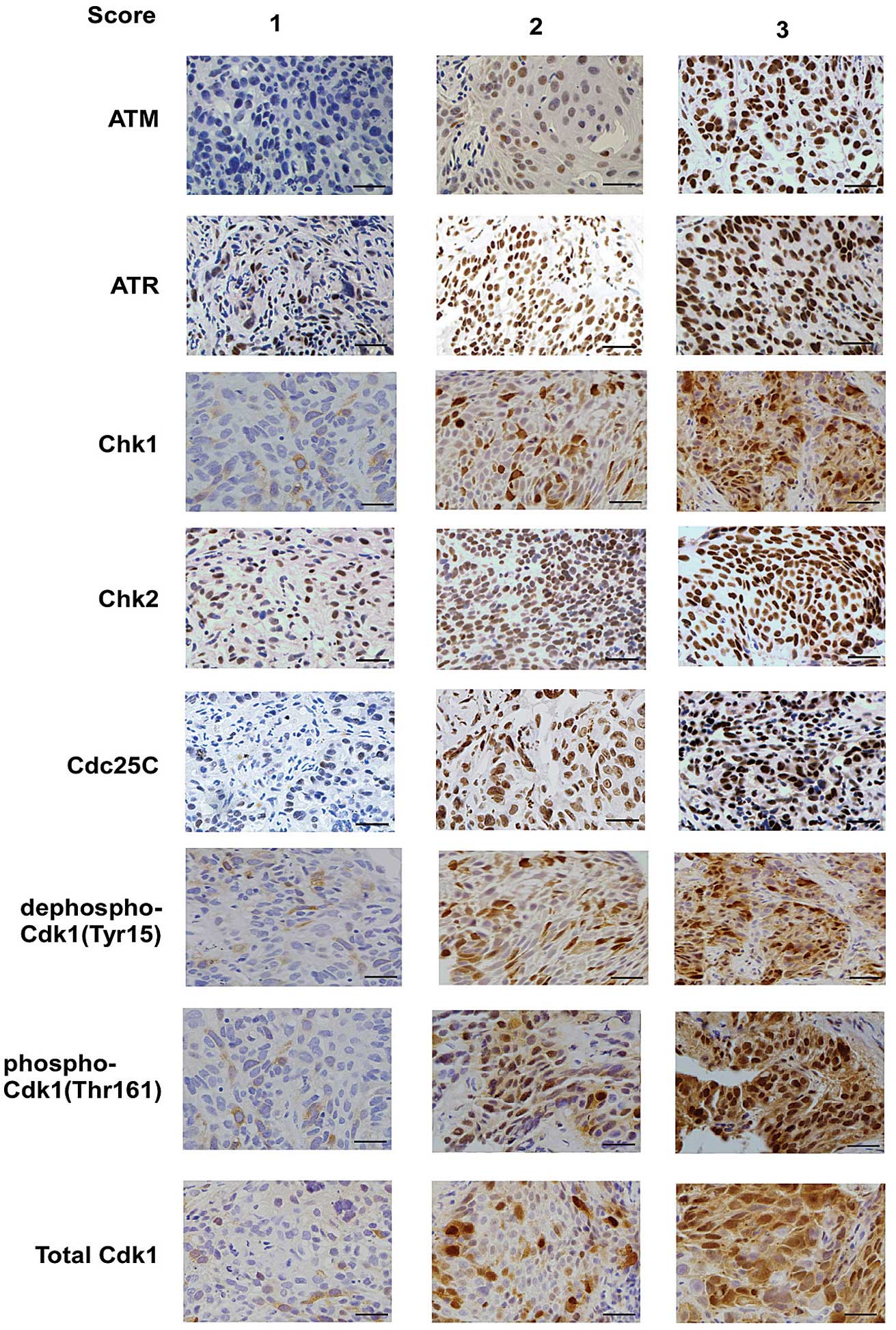
Two pathologists who were blinded to the clinical
data independently evaluated the IHC staining. The staining
intensity of each protein in the cancer cells was graded on a scale
of 1–3 (1, weak; 2, moderate; and 3, strong) (Fig. 1), and the percentage of cancer cells
positive for each protein was determined and assigned as a
proportion score (0, 0%; 0.1, 1–9%; 0.5, 10–49%; and 1.0, ≥50%), as
previously described (17). The
intensity and proportion scores were then multiplied to yield the
semiquantitative H-score. The median value of all the mean H scores
was selected as the cutoff value for each protein to separate the
cancer cells with high and low expression levels (18). Furthermore, OS was calculated using
the day of the lung cancer diagnosis as the first day and the day
of mortality as the final day.
Statistical analysis
The cases were evaluated for demographic and
pathological variables, and the expression of the proteins was
dichotomized as low versus high. Patient cumulative survival was
analyzed using the Kaplan-Meier method, with the date of
pathological diagnosis defined as time zero and mortality as the
end-point. Differences in survival were determined by performing a
log-rank test in the univariate analyses and by using a Cox
proportional hazards regression model with backward Wald for
prognostic factors in the multivariate analyses. All analyses were
performed using SPSS software (version 13.0; SPSS, Inc., Chicago,
IL, USA) and P<0.05 was considered to indicate a statistically
significant difference.
Results
Patient characteristics and univariate
analysis
Table I indicates
the clinical characteristics of the 144 patients in the present
study, including 64 cases of squamous cell carcinoma, 69 cases of
adenocarcinoma and 11 cases of other types of NSCLC. The median age
of the patients was 58.4 years. The patients included 109 males and
35 females, 93 (64.58%) of whom were ever-smokers and 51 (35.42%)
of whom were never-smokers. The predominant pathological
tumor-node-metastasis (TNM) stages of the patients were stage III
(45 patients; 31.25%) and stage IV (99 patients; 68.75%).
Furthermore, according to univariate analysis, determined by
log-rank test, the parameters of age, gender, smoking habit,
histology, tumor size (T) and extent of lymphatic metastasis (N)
were not significantly associated with the survival of the patients
with advanced NSCLC (Table I);
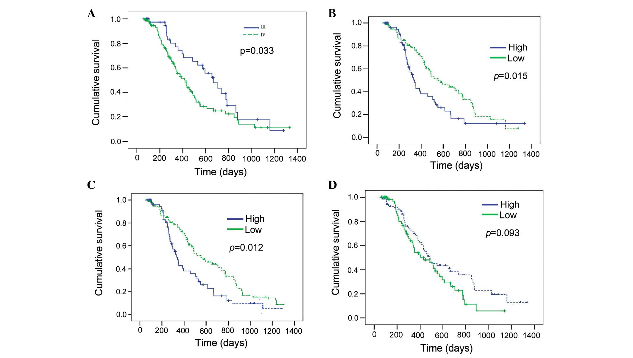
however, as expected, the pathological TNM stage was a significant
prognostic factor. The one-year survival rate was 74.20% for stage
III patients (median survival, 666 days) and 55.60% for stage IV
patients (median survival, 415 days) (P=0.033; Table I). Additionally, stage III patients
exhibited a more favorable prognosis compared with stage IV
patients (Fig. 2A).
 | Table IUnivariate analysis of prognostic
factors in advanced non-small cell lung cancer patients
(n=144). |
Table I
Univariate analysis of prognostic
factors in advanced non-small cell lung cancer patients
(n=144).
| Parameter | Patients, n (%) | One-year survival
rate, % | Median survival,
days | P-valuea |
|---|
| Gender | | | | 0.676 |
| Male | 109 (75.69) | 62.30 | 465 | |
| Female | 35 (24.31) | 58.30 | 430 | |
| Smoking habit | | | | 0.608 |
| Never | 51 (35.42) | 62.40 | 490 | |
| Ever | 93 (64.58) | 62.40 | 465 | |
| Histology | | | | 0.527 |
| Squamous cell | 64 (44.44) | 59.90 | 430 | |
| Adenocarcinoma | 69 (47.92) | 61.90 | 552 | |
| Other | 11 (7.64) | 55.60 | 389 | |
| T classification | | | | 0.998 |
| T1 | 12 (8.34) | 50 | 350 | |
| T2 | 48 (33.33) | 62.30 | 487 | |
| T3 | 25 (17.36) | 72.60 | 503 | |
| T4 | 59 (40.97) | 58.80 | 437 | |
| N classification | | | | 0.173 |
| N0 | 38 (26.38) | 66.50 | 550 | |
| N1 | 13 (9.03) | 66.70 | 773 | |
| N2 | 62 (43.06) | 56.20 | 428 | |
| N3 | 31 (21.53) | 60.40 | 430 | |
| Pathological TNM
stage | | | | 0.033 |
| III | 45 (31.25) | 74.20 | 666 | |
| IV | 99 (68.75) | 55.60 | 415 | |
| ATM expression | | | | 0.843 |
| High | 66 (45.83) | 57.00 | 389 | |
| Low | 78 (54.17) | 64.20 | 514 | |
| ATR expression | | | | 0.245 |
| High | 60 (41.67) | 58.90 | 415 | |
| Low | 84 (58.33) | 63.00 | 503 | |
| Chk1
expression | | | | 0.341 |
| High | 90 (62.50) | 71.50 | 487 | |
| Low | 54 (37.50) | 58.50 | 465 | |
| Chk2
expression | | | | 0.559 |
| High | 100 (69.44) | 62.20 | 465 | |
| Low | 44 (30.56) | 59.90 | 457 | |
| Cdc25C
expression | | | | 0.649 |
| High | 97 (67.36) | 61.50 | 465 | |
| Low | 47 (32.64) | 60.60 | 457 | |
| Total Cdk1
expression | | | | 0.093 |
| High | 71 (49.31) | 68.10 | 487 | |
| Low | 73 (50.69) | 54.20 | 430 | |
| Dephospho-Cdk1
(Tyr15) expression | | | | 0.015 |
| High | 64 (44.44) | 42.90 | 339 | |
| Low | 80 (55.56) | 74.00 | 552 | |
| Phospho-Cdk1
(Thr161) expression | | | | 0.012 |
| High | 65 (45.14) | 41.32 | 334 | |
| Low | 79 (54.86) | 76.78 | 562 | |
Prognostic value of proteins involved in
the G2/M arrest signaling pathway
According to the univariate analysis determined by
log-rank test, dephospho-Cdk1 (Tyr15; P=0.015) and phospho-Cdk1
(Thr161; P=0.012) exhibited prognostic significance, while the
other proteins exhibited no significant difference in patient
survival (ATM, P=0.843; ATR, P=0.245; Chk1, P=0.341; Chk2, P=0.559;
Cdc25C, P=0.649; total Cdk1, P=0.093) (Table I; Fig.
2B–D). Kaplan-Meier survival analysis indicated that high
expression levels of dephospho-Cdk1 (Tyr15) and phospho-Cdk1
(Thr161) correlated with a worse prognosis in the advanced NSCLC
patients, whereas the patients with tumors exhibiting low
dephospho-Cdk1 (Tyr15) and phospho-Cdk1 (Thr161) expression levels
exhibited a more favorable prognosis (Fig. 2B and C). Furthermore, the one-year
survival rates of the patients were 42.90% for high dephospho-Cdk1
(Tyr15) expression (median survival, 339 days) and 74.00% for low
dephospho-Cdk1 (Tyr15) expression (median survival, 552 days). The
same trend was observed in phospho-Cdk1 (Thr161), with one-year
survival rates of 41.32% for high phospho-Cdk1 (Thr161) expression
(median survival, 334 days) and 76.78% for low expression (median
survival, 562 days) (Table I;
Fig. 2B and C). Factors that were
determined to affect the survival rate in the univariate analysis
(smoking habit, N classification, pathological TNM stage and Cdk1
expression) were analyzed in a multivariate Cox regression analysis
of factors that may affect the survival rate. This analysis
demonstrated that the expression of dephospho-Cdk1 [Tyr15; odds
ratio (OR), 0.619; 95% confidence interval (CI), (0.458–0.925);
P=0.032] and phospho-Cdk1 (Thr161; OR, 0.631; 95% CI, 0.412–0.961;
P=0.026) were independent prognostic factors of NSCLC (Table II). In addition, the prognostic
role of Cdk1 in advanced NSCLC was validated by combining the
expression levels of dephospho- and phospho-Cdk1; Cox regression
analysis of this variant (active Cdk1) determined that active Cdk1
expression (OR, 0.624; 95% CI, 0.400–0.973; P=0.038) was also an
independent prognostic factor of NSCLC (Table II). As expected, the pathological
TNM stage (OR, 0.515; 95% CI, 0.297–0.894; P=0.018) was identified
as an independent prognostic factor, however, smoking habit and N
classification exhibited no significance with regard to the
prognosis of NSCLC (Table II).
| Table IIPrognostic significance of
pathological TNM stage and Cdk1 expression revealed by multivariate
analyses in advanced non-small cell lung cancer patients
(n=144). |
Table II
Prognostic significance of
pathological TNM stage and Cdk1 expression revealed by multivariate
analyses in advanced non-small cell lung cancer patients
(n=144).
| Multivariate
P-value | Odds ratio | 95% confidence
incidence |
|---|
| Smoking habit | 0.066 | 0.619 | 0.372–1.033 |
| N
classificationa | 0.094 | | |
| N1 | 0.441 | 0.648 | 0.215–1.950 |
| N2 | 0.104 | 1.569 | 0.912–2.700 |
| N3 | 0.07 | 1.909 | 0.949–3.838 |
| Pathological TNM
stage | 0.018 | 0.515 | 0.297–0.894 |
| Dephospho-Cdk1
(Tyr15) expression | 0.032 | 0.619 | 0.458–0.925 |
| Phospho-Cdk1
(Thr161) expression | 0.026 | 0.631 | 0.412–0.961 |
| Active Cdk1
expression | 0.038 | 0.624 | 0.400–0.973 |
Discussion
Early-stage NSCLC patients typically exhibit a high
five-year survival rate following curative surgery plus adjuvant
chemotherapy and radiotherapy (19). However, numerous patients with
advanced NSCLC (stages III and IV) succumb quickly due to disease
relapse, despite the administration of a combination of
multidisciplinary treatments (20).
Pathological TNM staging aids in the prediction of the OS of a
group of patients, however, it cannot provide a molecular target
for subsequent treatment. Thus, independent prognostic molecular
markers, which may additionally serve as treatment targets, must be
identified for advanced NSCLC. A number of studies for
molecular-targeted treatment in advanced NSCLC have been
successful. For example, epidermal growth factor (EGFR) mutations
(21) and anaplastic lymphoma
kinase (ALK) rearrangements (22)
were identified as independent prognostic factors for advanced
NSCLC, thus, EGFR tyrosine kinase inhibitors (23) and ALK inhibitors (24) were developed to target these two
genes, and proved successful in the treatment of advanced NSCLC.
The present study included patients with advanced-stage tumors who
were treated with multidisciplinary modalities. Additionally,
factors that may affect the prognosis of a group of patients, such
as age, gender, smoking habit, N classification, pathological TNM
stage and histological type, were included in the statistical
analysis. This was expected to reveal the value of G2/M signaling
pathway proteins as prognostic biomarkers of advanced NSCLC.
In the present study, univariate analysis determined
that age, gender, smoking habit, histology, and T and N
classification were not significantly associated with survival in
the advanced NSCLC patients. This may be attributed to the advanced
stage (stage III and IV) of the patients included in the present
study, which diminishes the effect of these factors on patient
prognosis. However, pathological TNM stage remained a strong
prognostic factor (P=0.033; Table
I) and Cox regression analysis demonstrated that pathological
TNM stage was an independent prognostic factor for the advanced
NSCLC patients (P=0.018; Table
II); this prognostic significance may indicate the reliability
of the current study.
In addition, univariate analysis demonstrated that
the protein expression levels of ATM, ATR, Chk1, Chk2, Cdc25C and
total Cdk1 were not significantly associated with a difference in
the survival of advanced NSCLC patients (P>0.05). The prognostic
role of a number of the aforementioned proteins has previously been
studied in early-stage NSCLC. For example, Choi et al
(25) reported that the protein
expression level of ATM and Chk2 had no effect on the OS of stage I
NSCLC patients, and Grabauskiene et al (26) identified that elevated Chk1
expression in early-stage primary lung adenocarcinoma (442 resected
specimens) correlated with poor tumor differentiation and
significantly diminished patient survival. However, the prognostic
role of the Chk1 expression level could not be validated in the
present study, possibly due to the advanced stage of the included
NSCLC patients. Wu et al (27) analyzed primary tumors and
corresponding healthy lung tissues from 40 NSCLC patients and
reported no Cdc25C overexpression and no association with patient
survival. Furthermore, Abdulkader et al (28) investigated a series of 205
carcinomas of the large bowel, breast, lung and prostate, and
determined that Cdk1 expression was not associated with the
prognosis of early-stage NSCLC. Cdk1 is located at the end of the
G2/M signaling pathway, and is therefore key in the G2/M arrest and
cell apoptosis induced by chemotherapy and radiotherapy in tumor
cells (29). Therefore, to identify
the prognostic role of Cdk1 protein, active dephospho-Cdk1 (Tyr15)
and phospho-Cdk1 (Thr161) were investigated.
In the present study, the log-rank test identified
that dephospho-Cdk1 (Tyr15) and phospho-Cdk1 (Thr161) exhibit
prognostic significance in advanced NSCLC patients. In addition,
the Cox regression model revealed active Cdk1 to be an independent
prognostic factor for NSCLC patients. Patients with high active
Cdk1-expression tumors exhibited a significantly shorter survival
time compared with low active Cdk1-expressing tumors, indicating
that advanced NSCLC patients may benefit from Cdk1 inhibitory
treatment. In the cell cycle, Cdk1 is a master modulator of
initiation and transition through mitosis, with high active Cdk1
expression levels able to promote G2/M transition and accelerate
tumor cell growth (30). Previous
studies have demonstrated that decreased phospho-Cdk1 (Thr161)
expression (30), as well as the
accumulation of phospho-Cdk1 (Tyr15) (31), are involved in G2/M arrest and
apoptosis in lung cancer, thus validating Cdk1 as a possible
therapeutic target. Furthermore, Vassilev et al (32) previously identified a selective
small-molecule inhibitor of Cdk1 that reversibly arrests human
cells at the G2/M phase and induces apoptosis in tumor cells,
indicating that selective Cdk1 inhibitors may have potential
clinical utility in cancer therapy. Moreover, Cdk1-regulated G2/M
arrest and cell apoptosis are involved in the molecular mechanisms
of a number of chemotherapeutic agents and radiotherapy regimens
(33); thus, Cdk1 inhibitors may
serve to sensitize cells to chemotherapy and radiotherapy in cases
of advanced NSCLC that are specifically resistant to conventional
treatment strategies.
In conclusion, the OS of a patient with advanced
NSCLC appears to depend on numerous factors. The present study
indicates that active Cdk1 protein is an independent prognostic
factor for advanced NSCLC, with high active Cdk1-expressing tumors
correlating with a poor prognosis compared with low active
Cdk1-expressing tumors. These results may validate the use of Cdk1
as a therapeutic target for advanced NSCLC patients.
Acknowledgements
The present study was supported by The Chinese
National Natural Science Foundation (grant no. 81272586).
References
|
1
|
Siegel R, Naishadham D and Jemal A: Cancer
statistics, 2013. CA Cancer J Clin. 63:11–30. 2013. View Article : Google Scholar : PubMed/NCBI
|
|
2
|
Travis WD, Brambilla E and Riely GJ: New
pathologic classification of lung cancer: relevance for clinical
practice and clinical trials. J Clin Oncol. 31:992–1001. 2013.
View Article : Google Scholar : PubMed/NCBI
|
|
3
|
Dickhoff C, Hartemink KJ, van de Ven PM,
et al: Trimodality therapy for stage IIIA non-small cell lung
cancer: benchmarking multi-disciplinary team decision-making and
function. Lung Cancer. 85:218–223. 2014. View Article : Google Scholar : PubMed/NCBI
|
|
4
|
Somasundaram A, Socinski MA and Burns TF:
Personalized treatment of EGFR mutant and ALK-positive patients in
NSCLC. Expert Opin Pharmacother. 15:2693–2708. 2014. View Article : Google Scholar : PubMed/NCBI
Omolo B, Carson C, Chu H, Zhou Y, Simpson
DA, Hesse JE, Paules RS, Nyhan KC, Ibrahim JG and Kaufmann WK: A
prognostic signature of G(2) checkpoint function in melanoma cell
lines. Cell Cycle. 12:1071–1082. 2013. View
Article : Google Scholar : PubMed/NCBI
|
|
5
|
Smith J, Tho LM, Xu N and Gillespie DA:
The ATM-Chk2 and ATR-Chk1 pathways in DNA damage signaling and
cancer. Adv Cancer Res. 108:73–112. 2010. View Article : Google Scholar : PubMed/NCBI
|
|
6
|
Nieborowska-Skorska M, Stoklosa T, Datta
M, Czechowska A, Rink L, Slupianek A, et al: ATR-Chk1 axis protects
BCR/ABL leukemia cells from the lethal effect of DNA double-strand
breaks. Cell Cycle. 5:994–1000. 2006. View Article : Google Scholar : PubMed/NCBI
|
|
7
|
Gatei M, Sloper K, Sorensen C, Syljuäsen
R, Falck J, Hobson K, et al: Ataxia-telangiectasia-mutated (ATM)
and NBS1-dependent phosphorylation of Chk1 on Ser-317 in response
to ionizing radiation. J Biol Chem. 278:14806–14811. 2003.
View Article : Google Scholar : PubMed/NCBI
|
|
8
|
Huang M, Miao ZH, Zhu H, Cai YJ, Lu W and
Ding J: Chk1 and Chk2 are differentially involved in homologous
recombination repair and cell cycle arrest in response to DNA
double-strand breaks induced by camptothecins. Mol Cancer Ther.
7:1440–1449. 2008. View Article : Google Scholar : PubMed/NCBI
|
|
9
|
Sanchez Y, Wong C, Thoma RS, Richman R, Wu
Z, Piwnica-Worms H and Elledge SJ: Conservation of the Chk1
checkpoint pathway in mammals: linkage of DNA damage to Cdk
regulation through Cdc25. Science. 277:1497–1501. 1997. View Article : Google Scholar : PubMed/NCBI
|
|
10
|
Peter M, Le Peuch C, Labbé JC, Meyer AN,
Donoghue DJ and Dorée M: Initial activation of cyclin-B1-cdc2
kinase requires phosphorylation of cyclin B1. EMBO Rep. 3:551–556.
2002. View Article : Google Scholar : PubMed/NCBI
|
|
11
|
Fesquet D, Labbé JC, Derancourt J, Capony
JP, Galas S, Girard F, et al: The MO15 gene encodes the catalytic
subunit of a protein kinase that activates cdc2 and other
cyclin-dependent kinases (CDKs) through phosphorylation of Thr161
and its homologues. EMBO J. 12:3111–3121. 1993.PubMed/NCBI
|
|
12
|
Poon RY, Chau MS, Yamashita K and Hunter
T: The role of Cdc2 feedback loop control in the DNA damage
checkpoint in mammalian cells. Cancer Res. 57:5168–5178.
1997.PubMed/NCBI
|
|
13
|
Edge SB, Byrd DR, Compton CC, et al: Lung.
AJCC Cancer Staging Manual. Seventh Edition. Springer; New York,
NY: pp. 299–324. 2010
|
|
14
|
Travis WD, Brambilla E, Muller-Hermelink
HK and Harris CC: Tumors of the lung. Pathology and Genetics of
Tumours of the Lung, Pleura, Thymus and Heart. IARC Press; Lyon:
pp. 1–68. 2004
|
|
15
|
Fisher D: Control of DNA replication by
cyclin-dependent kinases in development. Results Probl Cell Differ.
53:201–217. 2011. View Article : Google Scholar : PubMed/NCBI
|
|
16
|
Lee KH, Min HS, Han SW, Oh DY, Lee SH, Kim
DW, et al: ERCC1 expression by immunohistochemistry and EGFR
mutations in resected non-small cell lung cancer. Lung Cancer.
60:401–407. 2008. View Article : Google Scholar
|
|
17
|
Koh Y, Jang B, Han SW, Kim TM, Oh DY, Lee
SH, et al: Expression of class III beta-tubulin correlates with
unfavorable survival outcome in patients with resected non-small
cell lung cancer. J Thorac Oncol. 5:320–325. 2010. View Article : Google Scholar : PubMed/NCBI
|
|
18
|
Chansky K, Sculier JP, Crowley JJ, Giroux
D, Van Meerbeeck J and Goldstraw P: International Staging Committee
and Participating Institutions: The International Association for
the Study of Lung Cancer Staging Project: prognostic factors and
pathologic TNM stage in surgically managed non-small cell lung
cancer. J Thorac Oncol. 4:792–801. 2009. View Article : Google Scholar : PubMed/NCBI
|
|
19
|
Buccheri G and Ferrigno D: Prognostic
value of stage grouping and TNM descriptors in lung cancer. Chest.
117:1247–1255. 2000. View Article : Google Scholar : PubMed/NCBI
|
|
20
|
Wu M, Zhao J, Song SW, Zhuo M, Wang X, Bai
H, et al: EGFR mutations are associated with prognosis but not with
the response to front-line chemotherapy in the Chinese patients
with advanced non-small cell lung cancer. Lung Cancer. 67:343–347.
2010. View Article : Google Scholar
|
|
21
|
Yang P, Kulig K, Boland JM,
Erickson-Johnson MR, Oliveira AM, Wampfler J, et al: Worse
disease-free survival in never-smokers with ALK+lung
adenocarcinoma. J Thorac Oncol. 7:90–97. 2012. View Article : Google Scholar
|
|
22
|
Asahina H, Yamazaki K, Kinoshita I, Sukoh
N, Harada M, Yokouchi H, et al: A phase II trial of gefitinib as
first-line therapy for advanced non-small cell lung cancer with
epidermal growth factor receptor mutations. Br J Cancer.
95:998–1004. 2006. View Article : Google Scholar : PubMed/NCBI
|
|
23
|
Shaw AT, Kim DW, Nakagawa K, Seto T, Crinó
L, Ahn MJ, et al: Crizotinib versus chemotherapy in advanced
ALK-positive lung cancer. N Engl J Med. 368:2385–2394. 2013.
View Article : Google Scholar : PubMed/NCBI
|
|
24
|
Choi CM, Yang SC, Jo HJ, Song SY, Jeon YJ,
Jang TW, et al: Proteins involved in DNA damage response pathways
and survival of stage I non-small-cell lung cancer patients. Ann
Oncol. 23:2088–2093. 2012. View Article : Google Scholar : PubMed/NCBI
|
|
25
|
Grabauskiene S, Bergeron EJ, Chen G, Chang
AC, Lin J, Thomas DG, et al: CHK1 levels correlate with
sensitization to pemetrexed by CHK1 inhibitors in non-small cell
lung cancer cells. Lung Cancer. 82:477–484. 2013. View Article : Google Scholar : PubMed/NCBI
|
|
26
|
Wu W, Fan YH, Kemp BL, Walsh G and Mao L:
Overexpression of cdc25A and cdc25B is frequent in primary
non-small cell lung cancer but is not associated with
overexpression of c-myc. Cancer Res. 58:4082–4085. 1998.PubMed/NCBI
|
|
27
|
Abdulkader I, Sánchez L,
Cameselle-Teijeiro J, Gude F, Chávez JE, López-López R, et al:
Cell-cycle-associated markers and clinical outcome in human
epithelial cancers: a tissue microarray study. Oncol Rep.
14:1527–1531. 2005.PubMed/NCBI
|
|
28
|
Lamberto I, Plano D, Moreno E, Font M,
Palop JA, Sanmartín C and Encío I: Bisacylimidoselenocarbamates
cause G2/M arrest associated with the modulation of CDK1 and Chk2
in human breast cancer MCF-7 cells. Curr Med Chem. 20:1609–1619.
2013. View Article : Google Scholar : PubMed/NCBI
|
|
29
|
Chow JP and Poon RY: The CDK1 inhibitory
kinase MYT1 in DNA damage checkpoint recovery. Oncogene.
32:4778–4788. 2013. View Article : Google Scholar
|
|
30
|
Chang HY, Shih MH, Huang HC, Tsai SR, Juan
HF and Lee SC: Middle infrared radiation induces G2/M cell cycle
arrest in A549 lung cancer cells. PLoS One. 8:e541172013.
View Article : Google Scholar : PubMed/NCBI
|
|
31
|
Xiao D, Zeng Y, Hahm ER, Kim YA,
Ramalingam S and Singh SV: Diallyl trisulfide selectively causes
Bax- and Bak-mediated apoptosis in human lung cancer cells. Environ
Mol Mutagen. 50:201–212. 2009. View
Article : Google Scholar :
|
|
32
|
Vassilev LT, Tovar C, Chen S, Knezevic D,
Zhao X, Sun H, et al: Selective small-molecule inhibitor reveals
critical mitotic functions of human CDK1. Proc Natl Acad Sci USA.
103:10660–10665. 2006. View Article : Google Scholar : PubMed/NCBI
|
|
33
|
Raghavan P, Tumati V, Yu L, Chan N,
Tomimatsu N, Burma S, et al: AZD5438, an inhibitor of Cdk1, 2, and
9, enhances the radiosensitivity of non-small cell lung carcinoma
cells. Int J Radiat Oncol Biol Phys. 84:e507–e514. 2012. View Article : Google Scholar : PubMed/NCBI
|