Spandidos Publications Logo
  • About
    • About Spandidos
    • Aims and Scopes
    • Abstracting and Indexing
    • Editorial Policies
    • Reprints and Permissions
    • Job Opportunities
    • Terms and Conditions
    • Contact
  • Journals
    • All Journals
    • Oncology Letters
      • Oncology Letters
      • Information for Authors
      • Editorial Policies
      • Editorial Board
      • Aims and Scope
      • Abstracting and Indexing
      • Bibliographic Information
      • Archive
    • International Journal of Oncology
      • International Journal of Oncology
      • Information for Authors
      • Editorial Policies
      • Editorial Board
      • Aims and Scope
      • Abstracting and Indexing
      • Bibliographic Information
      • Archive
    • Molecular and Clinical Oncology
      • Molecular and Clinical Oncology
      • Information for Authors
      • Editorial Policies
      • Editorial Board
      • Aims and Scope
      • Abstracting and Indexing
      • Bibliographic Information
      • Archive
    • Experimental and Therapeutic Medicine
      • Experimental and Therapeutic Medicine
      • Information for Authors
      • Editorial Policies
      • Editorial Board
      • Aims and Scope
      • Abstracting and Indexing
      • Bibliographic Information
      • Archive
    • International Journal of Molecular Medicine
      • International Journal of Molecular Medicine
      • Information for Authors
      • Editorial Policies
      • Editorial Board
      • Aims and Scope
      • Abstracting and Indexing
      • Bibliographic Information
      • Archive
    • Biomedical Reports
      • Biomedical Reports
      • Information for Authors
      • Editorial Policies
      • Editorial Board
      • Aims and Scope
      • Abstracting and Indexing
      • Bibliographic Information
      • Archive
    • Oncology Reports
      • Oncology Reports
      • Information for Authors
      • Editorial Policies
      • Editorial Board
      • Aims and Scope
      • Abstracting and Indexing
      • Bibliographic Information
      • Archive
    • Molecular Medicine Reports
      • Molecular Medicine Reports
      • Information for Authors
      • Editorial Policies
      • Editorial Board
      • Aims and Scope
      • Abstracting and Indexing
      • Bibliographic Information
      • Archive
    • World Academy of Sciences Journal
      • World Academy of Sciences Journal
      • Information for Authors
      • Editorial Policies
      • Editorial Board
      • Aims and Scope
      • Abstracting and Indexing
      • Bibliographic Information
      • Archive
    • International Journal of Functional Nutrition
      • International Journal of Functional Nutrition
      • Information for Authors
      • Editorial Policies
      • Editorial Board
      • Aims and Scope
      • Abstracting and Indexing
      • Bibliographic Information
      • Archive
    • International Journal of Epigenetics
      • International Journal of Epigenetics
      • Information for Authors
      • Editorial Policies
      • Editorial Board
      • Aims and Scope
      • Abstracting and Indexing
      • Bibliographic Information
      • Archive
    • Medicine International
      • Medicine International
      • Information for Authors
      • Editorial Policies
      • Editorial Board
      • Aims and Scope
      • Abstracting and Indexing
      • Bibliographic Information
      • Archive
  • Articles
  • Information
    • Information for Authors
    • Information for Reviewers
    • Information for Librarians
    • Information for Advertisers
    • Conferences
  • Language Editing
Spandidos Publications Logo
  • About
    • About Spandidos
    • Aims and Scopes
    • Abstracting and Indexing
    • Editorial Policies
    • Reprints and Permissions
    • Job Opportunities
    • Terms and Conditions
    • Contact
  • Journals
    • All Journals
    • Biomedical Reports
      • Information for Authors
      • Editorial Policies
      • Editorial Board
      • Aims and Scope
      • Abstracting and Indexing
      • Bibliographic Information
      • Archive
    • Experimental and Therapeutic Medicine
      • Information for Authors
      • Editorial Policies
      • Editorial Board
      • Aims and Scope
      • Abstracting and Indexing
      • Bibliographic Information
      • Archive
    • International Journal of Epigenetics
      • Information for Authors
      • Editorial Policies
      • Editorial Board
      • Aims and Scope
      • Abstracting and Indexing
      • Bibliographic Information
      • Archive
    • International Journal of Functional Nutrition
      • Information for Authors
      • Editorial Policies
      • Editorial Board
      • Aims and Scope
      • Abstracting and Indexing
      • Bibliographic Information
      • Archive
    • International Journal of Molecular Medicine
      • Information for Authors
      • Editorial Policies
      • Editorial Board
      • Aims and Scope
      • Abstracting and Indexing
      • Bibliographic Information
      • Archive
    • International Journal of Oncology
      • Information for Authors
      • Editorial Policies
      • Editorial Board
      • Aims and Scope
      • Abstracting and Indexing
      • Bibliographic Information
      • Archive
    • Medicine International
      • Information for Authors
      • Editorial Policies
      • Editorial Board
      • Aims and Scope
      • Abstracting and Indexing
      • Bibliographic Information
      • Archive
    • Molecular and Clinical Oncology
      • Information for Authors
      • Editorial Policies
      • Editorial Board
      • Aims and Scope
      • Abstracting and Indexing
      • Bibliographic Information
      • Archive
    • Molecular Medicine Reports
      • Information for Authors
      • Editorial Policies
      • Editorial Board
      • Aims and Scope
      • Abstracting and Indexing
      • Bibliographic Information
      • Archive
    • Oncology Letters
      • Information for Authors
      • Editorial Policies
      • Editorial Board
      • Aims and Scope
      • Abstracting and Indexing
      • Bibliographic Information
      • Archive
    • Oncology Reports
      • Information for Authors
      • Editorial Policies
      • Editorial Board
      • Aims and Scope
      • Abstracting and Indexing
      • Bibliographic Information
      • Archive
    • World Academy of Sciences Journal
      • Information for Authors
      • Editorial Policies
      • Editorial Board
      • Aims and Scope
      • Abstracting and Indexing
      • Bibliographic Information
      • Archive
  • Articles
  • Information
    • For Authors
    • For Reviewers
    • For Librarians
    • For Advertisers
    • Conferences
  • Language Editing
Login Register Submit
  • This site uses cookies
  • You can change your cookie settings at any time by following the instructions in our Cookie Policy. To find out more, you may read our Privacy Policy.

    I agree
Search articles by DOI, keyword, author or affiliation
Search
Advanced Search
presentation
Oncology Letters
Join Editorial Board Propose a Special Issue
Print ISSN: 1792-1074 Online ISSN: 1792-1082
Journal Cover
May-2015 Volume 9 Issue 5

Full Size Image

Sign up for eToc alerts
Recommend to Library

Journals

International Journal of Molecular Medicine

International Journal of Molecular Medicine

International Journal of Molecular Medicine is an international journal devoted to molecular mechanisms of human disease.

International Journal of Oncology

International Journal of Oncology

International Journal of Oncology is an international journal devoted to oncology research and cancer treatment.

Molecular Medicine Reports

Molecular Medicine Reports

Covers molecular medicine topics such as pharmacology, pathology, genetics, neuroscience, infectious diseases, molecular cardiology, and molecular surgery.

Oncology Reports

Oncology Reports

Oncology Reports is an international journal devoted to fundamental and applied research in Oncology.

Experimental and Therapeutic Medicine

Experimental and Therapeutic Medicine

Experimental and Therapeutic Medicine is an international journal devoted to laboratory and clinical medicine.

Oncology Letters

Oncology Letters

Oncology Letters is an international journal devoted to Experimental and Clinical Oncology.

Biomedical Reports

Biomedical Reports

Explores a wide range of biological and medical fields, including pharmacology, genetics, microbiology, neuroscience, and molecular cardiology.

Molecular and Clinical Oncology

Molecular and Clinical Oncology

International journal addressing all aspects of oncology research, from tumorigenesis and oncogenes to chemotherapy and metastasis.

World Academy of Sciences Journal

World Academy of Sciences Journal

Multidisciplinary open-access journal spanning biochemistry, genetics, neuroscience, environmental health, and synthetic biology.

International Journal of Functional Nutrition

International Journal of Functional Nutrition

Open-access journal combining biochemistry, pharmacology, immunology, and genetics to advance health through functional nutrition.

International Journal of Epigenetics

International Journal of Epigenetics

Publishes open-access research on using epigenetics to advance understanding and treatment of human disease.

Medicine International

Medicine International

An International Open Access Journal Devoted to General Medicine.

Journal Cover
May-2015 Volume 9 Issue 5

Full Size Image

Sign up for eToc alerts
Recommend to Library

  • Article
  • Citations
    • Cite This Article
    • Download Citation
    • Create Citation Alert
    • Remove Citation Alert
    • Cited By
  • Similar Articles
    • Related Articles (in Spandidos Publications)
    • Similar Articles (Google Scholar)
    • Similar Articles (PubMed)
  • Download PDF
  • Download XML
  • View XML
Case Report

Imaging features of primary hepatic leiomyosarcoma: A case report and review of literature

  • Authors:
    • Wei‑Fu Lv
    • Jian‑Kui Han
    • De‑Lei Cheng
    • Wen‑Jing Tang
    • Dong Lu
  • View Affiliations / Copyright

    Affiliations: Positron Emission Tomography/Computed Tomography Center, Qilu Hospital, First Affiliated Hospital of Shandong University, Jinan, Shandong 250012, P.R. China, Department of Radiology, Affiliated Anhui Provincial Hospital of Anhui Medical University, Hefei, Anhui 230001, P.R. China
  • Pages: 2256-2260
    |
    Published online on: March 3, 2015
       https://doi.org/10.3892/ol.2015.3006
  • Expand metrics +
Metrics: Total Views: 0 (Spandidos Publications: | PMC Statistics: )
Metrics: Total PDF Downloads: 0 (Spandidos Publications: | PMC Statistics: )
Cited By (CrossRef): 0 citations Loading Articles...

This article is mentioned in:


Abstract

Primary hepatic leiomyosarcoma (PHL) is an extremely rare tumour. This tumour is difficult to diagnose by imaging examinations due to its rarity, and non‑specific conventional imaging manifestations and clinical presentation. The present study reports the case of a 42‑year‑old male with PHL that was confirmed by histopathological and immunohistochemical examinations. Multimodal imaging examinations, including ultrasound, computed tomography (CT), magnetic resonance imaging (MRI), positron emission tomography‑CT and digital subtraction angiography, were performed. The imaging manifestations were analysed and the associated literature was reviewed. The results found that no characteristic imaging appearance was present on ultrasound or plain CT scan. However, on unenhanced MRI, the tumours presented with a heterogeneous low signal density on T1‑weighted imaging (WI) and a high signal density on T2WI and diffusion‑WI. On gadopentetate dimeglumine enhanced MRI, the lesions were not enhanced during the arterial and portal venous phases; by contrast, these lesions were evidently enhanced during the 5‑min delayed phase. Therefore, the delayed imaging of enhanced MRI is likely to be used to differentiate PHL from other hepatic tumours.

Introduction

Primary hepatic leiomyosarcoma (PHL) is an exceedingly rare tumour. In total, <50 patients with PHL were reported in the English literature up until 2011. Among these patients, only five cases were involved in radiological studies (1–5). PHL may develop from the smooth muscle cells of intrahepatic vascular structures, bile ducts or ligamentum teres (1,3). It is difficult to obtain an early diagnosis due to the rarity, and non-specific conventional imaging manifestations and clinical presentation (6). Imaging examinations play a significant role in detecting and differentiating the hepatic masses (7). The present study reports the case of a middle-aged male with PHL. The patient was subjected to multimodal imaging examinations, including ultrasound, computed tomography (CT), magnetic resonance imaging (MRI), positron emission tomography-CT (PET-CT) and digital subtraction angiography (DSA). The imaging manifestations were analysed and the associated literature was reviewed.

Case report

In July 2013, a 42-year-old male presented to the First Affiliated Hospital of Shandong University (Jinan, China) with abdominal pain, marasmus and weakness that had persisted for two months. A physical examination revealed a notable hepatomegaly extending ~3 cm below the right costal margin. The patient had no medical history of previous liver diseases or alcohol abuse. Laboratory analysis showed normal liver and kidney functions. Other laboratory tests, including red blood cell, white blood cell and platelet counts, and hepatitis B surface antigen, hepatitis C virus antibody, α-fetoprotein, carbohydrate antigen 19-9 and carcinoembryonic antigen analysis, were normal.

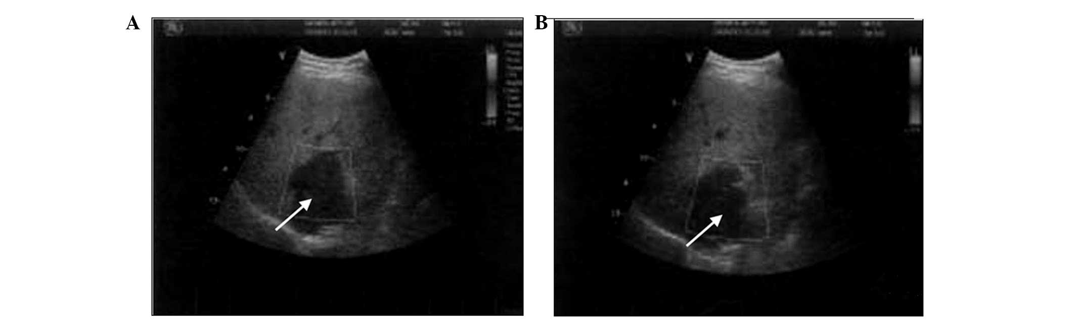
Abdominal ultrasonography (ClearVue 350; Philips Medical Systems, Best, the Netherlands) revealed two well-defined heterogeneous hypoechoic masses in the caudate lobe and the left lobe of the liver. The sizes of the two masses were 9.1×7.4×6.6 and 3.8×3.5×3.8 cm, respectively (Fig. 1).

Figure 1.

Ultrasound findings of primary liver leiomyosarcoma. (A) Two dimensional scanning showing a large heterogeneous hypoechoic mass (white arrow) in the caudate lobe of the liver. (B) Doppler flow imaging showing no blood flow signal.

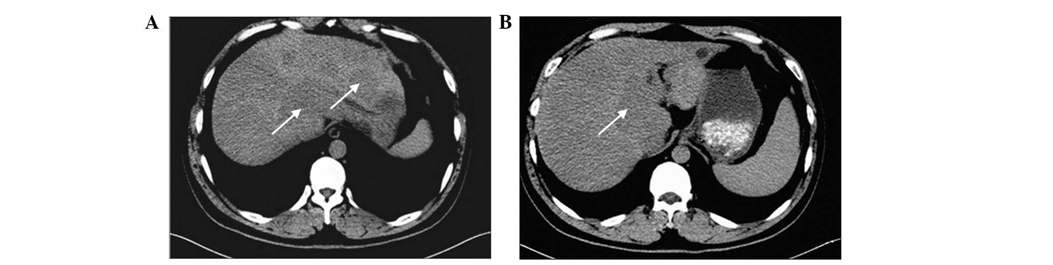
A plain CT (Discovery CT750 HD; GE Medical Systems Ltd., Milwaukee, WI, USA) scan disclosed two slight hypodense lesions with a definite margin, arising from the caudate lobe and the left lobe of the liver. Moreover, multiple hypodense lesions around the two larger lesions were observed. The inferior vena cava was compressed by the mass of the caudate lobe, but no enlarged lymph nodes were found (Fig. 2).

Figure 2.

Computed tomography (CT) findings of primary liver leiomyosarcoma. (A) Plain CT showing slightly low-density lesions (white arrow) with definite borders in the caudate and left lobes of the liver. (B) lower scan slice revealing a definite tumour border.

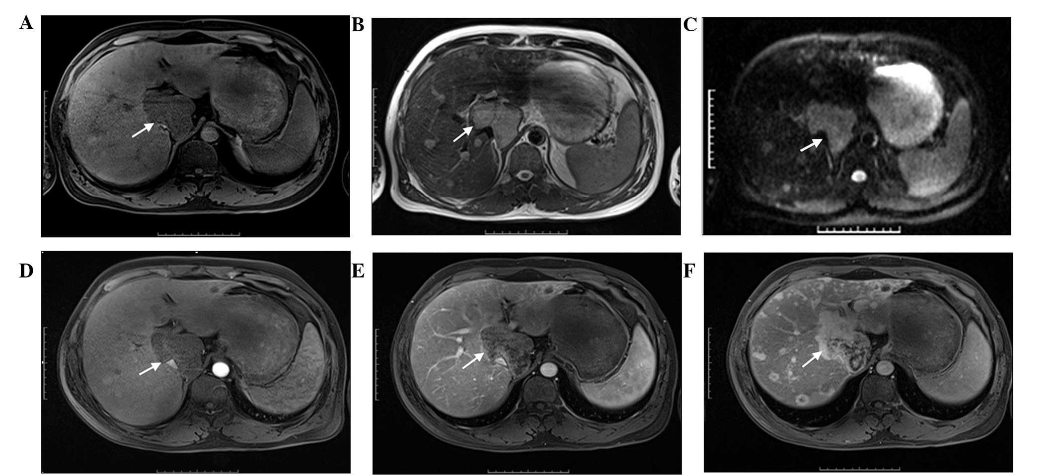
MRI was performed using a 3.0 Tesla whole-body MR hybrid system (Trio Time, Siemens, Germany) with regular pulse sequences. T1-weighted imaging (WI), T2WI, diffusion-WI (DWI) and gadopentetate dimeglumine-enhanced dynamic MRI were performed. The tumours presented with a heterogeneous low signal density on T1W1 (Fig. 3A) and a high signal density on T2WI (Fig. 3B). Furthermore, these tumours exhibited well-defined margins, but no evident capsule was observed. Satellite lesions were clearly shown on T2WI and DWI (Fig. 3B and C). On enhanced MRI (Fig. 3D–F), the lesions were not enhanced during the arterial and portal venous phases; by contrast, these lesions were evidently enhanced during the 5-min delayed phase.

Figure 3.

MRI findings of primary liver leiomyosarcoma. (A) On T1-weighted imaging (WI), the masses (white arrow) are well defined and slightly heterogeneous hypointensities. (B) On T2WI, the masses (white arrow) are heterogeneous hyperintensities. (C) On diffusion-WI, the masses (white arrow) display hyperintensity. (D) In the arterial phase of dynamic contrast-enhanced imaging, the masses (white arrow) show no enhancement. (E) In the venous phase of dynamic contrast-enhanced image, no evident enhancement can be observed. (F) In the 5-min delayed imaging, the majority of areas of the mass (white arrow) show marked enhancement. The intrahepatic metastases are also enhanced.

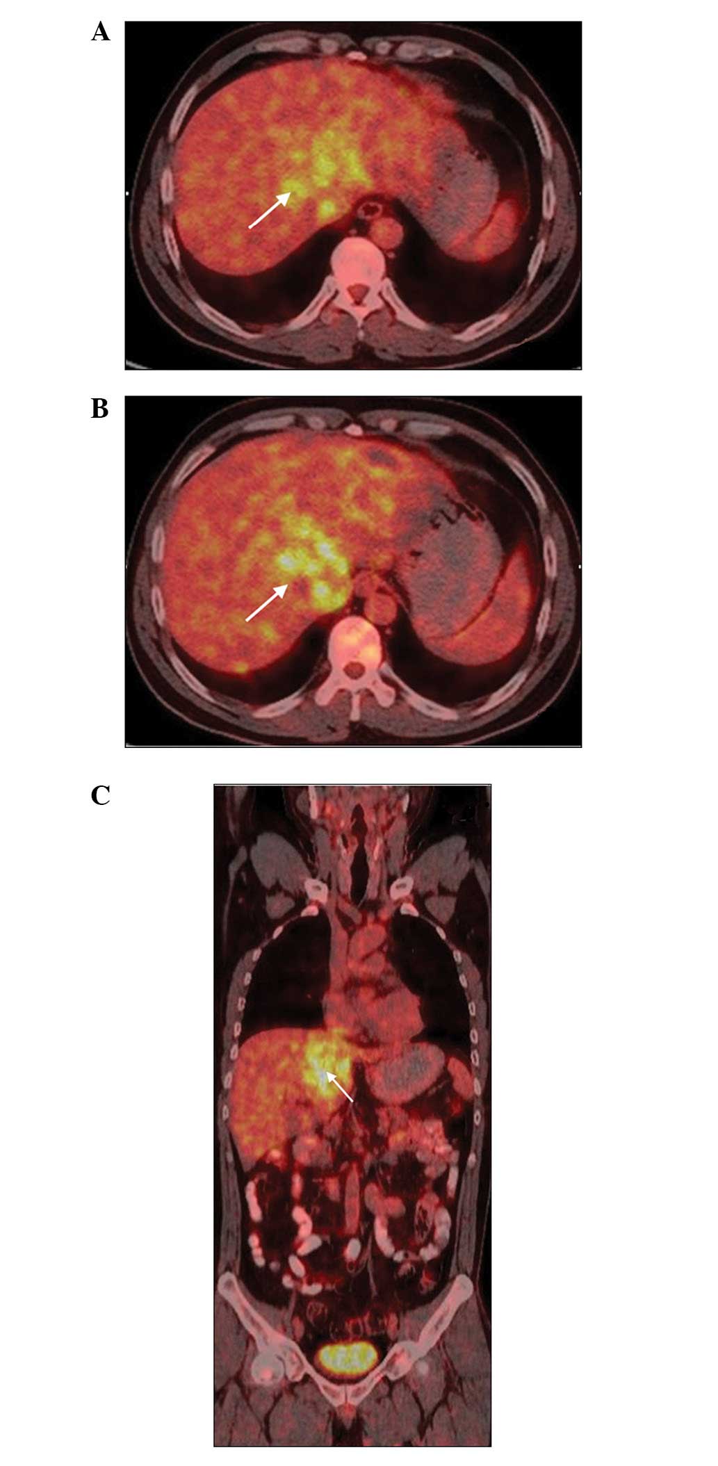
A whole body PET-CT (Biograph 16; Siemens Healthcare, Erlangen, Germany) scan was conducted to detect the masses and determine whether they were primary or metastatic. The scan spanned the base of the skull to the upper thighs. PET-CT showed multiple increased FDG uptake lesions in the liver; such lesions corresponded to the tumours revealed by CT and MRI. No evidence of metastasis from other organs was observed (Fig. 4).

Figure 4.

18F-fluorodeoxyglucose (FDG) positron emission tomography-computed tomography (PET-CT) findings of primary liver leiomyosarcoma. (A) PET-CT shows higher 18F-FDG uptake lesions on the liver (white arrow), corresponding to the tumour revealed by CT and MRI. (B) On the lower scan slice, multiple intrahepatic metastases are observed (white arrow). (C) On the whole body scan, no evidence of remote metastases is observed.

On the basis of the findings, a specific type of hepatic malignant tumour was suspected. This tumour was then 
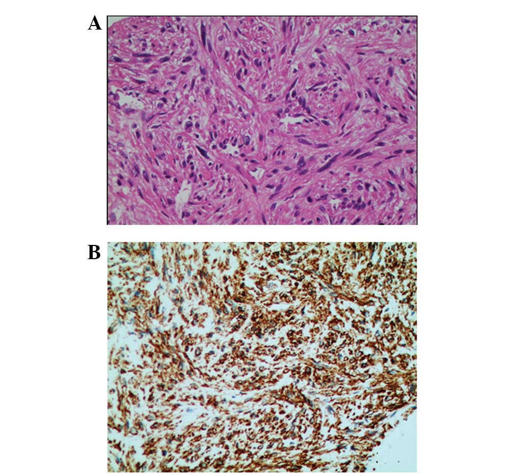
subjected to CT-guided transcutaneous puncture biopsy. Microscopic examination showed that the tumour was characterised by intersecting bundles of spindle-shaped cells, with eosinophilic cytoplasm and nuclear atypia. Immunohistochemical staining revealed that the tumour cells were positive for smooth muscle actin (SMA; Fig. 5) and desmin; by contrast, these tumour cells were negative for c-Kit (CD117), discovered on gastrointestinal stromal tumours 1 (DOG1), human melanoma black 45 (HMB45), PNL2 and Ki-67. These findings indicated the occurrence of PHL.

Figure 5.

Pathological findings of primary liver leiomyosarcoma. (A) Haematoxylin and eosin staining showing the proliferation of spindle cells, with hyperchromatic pleomorphic nuclei and eosinophilic cytoplasm, arranged in crosswise bundles (magnification, x400). (B) Immunohistochemical staining revealing tumour cells positive for smooth muscle actin (magnification, x400).

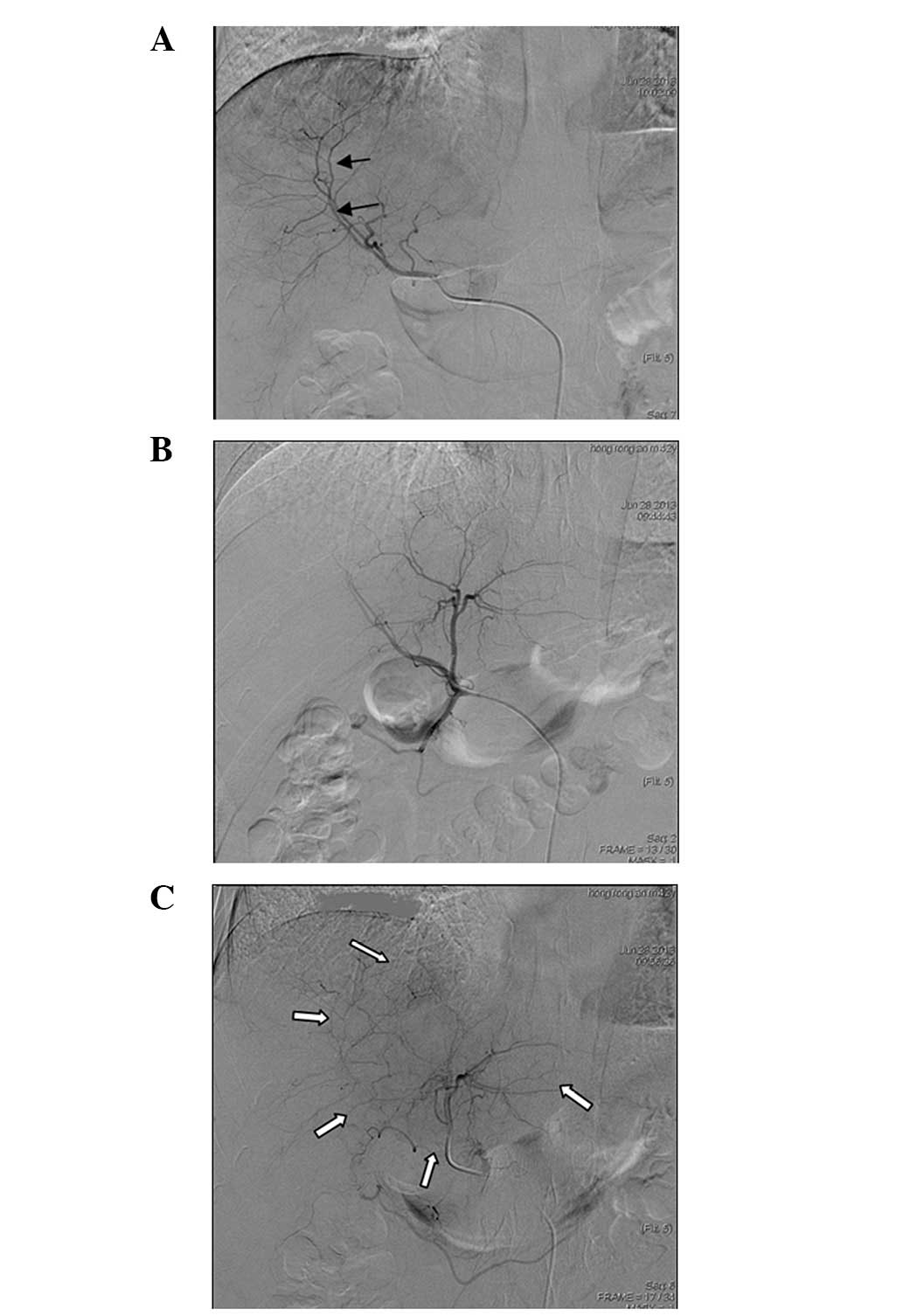
DSA (Innova 3100 system; GE Healthcare, Waukesha, WI, USA) of the hepatic artery was performed to administer chemotherapeutic drugs via transcatheter arterial infusion due to the loss of surgical removal indications. DSA showed that the branches of the hepatic artery that passed into the caudate lobe were slightly increased and depressed. No tumour stain was found during the arterial and portal venous phases, whereas weak stain was detected during the delayed phase (Fig. 6).

Figure 6.

Digital subtraction angiography findings of primary liver leiomyosarcoma. (A) Selective angiography of the hepatic artery shows no tumour stain in the caudate lobe, however compression and displacement of the vessel branches is observed (black arrow). (B) In the arterial phase of super-selective angiography, no obvious tumour stain is observed. (C) In the delayed phase, a weak tumour stain is observed (white arrows).

The patient underwent transarterial infusion (TAI) with 60 mg epirubicin and 800 mg carboplatin on day 1. On the second day, the patient recieved a 24 h intravenous infusion of 5 g/m2 ifosfamide and 5 g/m2 mesna. An additional 24 h infusion of 2 g/m2 mesna was administered on day 3. One cycle lasted for 7 days and a total of four cycles of treatment were administered over a period of four months. Follow-up examination eight months after the initation of TAI treatment revealed that the tumours had stabilized, according to the modified Response Evaluation Criteria in Solid Tumors guidelines (8). However, the patient succumbed to liver failure 384 days after the initiation of TAI treatment.

The present case report was approved by the Ethics Committee of the First Affiliated Hospital of Shandong University and written informed consent was obtained from the patient for publication of the case report and any accompanying images.

Discussion

The clinical manifestations of PHL are non-specific, and tumours are generally asymptomatic until they increase in size (9–11). Abdominal pain, weight loss, vomiting and jaundice are the common symptoms, while hepatomegaly and a palpable mass are frequently disclosed on physical examination. Acute bleeding is a rare symptom observed in patients with PHL (12). Liver function test results may be abnormal, but serological markers, such as α-fetoprotein, are normal. The absence of serological markers and non-specific clinical manifestations often delay diagnosis (2,7).

The CT findings of PHL reveal a large, well-defined and heterogeneous hypodensity mass with internal and peripheral enhancement (13), or a cystic mass with an enhanced thickened wall (1,14). In the present case, the plain CT scan showed two well-defined masses with heterogeneous hypodensity in the caudate lobe and the left lobe of the liver; multiple satellite lesions were also observed in the remaining portion of the liver. These findings are similar to those recorded in previously reported studies (1–4). However, an enhanced CT scan was not performed in the present case.

Soyer et al (5) reported the case of one patient with PHL who was subjected to MRI; the results showed that the tumours exhibited homogeneous or heterogeneous hypointensity in T1WI and hyperintensity in T2WI with occasional encapsulation. Similar MRI characteristics are also observed in the present case. In previous studies, the patients were subjected to unenhanced MRI. However, complete MRI sequences, including unenhanced MRI, DWI and dynamic contrast-enhanced MRI, were performed in the present case. The tumours in DWI showed hyperintensity, suggesting a limited diffusion of water molecules in the tumour. In the dynamic contrast-enhanced MRI, the masses were not evidently enhanced during the arterial and portal venous phases. However, the masses were markedly enhanced during the 5-min delayed imaging. This result could be a specific MRI characteristic of PHL.

No tumour stain was found during the arterial and portal venous phases, whereas weak stain was detected during the delayed phase of DSA. These findings are consistent with those of the enhanced MRI. Furthermore, these results could be used to differentiate PHL from hepatocellular carcinoma and liver haemangioma.

PET-CT can be applied to sensitively detect primary tumours and metastases. In the present case, multiple masses were found in the liver, with increased FDG metabolism. No lesions were detected in the other organs. Therefore, hepatic metastases could be excluded.

The histopathological diagnosis of PHL is characterised by the presence of uniform and diffuse infiltrates of spindle-shaped cells with hyperchromatic nuclei; the diagnosis is further confirmed by a positive immunohistochemistry reaction for SMA, desmin and vimentin, as well as a negative reaction for keratin and S-100 protein (15,16). In the present case, a microscopic examination showed intersecting bundles of spindle-shaped cells with eosinophilic cytoplasm and nuclear atypia. Moreover, immunohistochemistry revealed a positive reaction for SMA and desmin, and a negative reaction for c-kit receptor (CD117), DOG1, HMB45, PNL2 and Ki-67. Therefore, the diagnosis of PHL was confirmed.

In conclusion, in the present study, PHL was found to be a hypovascular tumour, which was not evidently enhanced during the arterial and portal venous phases on MRI, and exhibited no tumour stain on DSA. However, the lesions were evidently enhanced during the delayed imaging of enhanced MRI. These findings may reveal the imaging characteristics of PHL, however, further studies are required to confirm this. The microscopic characteristics of PHL included intersecting bundles of spindle-shaped cells with eosinophilic cytoplasm and nuclear atypia. Immunochemistry revealed a positive reaction for SMA, desmin, and vimentin.

References

1 

Ferrozzi F, Bova D, Zangrandi A and Garlaschi G: Primary liver leiomyosarcoma: CT appearance. Abdom Imaging. 21:157–160. 1996. View Article : Google Scholar : PubMed/NCBI

2 

Shamseddine A, Faraj W, Mukherji D, et al: Unusually young age distribution of primary hepatic leiomyosarcoma: case series and review of the adult literature. World J Surg Oncol. 8:562010. View Article : Google Scholar : PubMed/NCBI

3 

Civardi G, Cavanna L, Iovine E, et al: Diagnostic imaging of primary hepatic leiomyosarcoma: a case report. Ital J Gastroenterol. 28:98–101. 1996.PubMed/NCBI

4 

Gandhi MR, Wong DC and Wood DJ: Ultrasound and computed tomography appearances of primary hepatic leiomyosarcoma. Australas Radiol. 39:289–291. 1995. View Article : Google Scholar : PubMed/NCBI

5 

Soyer P, Blanc F, Vissuzaine C, et al: Primary leiomyosarcoma of the liver MR findings. Clin Imaging. 20:273–275. 1996. View Article : Google Scholar : PubMed/NCBI

6 

Chi M, Dudek AZ and Wind KP: Primary hepatic leiomyosarcoma in adults: analysis of prognostic factors. Onkologie. 35:210–214. 2012. View Article : Google Scholar : PubMed/NCBI

7 

Tsai PS, Yeh TC and Shih SL: Primary hepatic leiomyosarcoma in a 5-month-old female infant. Acta Radiol Short Rep. 2:20479816134987222013.PubMed/NCBI

8 

Lencioni R and Llovet JM: Modified RECIST (mRECIST) assessment for hepatocellular carcinoma. Semin Liver Dis. 30:52–60. 2010. View Article : Google Scholar : PubMed/NCBI

9 

Holloway H, Walsh CB, Thomas R and Fielding J: Primary hepatic leiomyosarcoma. J Clin Gastroenterol. 23:131–133. 1996. View Article : Google Scholar : PubMed/NCBI

10 

Rosas JS, Del Rosario A, Bui HX, et al: Primary hepatic leiomyosarcoma in a child with the acquired immunodeficiency syndrome. Hum Pathol. 23:69–72. 1992. View Article : Google Scholar : PubMed/NCBI

11 

Brichard B, Smets F, Sokal E, et al: Unusual evolution of an Epstein-Barr virus-associated leiomyosarcoma occurring after liver transplantation. Pediatr Transplant. 5:365–369. 2001. View Article : Google Scholar : PubMed/NCBI

12 

Jeong TY, Kim YS, Park KJ, et al: A case of primary leiomyosarcoma of the liver presenting with acute bleeding. Korean J Gastroenterol. 51:194–198. 2008.(Korean). PubMed/NCBI

13 

Gates LK Jr, Cameron AJ, Nagorney DM, et al: Primary leiomyosarcoma of the liver mimicking liver abscess. Am J Gastroenterol. 90:649–652. 1995.PubMed/NCBI

14 

Yu RS, Chen Y, Jiang B, et al: Primary hepatic sarcomas: CT findings. Eur Radiol. 18:2196–2205. 2008. View Article : Google Scholar : PubMed/NCBI

15 

Smith MB, Silverman JF, Raab SS, et al: Fine-needle aspiration cytology of hepatic leiomyosarcoma. Diagn Cytopathol. 11:321–327. 1994. View Article : Google Scholar : PubMed/NCBI

16 

Sprogøe-Jakobsen S and Hølund B: Immunohistochemistry (Ki-67 and p53) as a tool in determining malignancy in smooth muscle neoplasms (exemplified by a myxoid leiomyosarcoma of the uterus). APMIS. 104:705–708. 1996. View Article : Google Scholar : PubMed/NCBI

Related Articles

  • Abstract
  • View
  • Download
  • Twitter
Copy and paste a formatted citation
Spandidos Publications style
Lv WF, Han JK, Cheng DL, Tang WJ and Lu D: Imaging features of primary hepatic leiomyosarcoma: A case report and review of literature. Oncol Lett 9: 2256-2260, 2015.
APA
Lv, W., Han, J., Cheng, D., Tang, W., & Lu, D. (2015). Imaging features of primary hepatic leiomyosarcoma: A case report and review of literature. Oncology Letters, 9, 2256-2260. https://doi.org/10.3892/ol.2015.3006
MLA
Lv, W., Han, J., Cheng, D., Tang, W., Lu, D."Imaging features of primary hepatic leiomyosarcoma: A case report and review of literature". Oncology Letters 9.5 (2015): 2256-2260.
Chicago
Lv, W., Han, J., Cheng, D., Tang, W., Lu, D."Imaging features of primary hepatic leiomyosarcoma: A case report and review of literature". Oncology Letters 9, no. 5 (2015): 2256-2260. https://doi.org/10.3892/ol.2015.3006
Copy and paste a formatted citation
x
Spandidos Publications style
Lv WF, Han JK, Cheng DL, Tang WJ and Lu D: Imaging features of primary hepatic leiomyosarcoma: A case report and review of literature. Oncol Lett 9: 2256-2260, 2015.
APA
Lv, W., Han, J., Cheng, D., Tang, W., & Lu, D. (2015). Imaging features of primary hepatic leiomyosarcoma: A case report and review of literature. Oncology Letters, 9, 2256-2260. https://doi.org/10.3892/ol.2015.3006
MLA
Lv, W., Han, J., Cheng, D., Tang, W., Lu, D."Imaging features of primary hepatic leiomyosarcoma: A case report and review of literature". Oncology Letters 9.5 (2015): 2256-2260.
Chicago
Lv, W., Han, J., Cheng, D., Tang, W., Lu, D."Imaging features of primary hepatic leiomyosarcoma: A case report and review of literature". Oncology Letters 9, no. 5 (2015): 2256-2260. https://doi.org/10.3892/ol.2015.3006
Follow us
  • Twitter
  • LinkedIn
  • Facebook
About
  • Spandidos Publications
  • Careers
  • Cookie Policy
  • Privacy Policy
How can we help?
  • Help
  • Live Chat
  • Contact
  • Email to our Support Team